Researchers Explore Regeneration in Critical Layer of Cornea
December 12, 2016
 On the back side of the cornea is a single layer of cells that play an all-important role, maintaining just the right fluid balance to keep the cornea transparent so light can enter the eye.
Until recently, it was believed this layer, called the corneal endothelium, is incapable of replacing its damaged cells. As more cells become damaged, the cornea becomes opaque, leading to loss of vision and, ultimately, to as many as 30,000 endothelium transplants a year in the United States alone.
University researchers, including a team from Prof. Jannick Rolland's lab at The Institute of Optics, is exploring the possibility that stem cells on the outer edges of the cornea, given the right stimulation, can migrate into the endothelium to replace damaged cells. (Undifferentiated stem cells develop into specialized cells.) The work raises the possibility of restoring vision without the need for transplants.
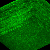
The image, from the lab of Jannick Rolland, the Brian J. Thompson Professor of Optical Engineering, shows a cross section of the cornea. The bottom layer is the corneal endothelium. The image was obtained using Gabor domain optical coherence microscopy, a novel imaging technology developed in the Rolland lab, which allows rapid, noninvasive imaging of cellular structures beneath the surface of the skin or within the human eye. (University photo / Jannick Rolland)
Read the full story at the UR Newscenter.