Spider Silk
An Electron Microscopic Study
Spider Silk is a natural fiber secreted by spiders for the prodcution
of webs and egg sacs as well as transportation. The silk is secreted from glands
inside the spiders spinnerets, located on the back of a spiders abdomen. Spider
silk is renouned for being stronger than steal by mass and is surprisingly elastic
and has generated interrest for an array of applications. Allegedly, these properties
are a result of both its structure and chemical make up. One source claims that
the structure is a combination of crystaline sections linked by irregular elastic
amino acids. (Wikipedia)
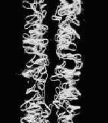
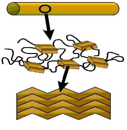
Figure 1:Spider Silk Structure Figure 2:Structure Schematic
(http://www.xs4all.nl/~ednieuw/Spiders/InfoNed/webthread.html)
http://en.wikipedia.org/wiki/Spider_silk#Properties
The most common amino acids are thought to be glycine (C2H5NO2
) and alanine (C3H7NO2).
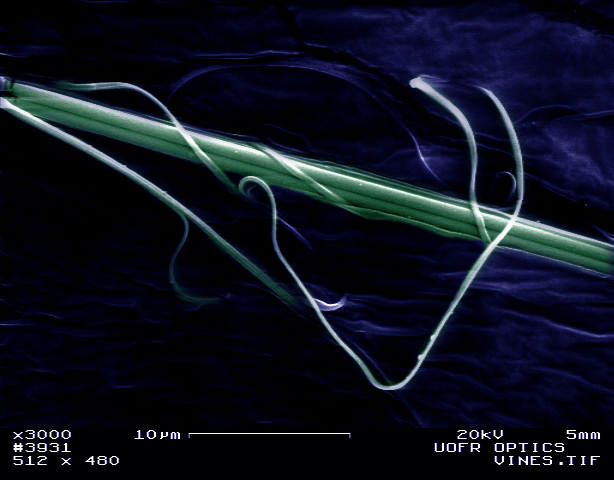
The size of spider silk, around 1 um in diameter, makes visual investigation
impossible. This project is an exploration of spider silk with an electron microscope.
Back to Top
Methods
I. Sample Collection
Silk was collectd from common spiders from around the University of Rochester
campus as well as near by parks. A sample stub was prepared with double sided
tape and then pushed through the webs. For comparrison, spider silk was also wrapped
around a human hair (special thanks to my roomate for his generous contributions
to science) before being placed on the stub. One of the spiders who produced
the web for this project was also collected.
II. Sample Preperation
Silk samples were first prepared by painting the edges of the stub with a conductive
carbon-based adhesive. Then, specimens were coated with a gold and palladium
alloy in a sputter coater for various lengths of time from thirty seconds to
two minutes.
The spider was first submerged in gluderaldehide
to fix the sample. Then the sample was submerged in Hexamethyldisilazane (HMDS),
a chemcial used to dry the sample without destroying soft tissues. After the
HMDS evaporated off, the sample was mounted on a sample stup and sputter coated
as described for the silk samples.
III. Data Collection [Click on a Section Title
to see those results]
The University of Rochester's LEO 982 FE-SEM was the
instrument used to collect the raw data for this project.
A. Secondary Electron Images were
taken with various microscope parameters. These parameters can be viewed in the display bar
of the micrograph images. In addition, the signal mixer was used to mix the in-chamber
and in-lens SE detectors which provides for more control in signal processing. Some
of the microscopes software was utilzed in making point-to-point measurements
of sample features.
B. Anaglyphs were
created of the spiders spinneretts. In order to create the anaglyphs, two images
need to be taken. The first image is taken and a distinct feature at the center
of the screen is marked with a dry erase marker. Then, the sample stage is tilted
about 4 degrees. Then, the sample is repositioned so that the distinct feature
lines back up with the mark made on the screen and a second image is captured. The
images were then processed in Photoshop in the following way. Both images are
converted into RGB color format and the zero tilt image is given a red hue and the
tilted image is given a blue hue. Then, the images were layered one on top of
the other and the opacity of the top image is adjusted to 50% so that the images
are superimposed.
C. Colorization
of micrographs was done using Adobe Photoshop. This is more artistic than anything
else.
D. X-ray Analysis
was conducted on the silk as well as a control group of just the sample stub and
tape. The SEM's EDAX detector was utilized along with the help of the software
included.
E. The Electron Flight Simulator program
was also used to provide some insight on the beam interaction with the sample. For
the purposes of the simulation, the spider silk was modelled as a carbon based
organic compound about a micron thick, as per the direction of Brian McIntyre. The model
also includes a 10 nanometer layer of Gold and Palladium alloy on top of the spider
silk model to simulate the sputter coating. Part of the reason for this anaylsis
was to invesitgate the problem with the webs being destroyed by the electron
beam.
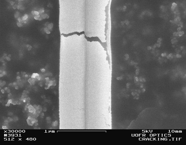
Figure3: Micrograph of a spider silk fiber being destroyed
by the electron beam.
Back to Top
Results and Discussion
[Click
on a Section Titles to go back to the corresponding Methods]
A. Secondary Electron Images
Taking images of just the spider silk gives a good idea of the structure
and size of the silk. During the course of the project I saw single-stranded
silk, double-stranded (above) silk and multi-stranded silk.
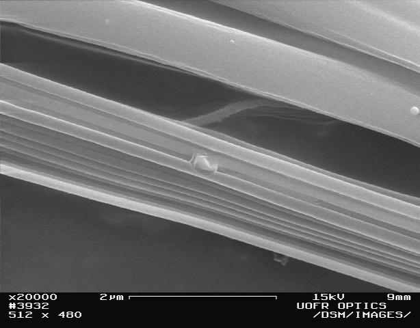
Figure 4: Multi-Standed and Single-Stranded Spider Silk (From
the Same Spider)
To be a little more quantitative...
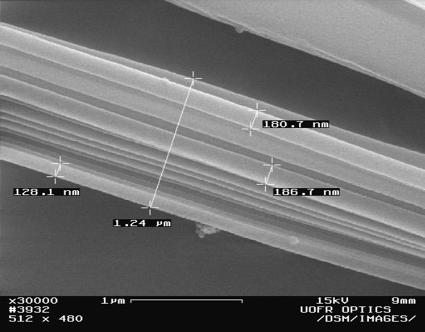
Figure 5: Multi-Standed
Fiber, Measured with SEM software
And my favorite:
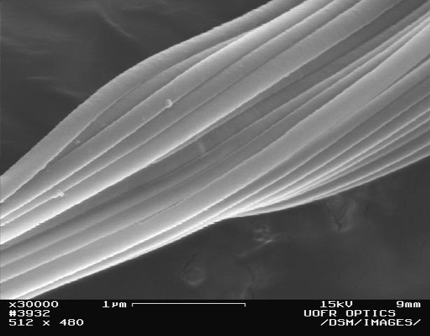
Figure 6: Multi-Stranded Silk. I count 17 strands that can be seen,
but there must be more.
To give you a better sense of the size, here
are micrographs of spider silk on human hair (thanks again to Jim Morphis):
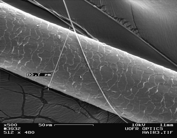
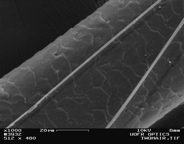
Figure 7: Spider Silk laid over human hair.
Now for where the silk comes from.
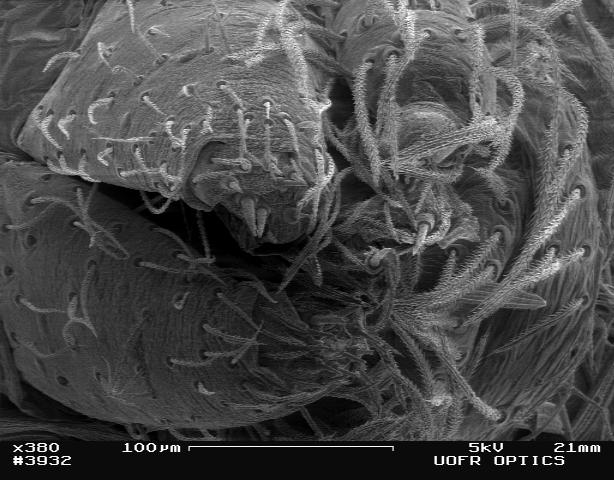
Figure 9: Spinnerrette Arrrays on the back of the spider's abdomen.
A little closer...
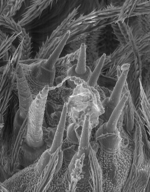
Figure 10: Several spinnerettes in an array.
And a little closer....
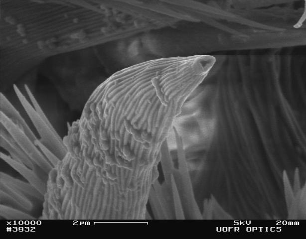
Figure 11: A single spinnerette
B. Anaglyphs
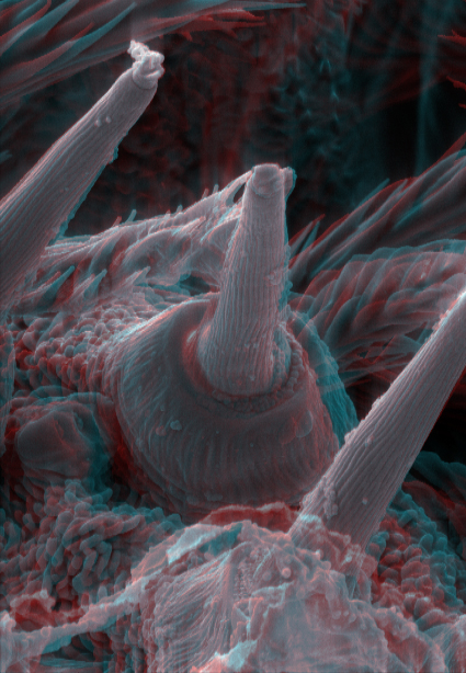
Figure 12: Spinnerette Anaglyph
After putting on 3-D glasses, try focusing on smaller objects in the background
until your brain superimposes the images, creating a3-D effect.
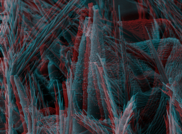
Figure 13 : Spinnerette Anaglyph 2
Cool. They work okay. This is trickey for high mag images.
C. Colorization
Figure 14: Spider Web Art
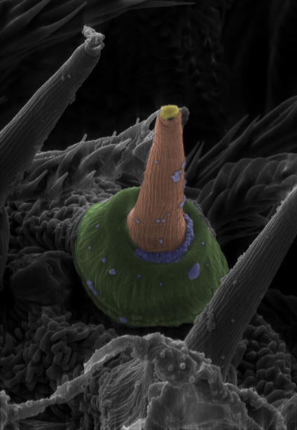
Figure 15: Spinnerette Colorized
D. X-ray Analysis
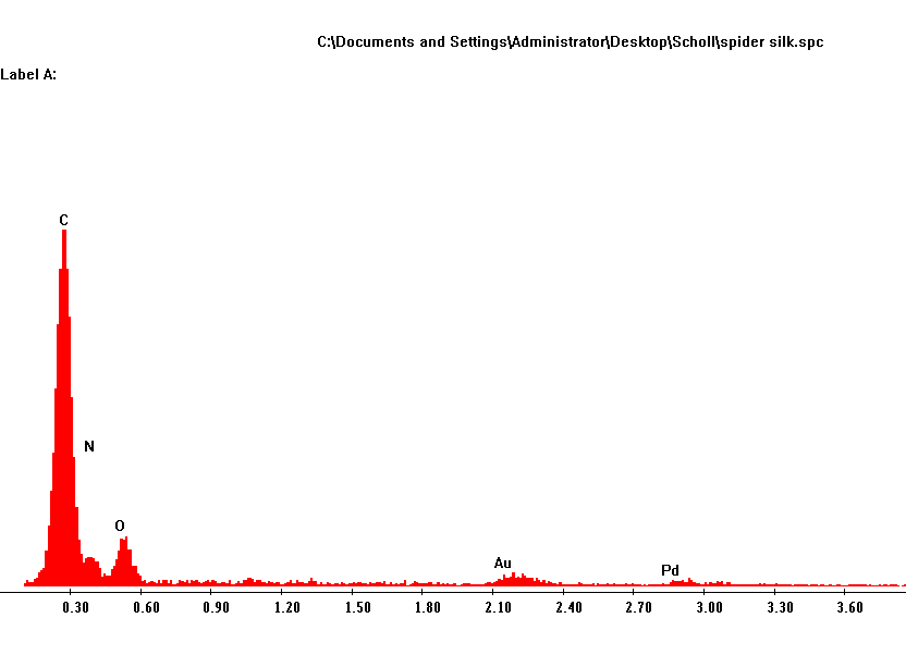
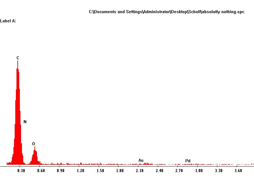
Figure 16: X-ray spectrum from Spider Silk Figure 17:
X-ray Spectrum from Control Group
By comparing the x-ray spectrum of the spider silk to that of the control group
we can see that the only significant difference is that the spider silk spectrum
has a small nitrogen signal at base of the carbon signal. The carbon, oxygen,
gold, and palladium signals can be considered to come mostly from the stub,
tape, and coatings. The presense of Nitrogen is in agreement with the background
information that the amino acids glycine (C2H5NO2
) and alanine (C3H7NO2) are present in spider
silk.
E. The Electron Flight Simulator
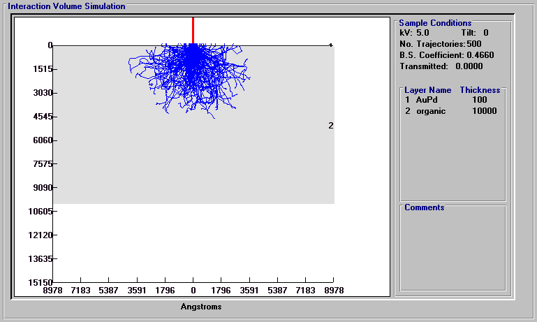
Figure 18: EFS, 5kV Accellerating Voltage
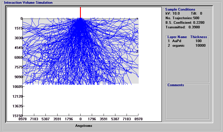
Figure 19: EFS, 10kV Accellerating Voltage
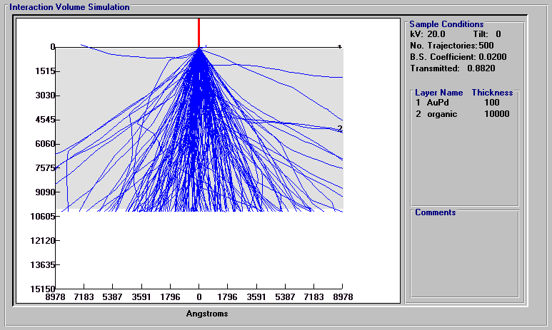
Figure 20: EFS, 20kV Accellerating Voltage
The analysis shows that a 5 kV accellerating voltage causes the beam to penetrate
into the sample only about 450 nm. The 10 kV beam penetrates all the way through
the sample, but a lot of the energy is dissipated into the silk. The 20 kV beam
penetrates through the sample again, but this time with much less difflection
into the sample and so much less energy dissipated into the spider silk.
Back to Top
Conclusion
Spider Silk is some pretty cool stuff. Its diameter varies
but it generally around a micron in diameter, about 80 times smaller than the human
hair donated by my roomate, Jim. It can be made up of a single strand or several
strands, each as small as 100 nanometers. X-ray analysis confimed the presense
of nitrogen, which is in agreement with the theory that the silk is composed
of the amino acids glycine and alanine. It's fragility may require selecting low
energy electrons to prevent damaging the sample or high energy electrons which
pass through the sample readily. Sputter coating, of course, is also useful. I
had also never seen the spinnerets of a spider before and did not realize how
many there were. There was a lot of interresting structure to see on the spide
itself.