
Banjos are one of the coolest instruments to play and to listen to. The tonal quality of the banjo is unique and awesome due partly to the strings that are used. Banjos use a total of five strings, four that are simply a round core and one that is a core surrounded by a winding, a wound string. There are a number of different types of strings used for banjo playing, differing in composition, coatings, winding types, and gauges. But what do these strings actually look like close-up and what exactly is their chemical composition and structure? Scanning electron microscopy (SEM) can be used to discover the answers to these questions.
SEM is a type of electron microscopy that provides images of a sample using a focused electron beam. The incident electrons interact with the sample to produce various signals whose detection leads to information about the sample surface and composition. SEM can produce high resolution and high magnification images, allowing for imaging of sub-micron features.
Secondary electron (SE) imaging uses low energy, inelastic secondary electrons that escape from the uppermost portion of the interaction volume. These electrons reveal details about the sample surface as they are generated from near the surface of the specimen. Conversely, backscattered electron (BSE) imaging uses higher energy, elastic backscattered electrons that escape from deeper in the interaction volume. Because these backscattered electrons are coming from deeper in the sample, BSE imaging can provide information about the atomic composition of the sample, mainly BSE intensity increasing with increasing atomic number. In addition to the secondary and backscattered electrons, x-rays can be used in SEM. Characteristic x-rays are formed when inner shell electrons are ejected by the primary beam and outer shell electrons then fall to fill the vacancies. The differential energy is released as an x-ray of characteristic energy defined by the energy level differences of the orbitals. X-rays are high energy and thus can escape from very deep in the interaction volume leading to identification of the different elemental components of the sample.
Using both SE and BSE detection along with x-ray detection, the structure and composition of banjo strings can be discovered. In this project, two types of banjo strings were studied with SEM, "Martin Vega Banjo Strings" (Martin strings) and "Elixir Polyweb Banjo Strings" (Elixir strings). The Martin strings are advertised as having silvered steel cores and nickel windings while the composition of the Elixir strings is not provided. Both types of strings have a reported gauge size of 0.023 in/ 0.58 mm for the wound string. Through this study, the structure and composition of both types of wound strings can be compared. SE imaging was used to image the surface structure of the strings, BSE imaging was used to image the compositional information of the strings, and x-ray analysis was used to identify the elements present in the strings. After imaging, image colorization was performed and 3D anaglyphs were created, both processes performed in Adobe Photoshop.

Two different types of banjo strings were studied. The first was a cheaper Martin brand while the second was a slightly more costly Elixir brand. The Elixir brand claims to provide strings that last longer and sound better. The Martin strings were reported to have a stainless steel core and nickel winding while the compositional information was not reported for the Elixir strings. The gauge sizes of the strings were reported as being the same for the two brands of strings.
Looking at a banjo string in the SEM is pretty simple- not much effort is needed in the sample preparation area. The strings are conductive and ready to go for SEM. The side view samples were simply prepared by cutting a piece of the string and sticking it side-down onto carbon tabe on a sample stub.
However, to take a look at the cross section of a string requires a bit more work. Short pieces of the strings were affixed vertically in epoxy within a metal cap, and then the ends were polished until smooth using the "Polisher" at the University of Rochester. Because the epoxy is not conductive, it was covered with carbon paint and then the whole cross section sample was sputter coated with a thin layer of gold using the Denton Vacuum sputter coater at the Institute of Optics at the University of Rochester.
Light microscopy was performed with the Olympus BX51 Light Microscope and all SEM imaging and x-ray analysis was peformed using the Zeiss Auriga Crossbeam SEM, both microscopes belonging to the Institute of Optics at the University of Rochester.
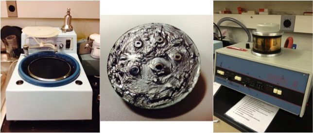
"The Polisher" (left) and the Denton Vacuum sputter coater (right) at the Institute of Optics at the University of Rochester and the cross section sample after polishing and painting with carbon paint (middle)

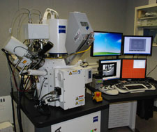
The Olympus BX51 Light Microscope (left) and the Zeiss Auriga SEM (right) at the Institute of Optics at the University of Rochester
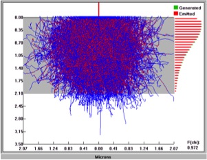
Electon flight simulation was performed using EDS Software owned by the Institute of Optics at the University of Rochester. As shown here, the electron and x-ray trajectories were simulated for a thick nickel coating on iron (side view of the string). As can be seen from the simulations, only a small number of iron x-rays are generated but a large number of nickel x-rays are created when the electron beam hits the side of the string (a 2.1 micron layer of nickel on iron). Simulations also showed the penetration depth changing depending on the material being studied. For example, simulations for bulk iron, bulk nickel, and bulk tin gave penetration depths of approximately 1.70 microns, 3.50 microns, and 4.10 microns, respectively. All simulations were performed with an accelerating voltage of 20.0 kV.

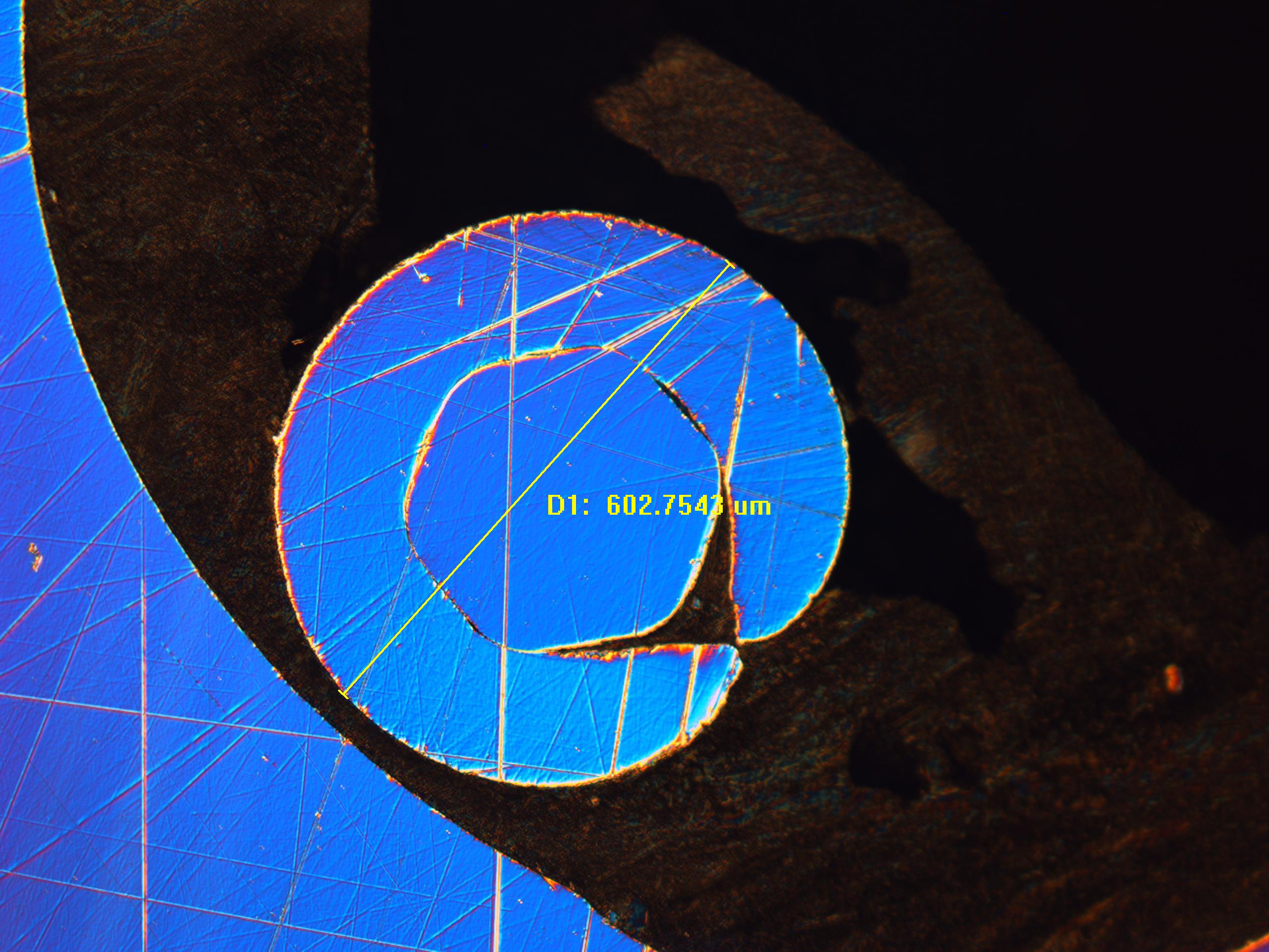
Light microscopy provided nice images of the cross section samples but proved to be insufficient for the side view of the strings. The depth of field was just not great enough to provide a clear image across the whole sample.
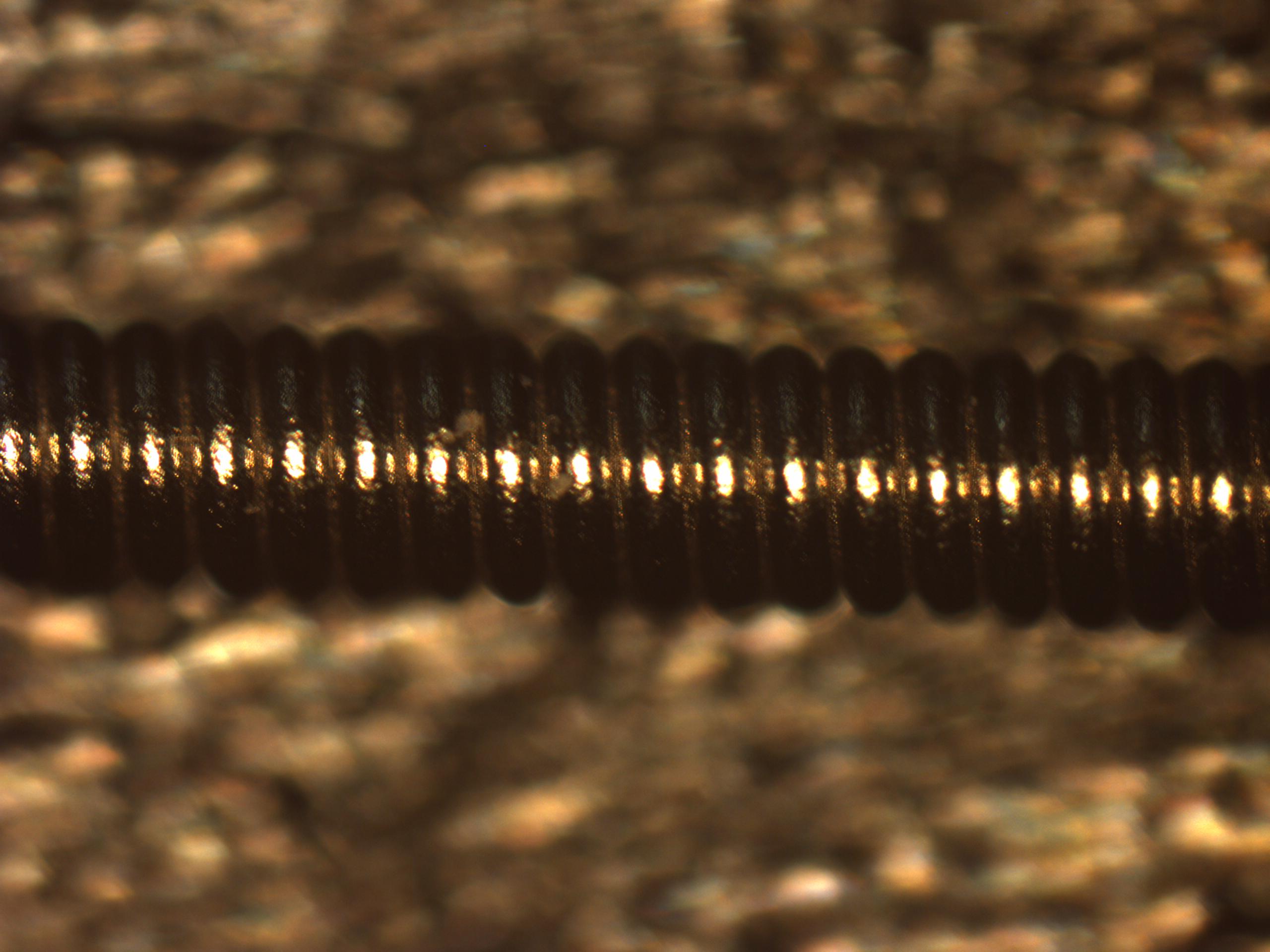
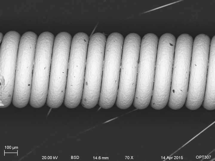
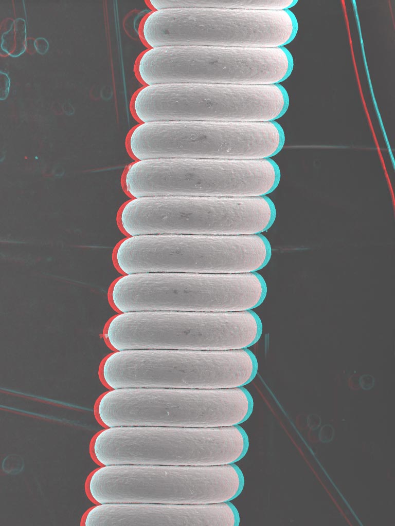
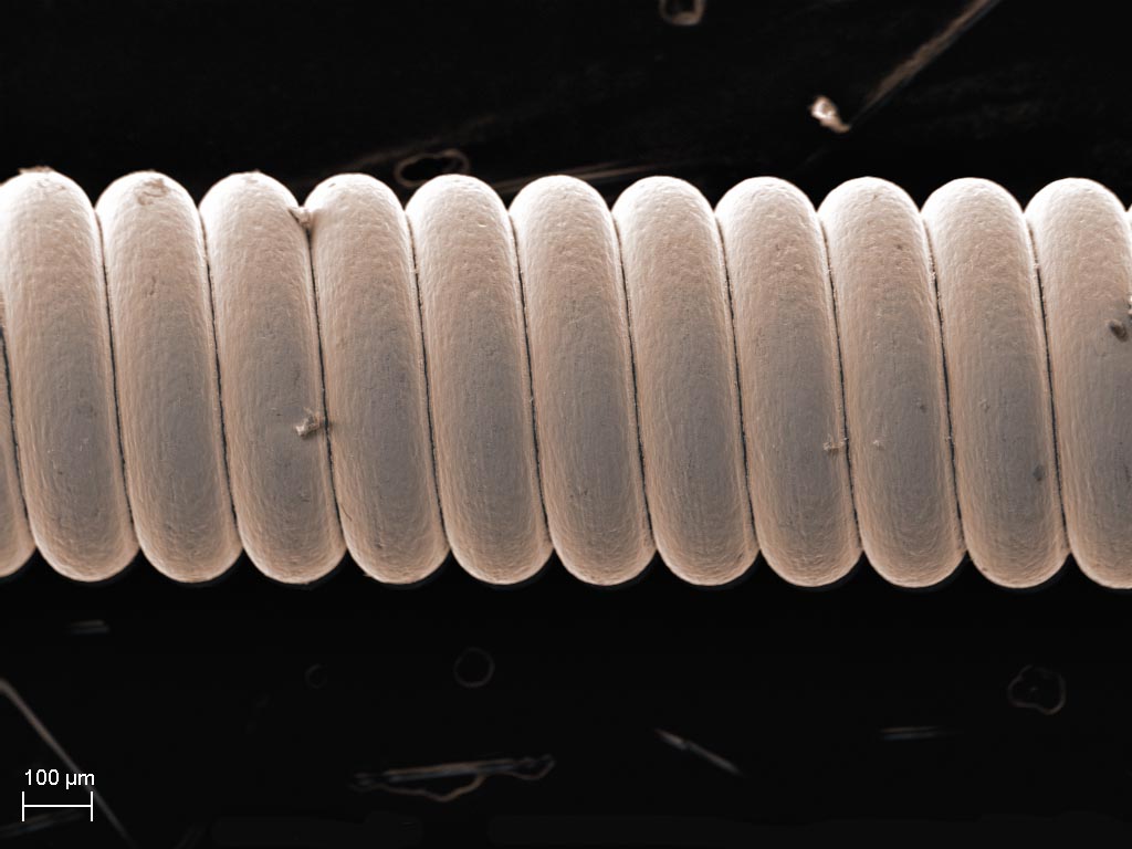
Side view
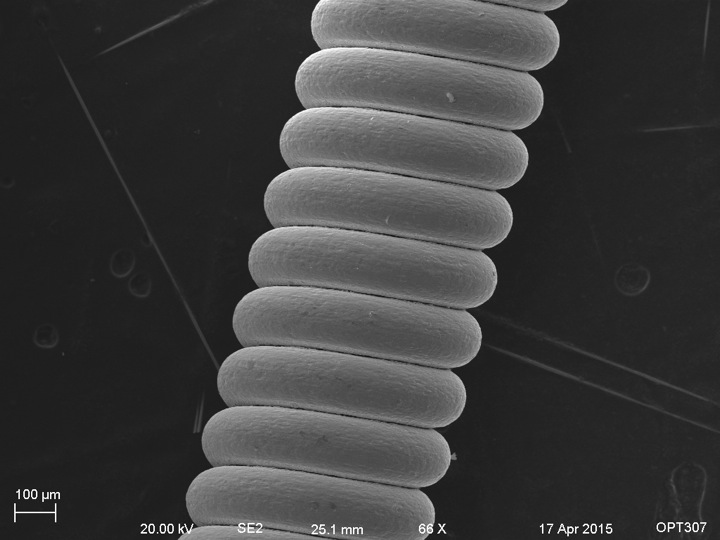
Side view of Elixir wound string
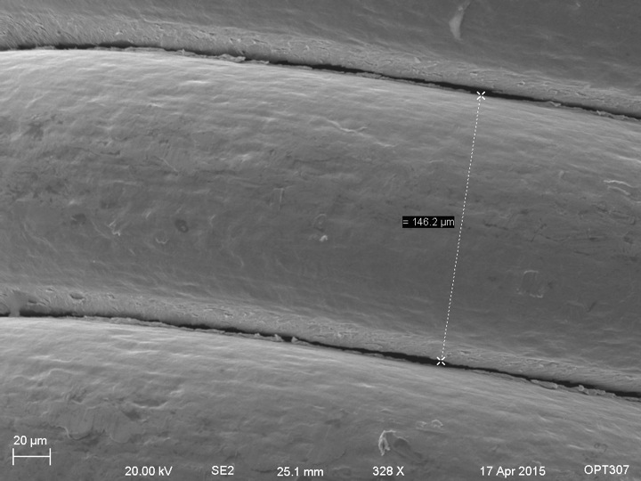
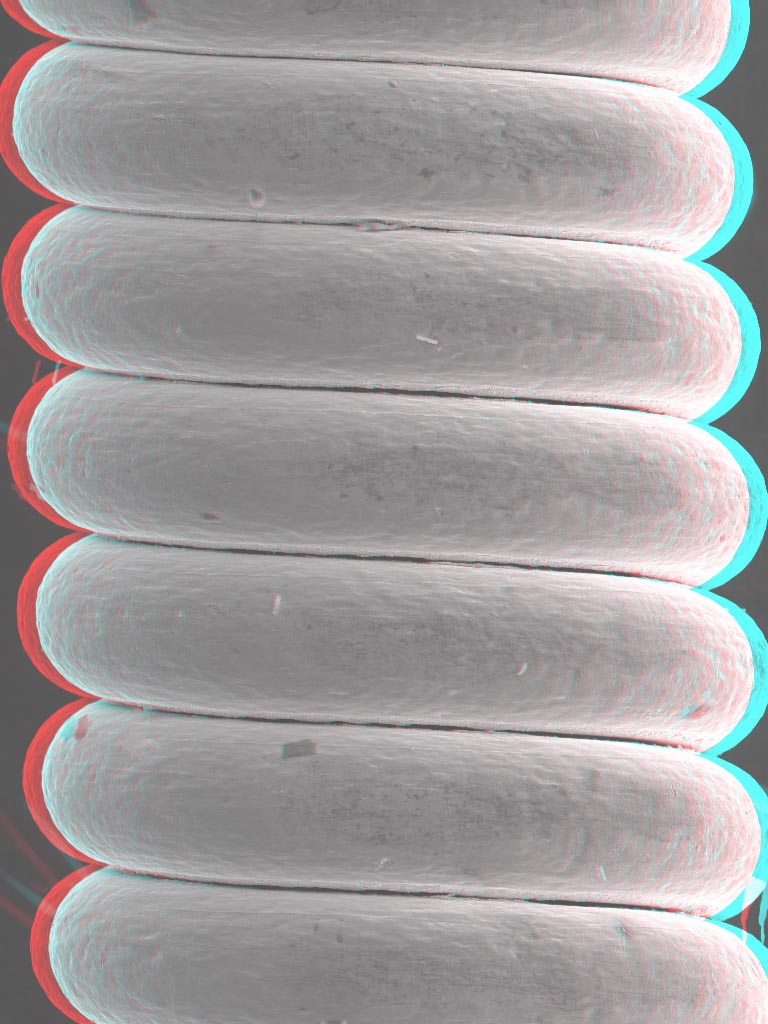
Closer look at the spaces between the windings
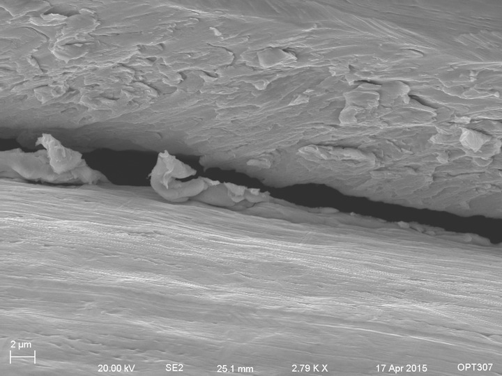
Even closer look at the spacing between windings
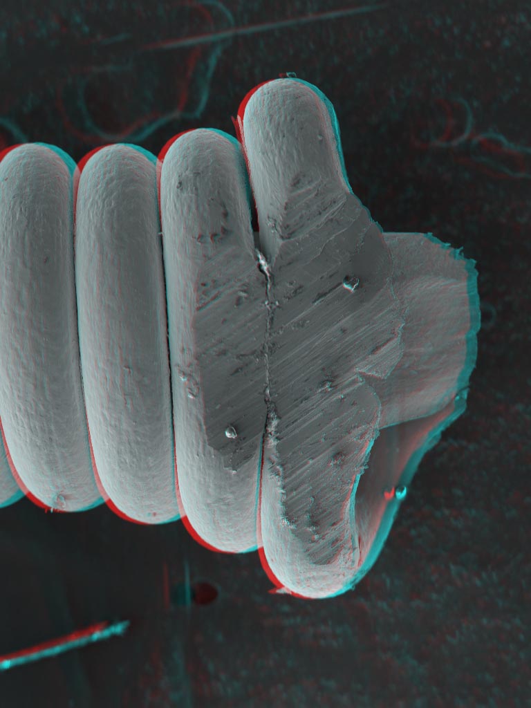
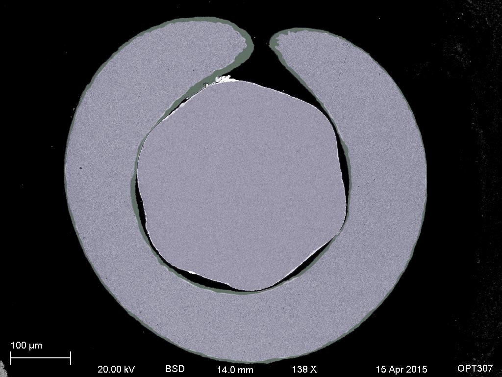
Cross section
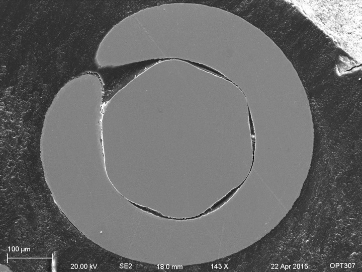
Cross section of Martin wound string
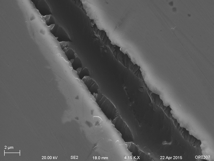
Close up view of the space between the core and the winding of the Martin wound string
BSE side view of Martin wound string
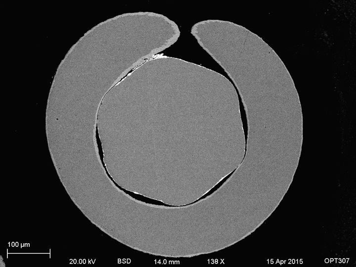
BSE image of Martin string cross section showing three different colors and thus three different materials
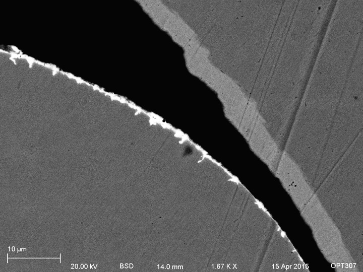
BSE closer look at the three different materials of the Martin string cross sectional sample
The x-ray spectra were obtained with the Zeiss Auriga SEM with attached EDAX energy dispersive x-ray spectrometer. X-ray analysis of the wound strings showed the cores were made of iron with a thin layer of tin and a bit of copper mixed in on the exterior and the windings were composed of iron with a coating of nickel on the outside. Interestingly, this was the case for both the Martin and the Elixir strings; the strings had nearly identical compositions.
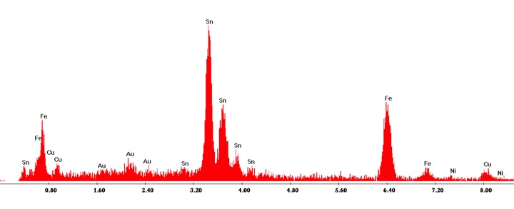
EDAX Analysis for outside portion of the core of the wound strings
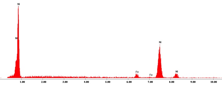
EDAX Analysis for side view of wound strings
Anaglyphic 3D images were created using two SE images differing only in a slight tilt, approximately 4 degrees, of the sample. The images were superimposed and then colorized with Adobe Photoshop to create the 3D effect.
Cool 3D effect!
Images were colorized using Adobe Photoshop.
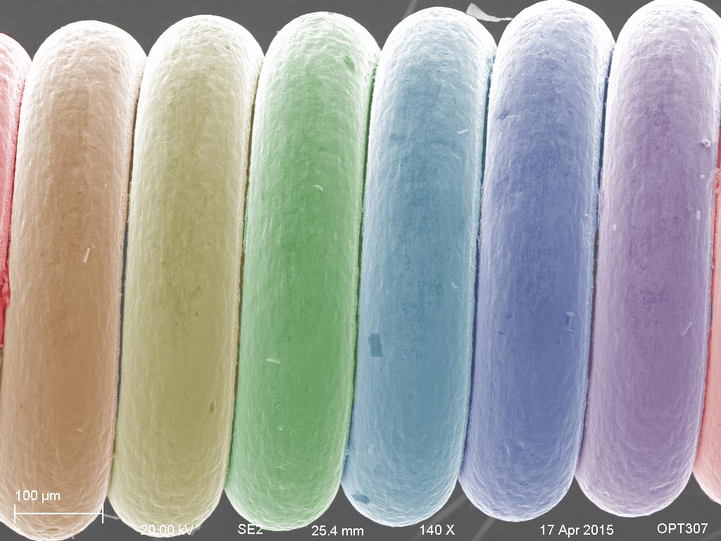
Pretty colors!