Methods and Results
1. Microtomy
Sample material was previously fixed within solid resin blocks following a routine method for microtomy. These blocks were then sliced via an ultramicrotome to approximately 100 nanometer thicknesses. Resulting slices were distrbuted onto TEM grids for observation.
2.HMDS
Selected sample material was preserved and prepared for TEM observation via HMDS techniques. To begin the process the sample was first fixed, after distributing onto TEM grids, using gluteraldehyde. Water in the sample was then replaced through a series of graded ethanol washes (95 and 100 percent ethanol). After the water was replaced, the samples were then placed under a fumehood and dried using HMDS.
3. Bright Field
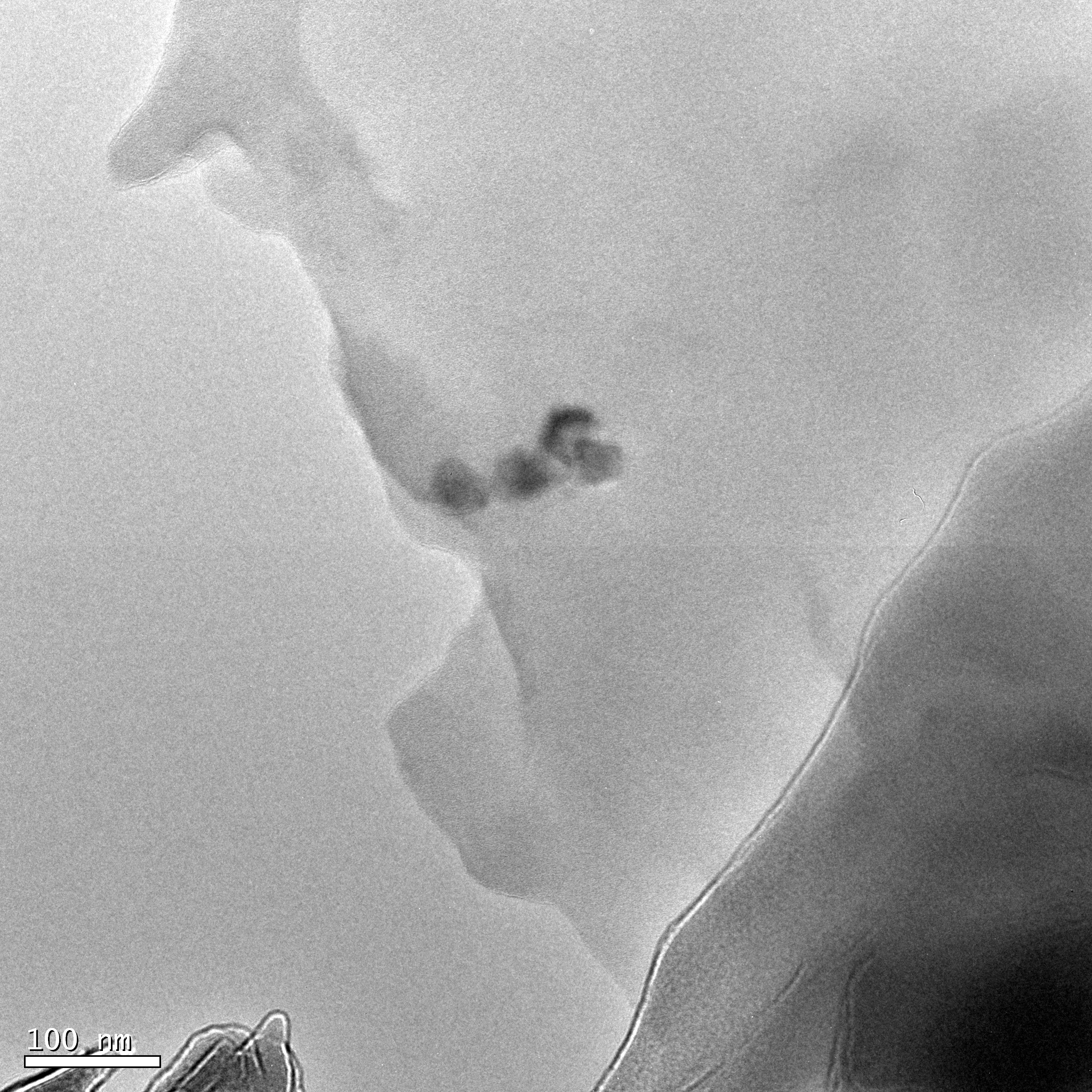
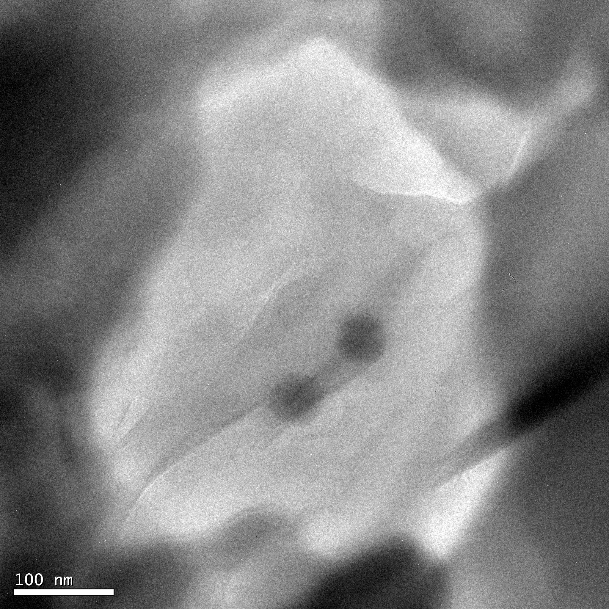
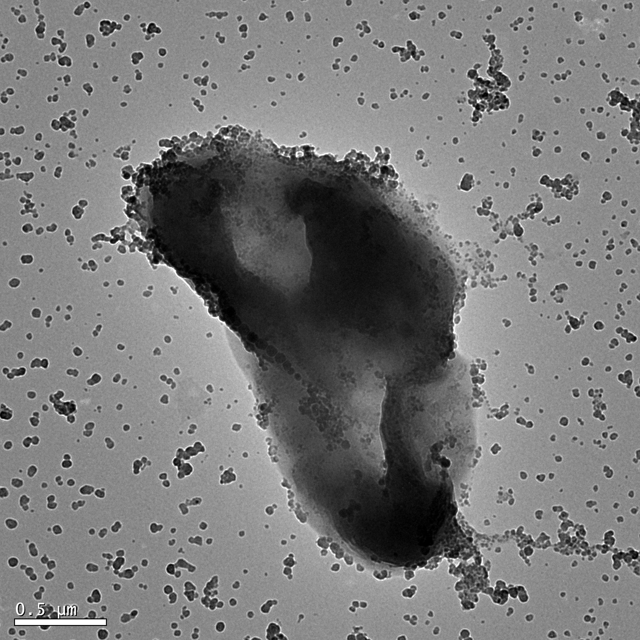
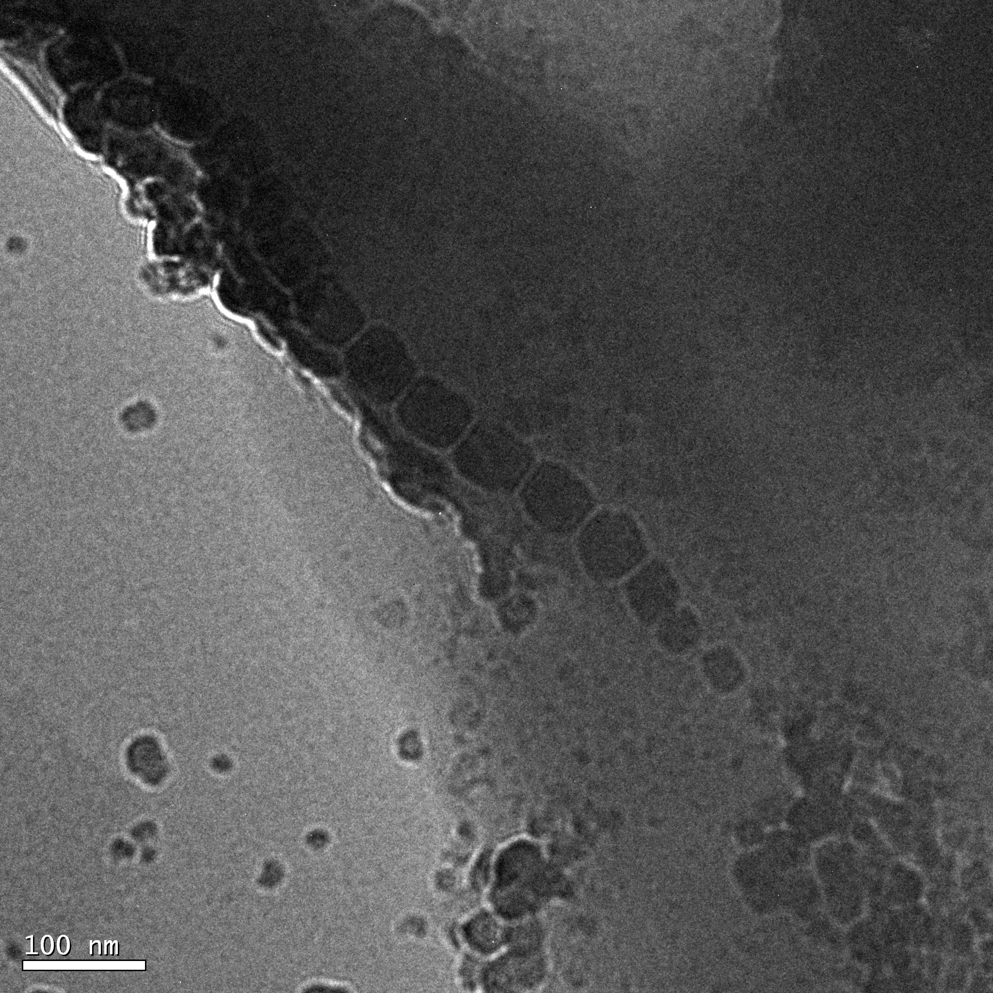
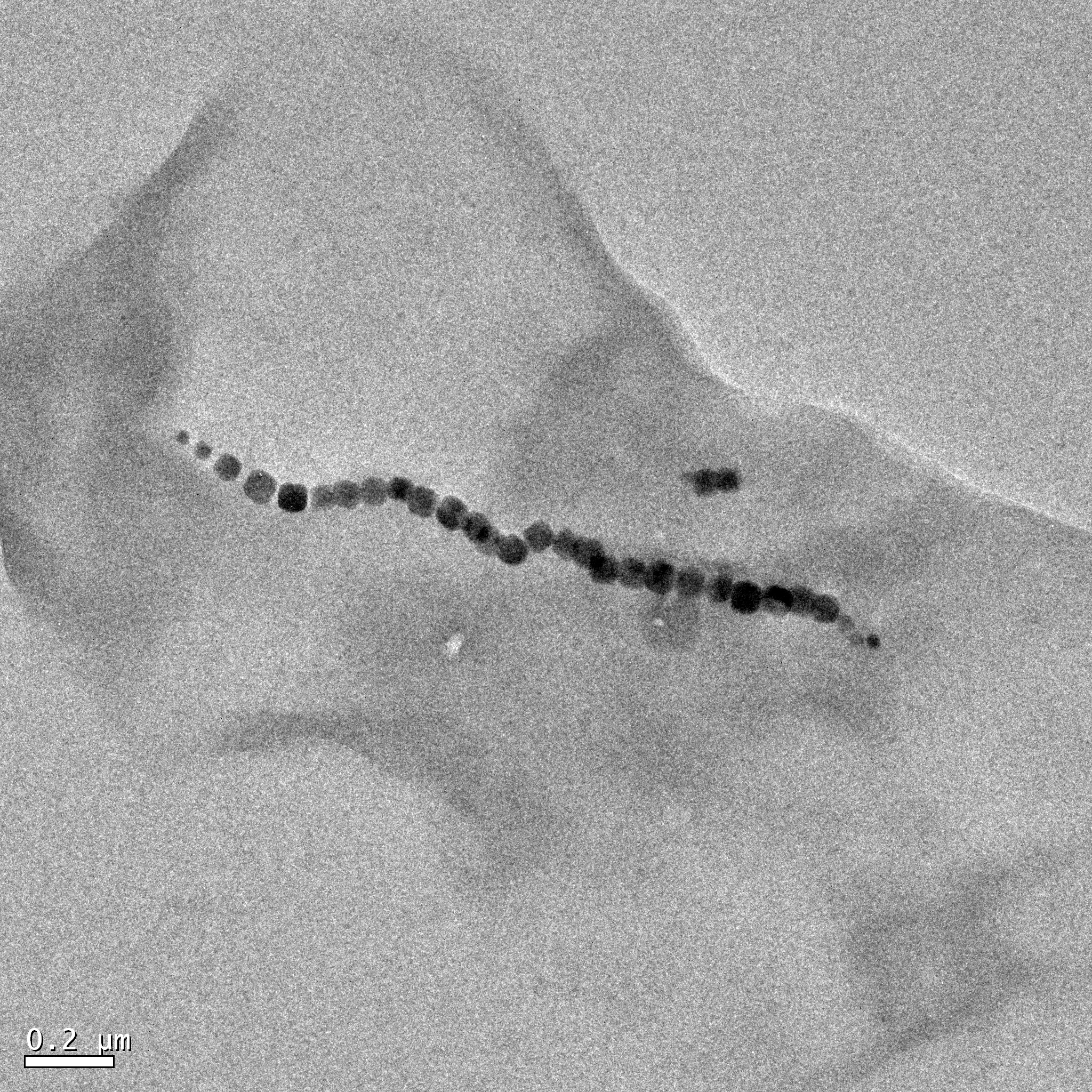
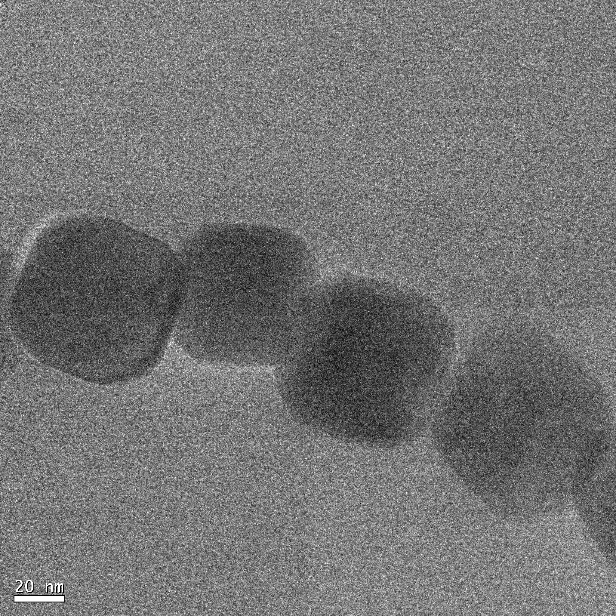
Figure 1: Bright Field TEM micrographs, A-F (Clockwise from top left image). (A) Four nanoparticles prepared via microtome. (B) Two nanoparticles prepared via microtome. (C) Dipplococcus MTB preserved and prepared via HMDS, reference to image D. (D) Higher magnification micrograph of the magnetosome chain from image C. (E) Dipplococcus MTB preserved and prepared via HMDS, reference to image F. (F) Higher magnification micrograph of the magnetosome chain from image E.
4.Dark Field 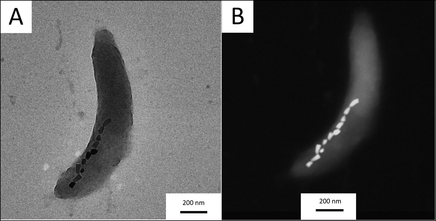
| |||
Figure 2: Bright field micrograph (left, A) and Dark Field micrograph (right, B) of the same vibrio MTB.
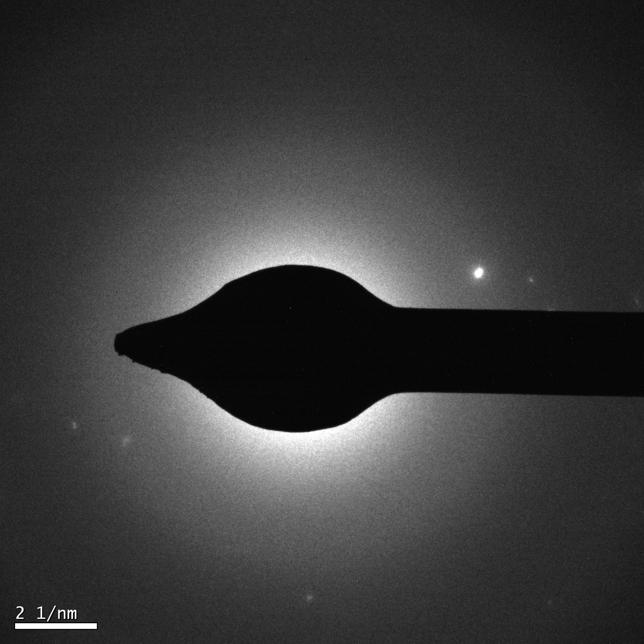
5.EDS
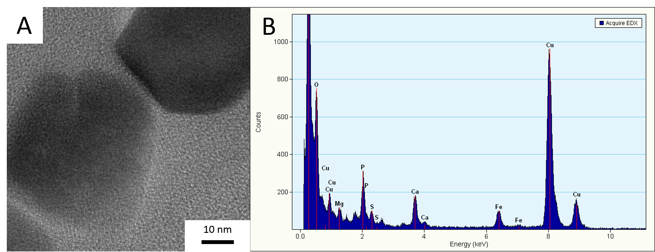
|
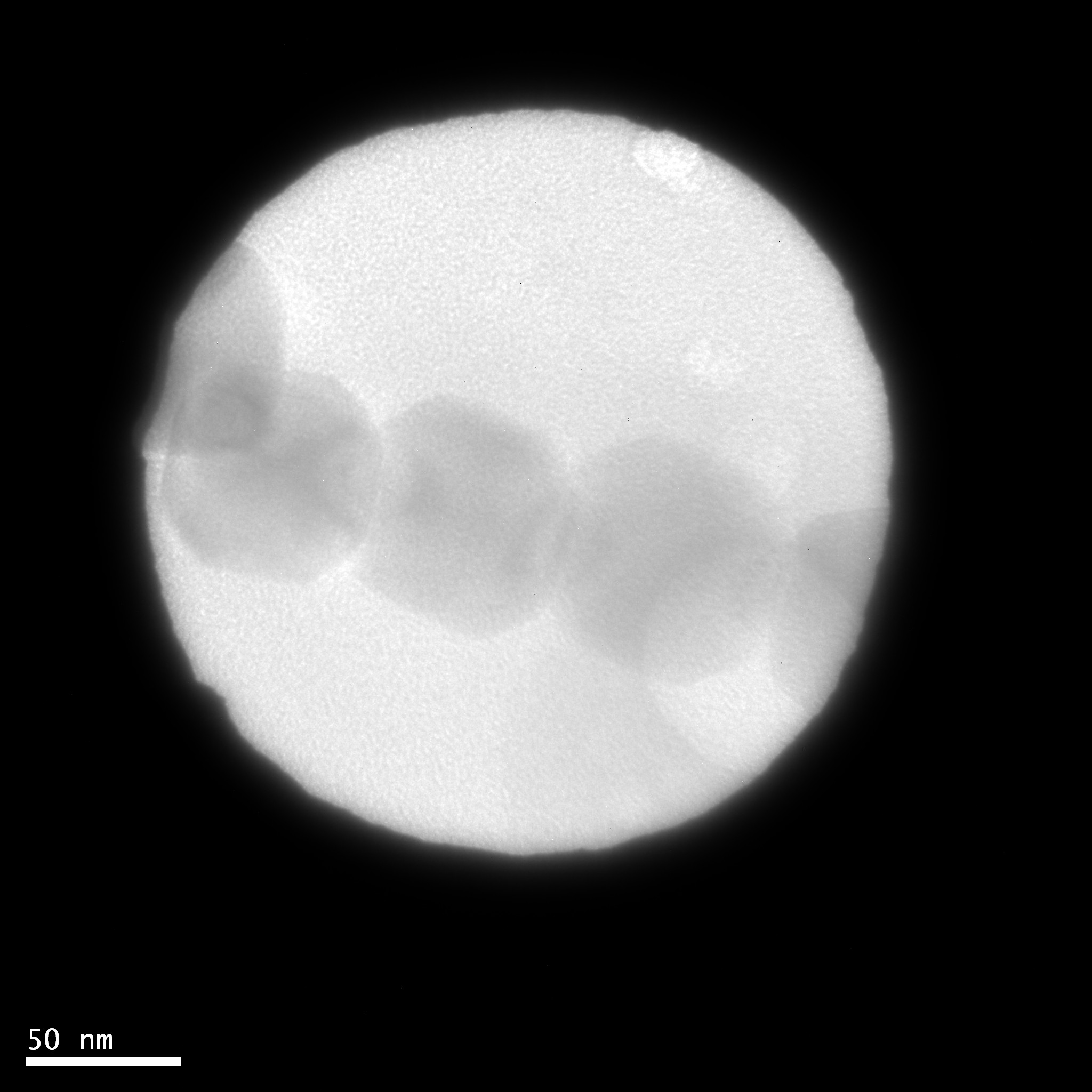
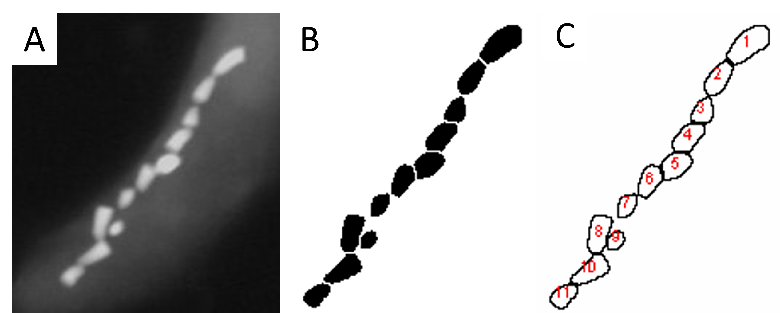
END REMARKS 1. Discussion and Conclusion Samples prepared by microtomy (Figures 1A and 1B) were unstable and difficult to image. Samples prepared via HMDS proved successful for imaging (Figures 1C-1F). Specimens such as in Figures 1E and 2 exhibit in-tact bacterial cells. Figure 1C shows that some samples may not have preserved well during the drying process, however.
Magnetosomes were imaged in situ for three MTB cells (Figures 1C, 1E, and 2) through bright field imaging. Dark field imaging was also utilized on selected specimens (Figure 2B). EDS was successful on targeted particles within one specimen (Figure 3). The spectra indicate high levels of Fe and Ca. A few other elements are also present (such as C, Cu, O, P, Mg, and S) likely due to preparation techniques and the presence of a biotic organism. 2. Acknowledgements I would like to acknowledge Brian McIntyre. He has taught me many techniques involving electron microscopy and has served as an excellent troubleshooting partner for this project. Also, I would like to acknowledge my senior thesis advisor, Dr. John Tarduno, for allowing me to work on this amazing project.
3. References [1]Bazylinski, D and Frankel R. 2004. Magnetosome formation in prokaryotes. Nat. Rev. Microbiol, 2, 217-230.
[2]Pan, Y et al.. 2005. Rock magnetic properties of uncultured magnetotactic bacteria. Earth and Planetary Science Letters, 237, 311-325.
[3]Lefevre, C et al.. 2011. A bacterial backbone: magnetosomes in magnetotactic bacteria. In: Metal nanoparticles in microbiology, Springer-Verlag. Berlin, 75-102.
[4]Pósfai, M et al.. 2013. Biominerals at the nanoscale: transmission electron microscopy methods for studying the special properties of biominerals, in Minerals at the Nanoscale, EMU Notes in Mineralogy, Nieto, F and K Livi (Ed), (London: European Mineralogical Union and Mineralogical Society of Great Britain and Ireland), 377435.
[5]Devouard, B et al.. 1998. Magnetite from Magnetotactic Bacteria: Size Distributions and Twinning. American Mineralogist, 83, 1387-1398.
Comments