University of Rochester, Department of Earth and Environmental Sciences
1. Introduction
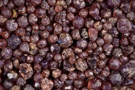
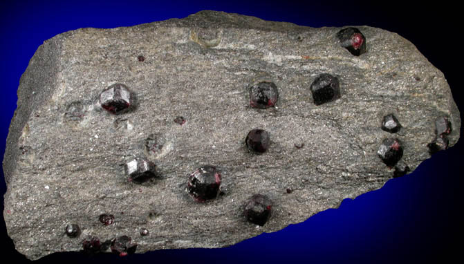
Garnets are a truly amazing mineral. Aside from being incredible examples of nature’s capacity for creating wonderful shapes, the garnet takes on a cubic or more interestingly a rhombic dodecahedral shape when it is euhedral. They are incredibly important to our experimental ability to understand our Earth.
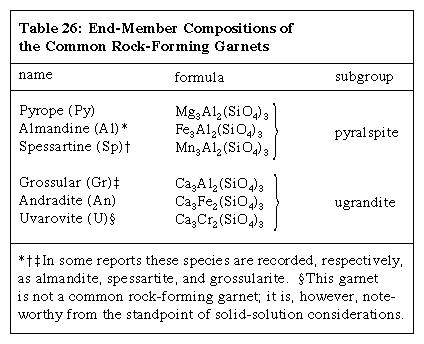
Table 1: Garnets exist naturally on two solid solutions series: pyrope-almandine-spessartine and uvarovite-grossular-andradite. The above table shows these series and associated chemical compositions.[1]
Garnets are one of the geologists most valuable tools and tell us a lot about the geological history of an area. A number of methods have been developed to date garnets and thus, the rock units they reside in. Not only is this important to building a complete history of an area it also can tell us about the nature of the unit and its potential value for mining.
Three samples were selected to be analyzed for this project. The three samples were collected from, Hooper mine, Adirondack region, New York; Coast near Scourie, Scotland; Unknown locality in Western Scotland. Garnets vary widely in their composition as a function of the miscibility of their endmembers. Thus, leading to garnet compositions generally being expressed in terms of percent of pure endmembers. A possible example being: Pyr63Alm30Grs7.
A number of techniques were utilized to try to understand the differences in garnets caused by composition. Scattered electron imaging (SE2) was used to gain a better understanding of the surface and near surface of the crystal, backscatter detection (BSD) for basic compositional differences, interaction volume modeling to understand how compositional differences would affect electron flight, X-ray spectrums and mapping to resolve inter-crystal spatial compositional differences if any, and colorization to highlight interesting features in SEM images.
2. Sample Preparation
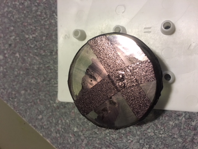
To prepare the specimens for the SEM, crystals were knocked off of the rocks and ensconced in epoxy. After the epoxy hardened the sample puck was sanded and then polished down. Afterwards, the sample had carbon tape and carbon paint applied to allow for charge dissipation. Finally, the sample was placed in the gold sputter coater and had 1.5um of gold applied to its surface.
3. Scanning Electron Microscopy
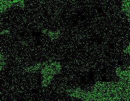
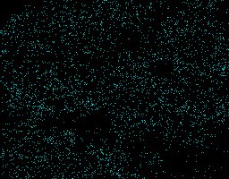
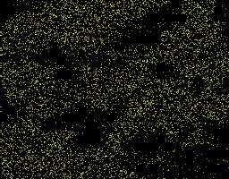
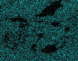
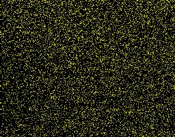
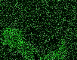
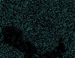
SEM imaging is predicated on interactions that electrons experience when they come in contact with a surface. The first set of images collected were done using the SE2 lens. This lens detects electrons that have inelastically collided with the specimen matter and because they are generally emitted from the uppermost layer of the interaction volume, and reflect the topographical information of the specimen. The BSD was also used, which functions through the use of elastically scattered electrons. BSD also allows for preliminary compositional information to be gathered, as areas with a lighter color contain heavier elements. All images were collected on a Zeiss Auriga CrossBeam SEM-FIB.
All three garnet samples were imaged using the SE2 and BS detectors.
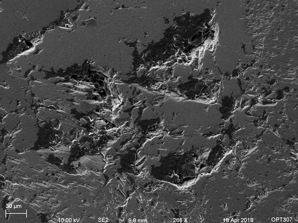
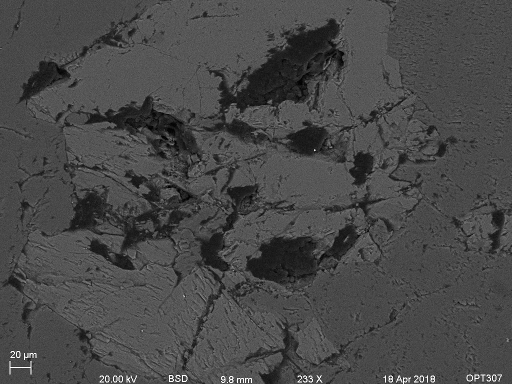
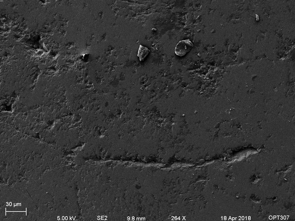
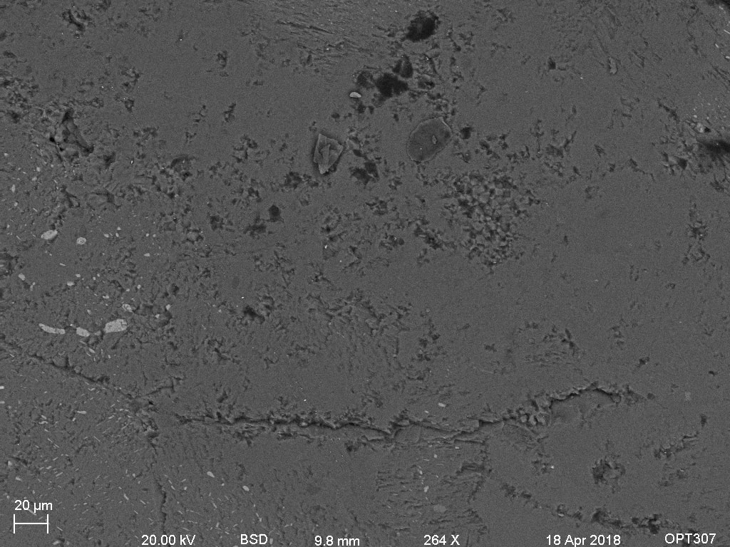
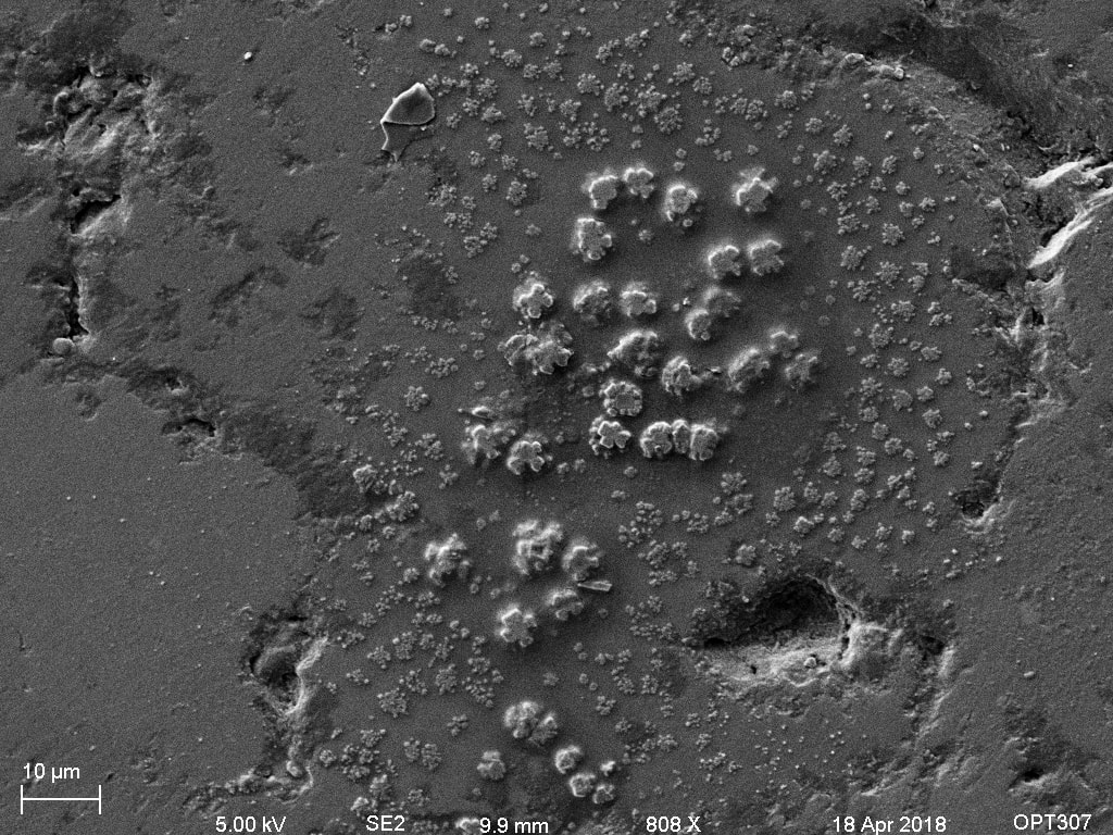
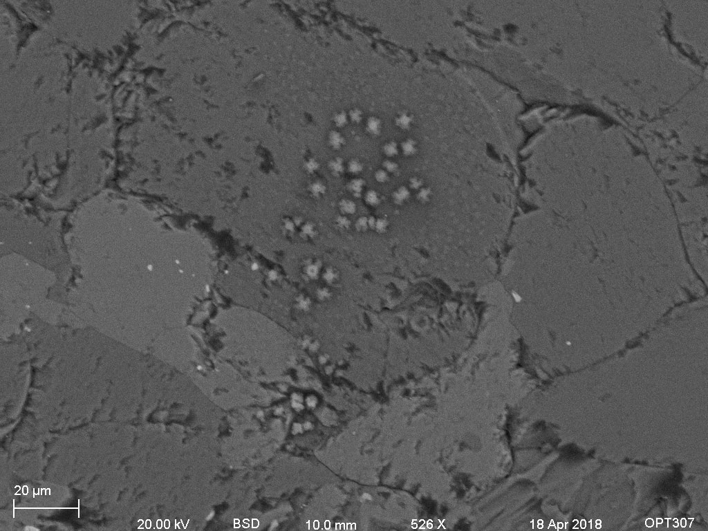
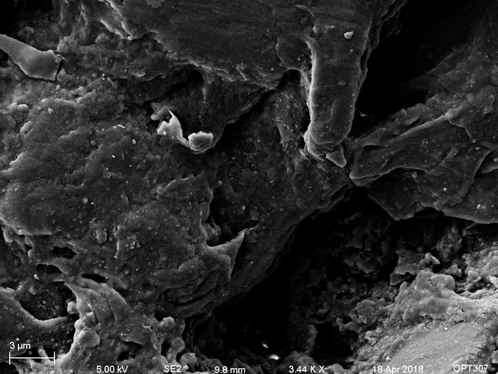
The differences between SE2 and BSD are fairly clear in these images. The SE2 images reveal topographical information about the crystals, while the BSD shows some possible compositional differences. The missing chunks in the first set of images is most likely grains that were ripped from the crystal during sanding. It is interesting however to note that in BSD this appears as a compositional difference when it is very likely to be a function of the depth gradient. Overall, the BSD images show small differences, although there is some evidence of inter-crystalline grains. This is shown in the first set of images and the last. The last set of images also exhibits possible contamination which seems to be of heavier elements than the surrounding crystal.
On first glance the garnets exhibit a few details that differentiate them, the removed grains feature was only observed in the Unknown Scottish sample and some kind of compositional differences were only seen in the previously mentioned sample and the Scourie shore sample.
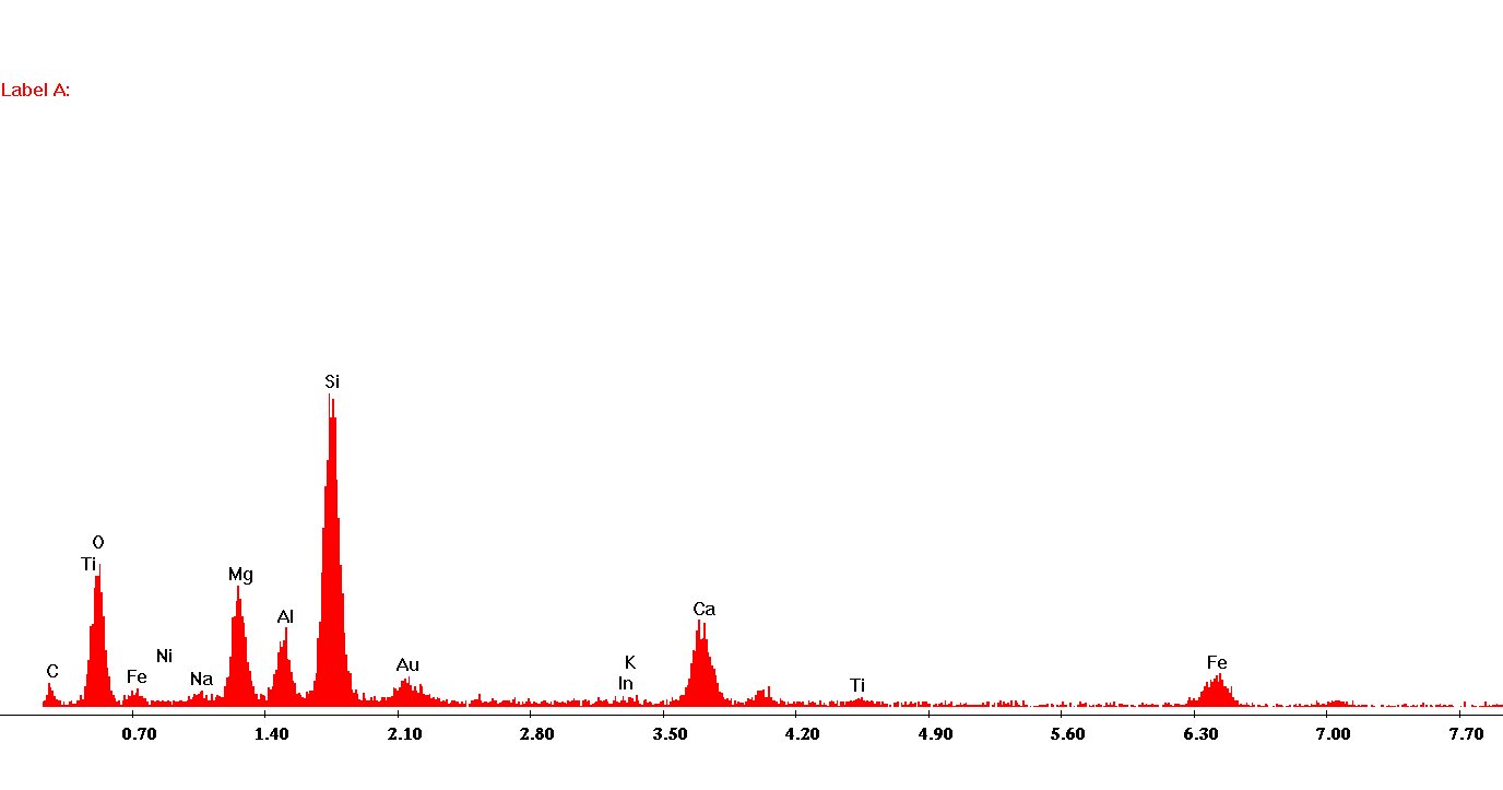
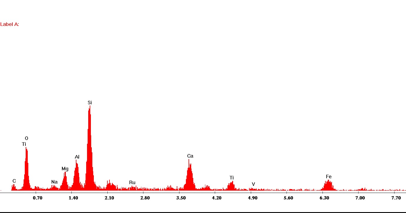
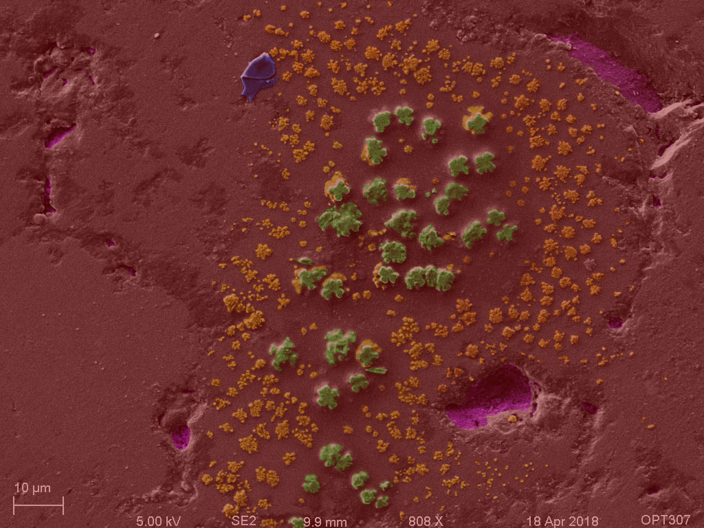
4. X-Ray Maps and Spectra
X-ray maps and spectra allow for a more detailed look at compositional variations within a sample. While BSD allows for relative comparisons based on mass of an element, energy dispersive spectroscopy, identifies actual energy peaks and is able to assign them to a specific element. Here, this is used to investigate inter-crystalline and inter-granular relationships. The spectra give a compositional analysis while the maps show composition in space.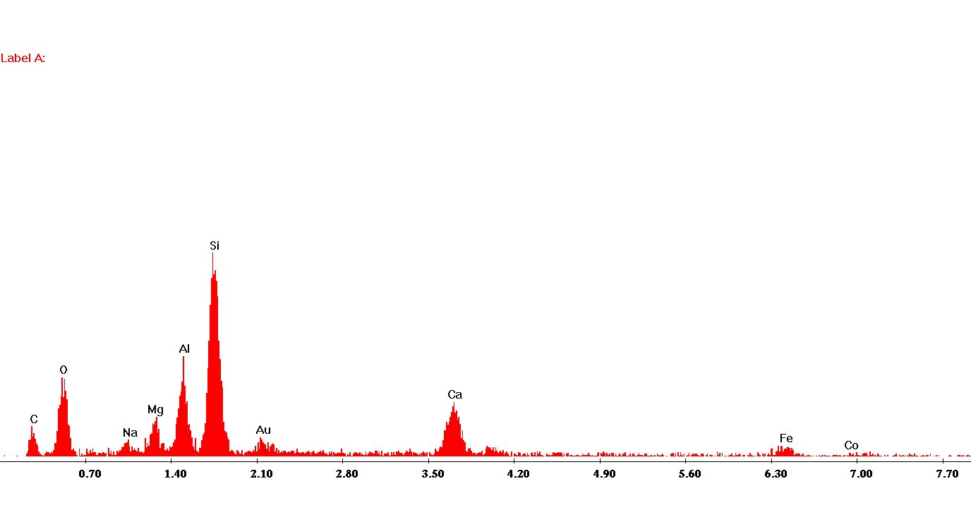

5. Interaction Modeling
Using a program called WinXRay (an electron flight simulator), the interaction volumes of each of the most common garnet sub-varieties and a unique synthesized garnet commonly known as YIG (Yttrium Iron Garnet), Y3Fe2(FeO4)3, was tested. Each simulation was run at 10kV, 0-degree tilt and 1E-7 amp current. The scale bars on each image represent 49nm in the Y-direction and 29nm in the X-direction.
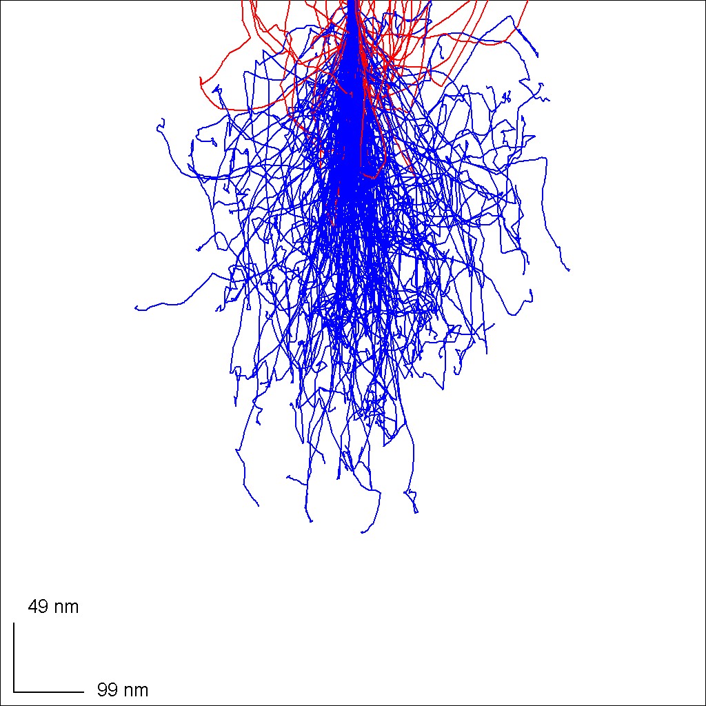
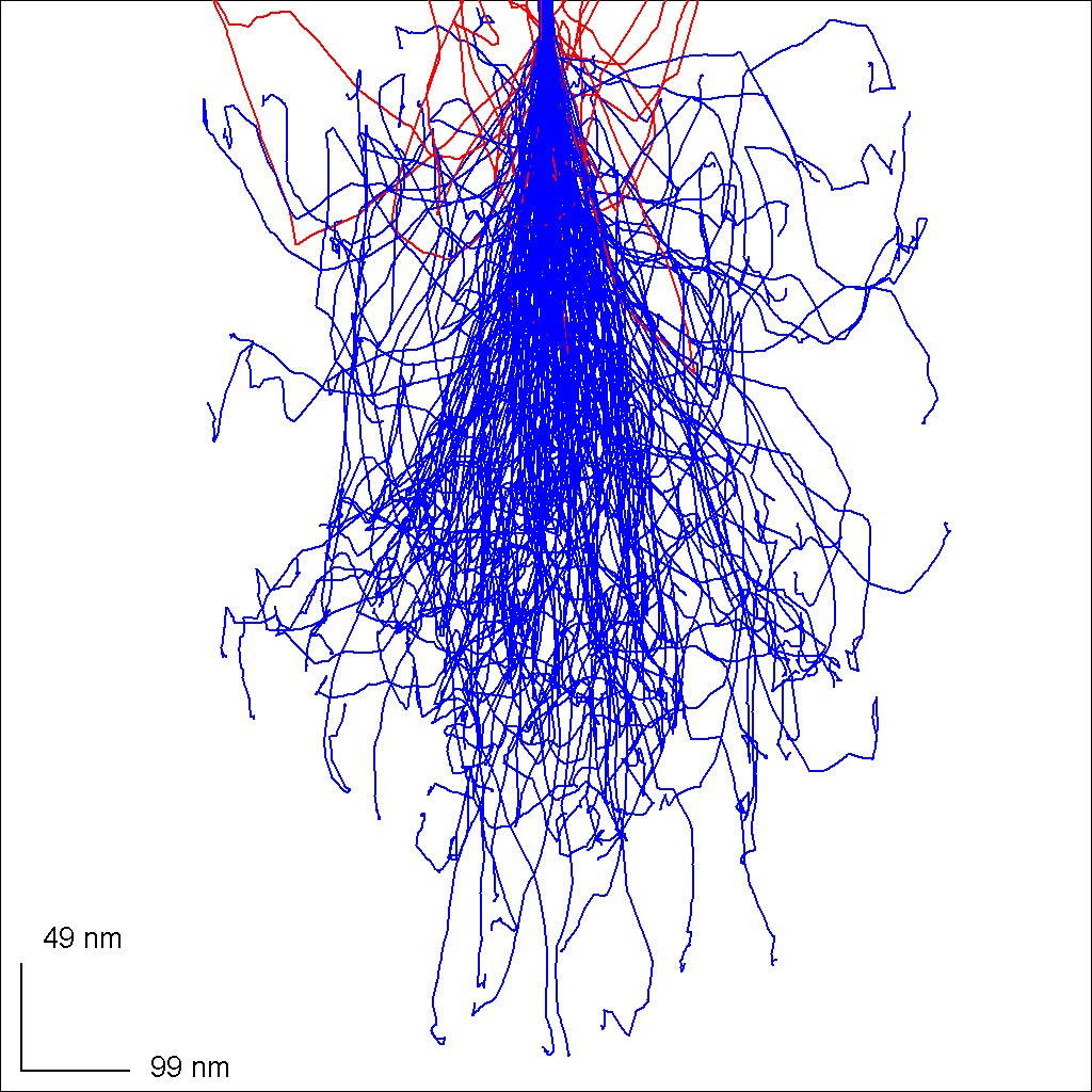
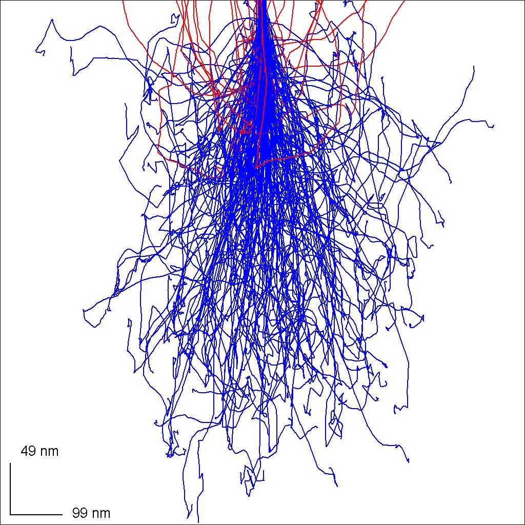
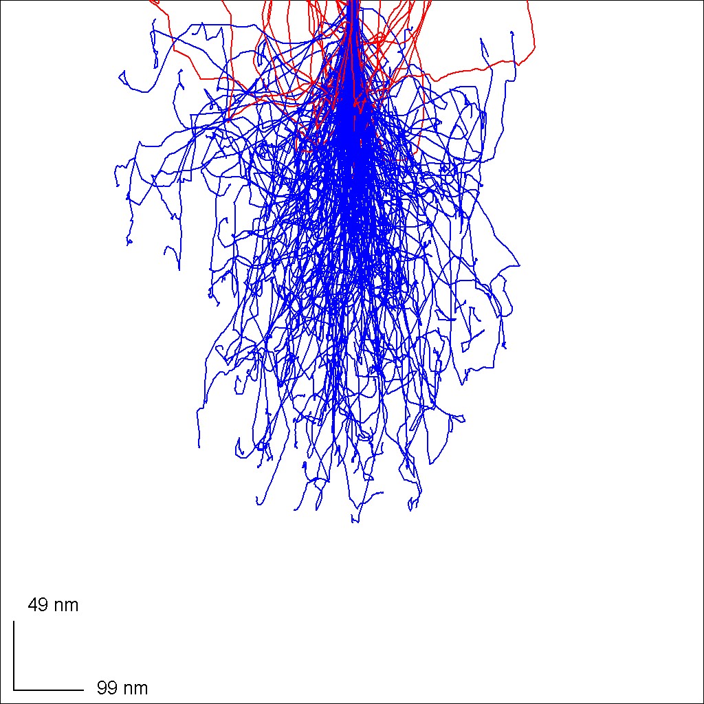

Pyrope, grossular, almandine and spessartine all exhibit relatively deep and laterally expansive electron paths. Unsurprisingly, the more dense and electronically potent YIG shows a more condensed set of electron paths.
6. Colorization
Adobe Photoshop was used to colorize an image of possible contamination on a the Scourie shore garnet.
7. Conclusions
The results showed that there were inter-crystalline compositional differences. These results were identified using BSD and then shown definitively through the use of X-ray maps and spectra. The most dramatic example can be seen with the images taken from the Scourie shore sample which shows what can be tentatively assumed to be a purer grossular grain in a more evenly mixed grossular-almandine-pyrope matrix. Overall, differences could be seen and this combination of techniques has proved itself to be a useful first step into more rigorous analysis.
Acknowledgments
I would like to thank Brian McIntyre for being a fantastic teacher and giving me a tangible skill. SEM work is not only useful but something I have found myself truly enjoying more and more. I would also like to thank my TA Caleb, who was a mentor and friend throughout this course.
References