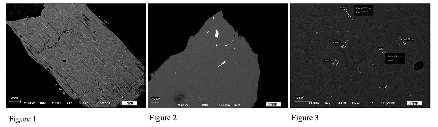
Figure 1: Backscattered image of the pyroxene grain, long white inclusions are dense iron oxide needles.
Figure 2 / 3: Backscattered images of the plagioclase feldspar grain. White inclusions are much larger, amorphous inclusions that are not oriented in any preferential fashion.
In Figure 1, the grain clearly showcases single domain behavior, as all of the well-formed needles are oriented the same direction, uniformly throughout the sample. With Figures 2/3, the un-oriented, jumbled nature of the iron oxide inclusions suggests multidomain properties. This supports our observations in the laboratory previously.
2. Sample Preparation
Light Microscopy sample preparation was completed in one stage: Thin section preparation by the Earth and Environmental Sciences department sample prepper. SEM sample preparation was completed in two stages: Sample polishing and mounting of the grains in epoxy, and sputter coating. TEM sample preparation was completed in one stage: Magnetic separation and mounting of the separation on a carbon grid.
I. Thin Section Preparation:
Each grain had a thin section made of its respective rock that was representative of the bulk sample and composition.II. Sample Polishing:
Each grain was cleaned of any silicon grease that may have been present from previous laboratory testing, and polished by the department sample prepper. The grains were then mounted in epoxy in order to more easily mount the sample to the SEM stage.III. Sputter Coating:
The mounted grains were coated in a layer of gold to ground the samples, as they are non-conductive. This presence of gold is seen in many of the EDAX elemental abundance analyses.
IV. Magnetic Separation:
In order to use TEM diffraction techniques on only the iron inclusions, the rocks the grains were taken from had a piece broken off, crushed, and magnetically separated. This process was done twice. This left a dust composed of almost entirely the iron inclusions within the samples. The dust was then spread across a carbon grid for TEM imaging. Since the small magnetic inclusions could hang off the side of the grid, the TEM was able to image the small particles well and obtain clear diffraction patterns.
3. Result of Microscopy and Discussion
In order to gain a wide variety of information about each of the grains and the respective samples they came from, three microscopes were used. These three microscopes are (i) a petrographic microscope (light microscopy), (ii) an SEM, and (iii) a TEM.
A. Light Microscopy:
Light microscopy (more specifically, petrographic microscope use), allows one to examine the sample in a broader scope and is useful for quick mineral identification, as well as more thorough rock and mineral identification through the Michel-Levy Technique. Figure 4 shows a thin section of a similar sample from the same site as the grains under transmitted, crossed polarized light taken at 10x.
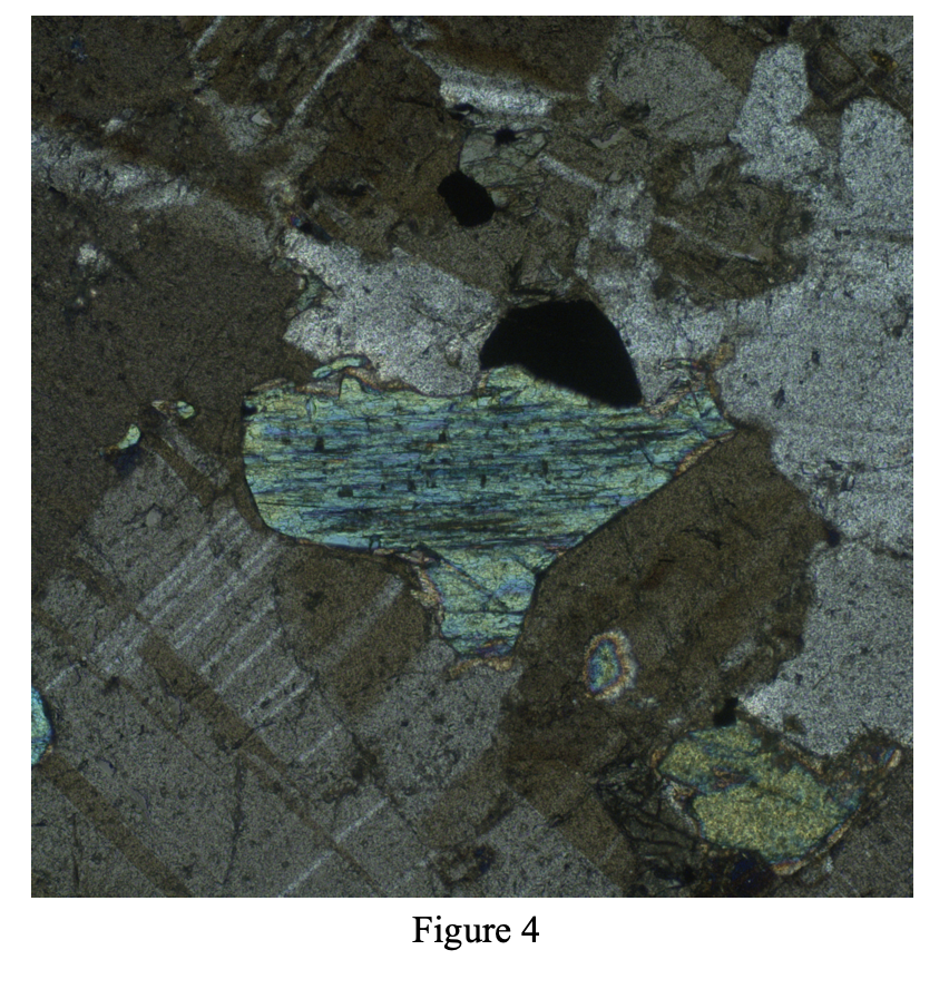
Figure 4: Centered, bright colored grain is a pyroxene surrounded by plagioclase feldspar. This clinopyroxene grain exhibits inclined extinction.
Due to this and all other pyroxene grains in the thin section exhibiting inclined extinction, I at first thought the rocks to be composed completely of clinopyroxene rather than orthopyroxene, which would show parallel extinction. However, later in the project, EDAX data suggested otherwise.
In order to determine the rock type of these anorthosites, the Michel-Levy Technique can be used, which examines the twinning angles of plagioclase feldspar and categorizes the rock into its proper name according to the average twinning angle. Six plagioclase feldspar grains were chosen for two representative samples and had their twinning angles measured, with the results plotted on Figures 5 and 6. Clearly, almost every feldspar grain plotted in the Labradorite region, confirming the identity of the rocks as labradorite.
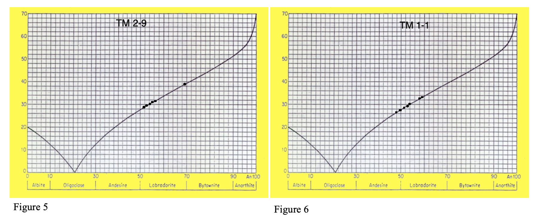
Figure 5: Michel-Levy Technique plot of TM 1-1 feldspars
Figure 6: Michel-Levy Technique plot of TM 2-9 feldspars
B. SEM:
Since with backscatter imaging, denser materials give off many more backscattered electrons, the contrast between materials of different densities is most easily seen with the BSE detector. As much of this project is searching for iron oxides within pyroxene or plagioclase feldspar, this made BSE the most optimal detector to use when trying to find iron oxide inclusions. See Figures 1, 2, and 3 to show the high contrast of the inclusions of the samples in BSE.
The high contrast and easily locatable inclusions allow for very efficient EDAX analysis of both the needles and the mineral ground mass they are located inside of. Ideally, the inclusions would be made out of only iron and oxygen, suggesting a possible composition of magnetite. However, if the inclusion’s EDAX data shows a peak of titanium, the grain could be titanomagnetite, or could be showing exsolution of magnetite to ilmenite (FeTiO3) which would be suboptimal for magnetic analysis. Figures 9 (a-e) are inclusions within the plagioclase feldspar grains with their respective EDAX analyses done at different point around the grains.
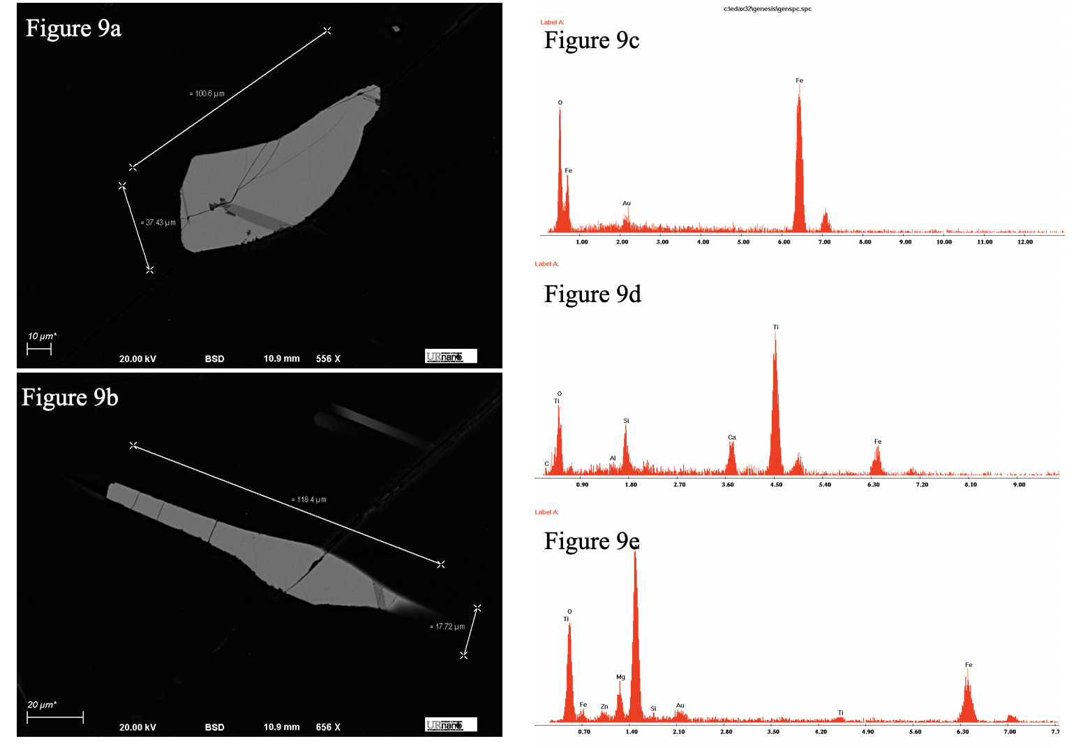
Figure 9 (a-e): a) Iron oxide inclusion within the plagioclase feldspar grain.
b) Another Iron oxide inclusion within the plagioclase feldspar grain.
c) A representative EDAX elemental abundance analysis of the lightest area of the inclusions (done on Figure 9a, but representative of Figure 9b as well).
d) A representative EDAX elemental abundance analysis of the medium colored area of the inclusions (done on Figure 9a, but representative of Figure 9b as well).
e) An EDAX elemental abundance analysis of the darkest portion of the inclusion, only found within Figure 9a.
In Figures 9 (a-e), there are three distinctly different colored areas throughout two of the grains in the plagioclase feldspar grain, which when analyzed to a point with EDAX show interesting results. The lightest colored, majority of the grain is almost exclusively iron and oxygen (Figure 9c), which is what we ideally want to see, suggesting a possible magnetite composition. However, the distinguished darker color shows a new peak of titanium not present in the rest of the inclusion (Figure 9d). This, coupled with the sharp, distinguished geometry of the discoloration suggests exsoultion of magnetite to ilmenite rather than simply a polishing artifact. This can cause issues while analyzing the samples magnetically, as ilmenite is not as magnetic as magnetite. However, this dissolution was only seen in the multidomain plagioclase grain, I could not find any examples of this dissoultion in the single domain pyroxene grain. Furthermore, there are even darker areas within one of the inclusions (Figure 9a). This elemental abundance showed a peak of aluminum, which is odd (Figure 9e). This may be due to slight impurities in the inclusion.
After examining the magnetic separated dust in the TEM, the carbon grid they were analyzed on was mounted on an SEM stub and looked at in the SEM as well, obtaining more EDAX elemental abundance data to further investigate the composition of the inclusions. Both sample’s magnetic separations were tested. Figures 10a – 10b show the feldspar sample’s magnetic inclusions and its respective elemental abundance plot, and Figures 11a – 11b show the pyroxene sample’s inclusions with its respective elemental abundance plot.
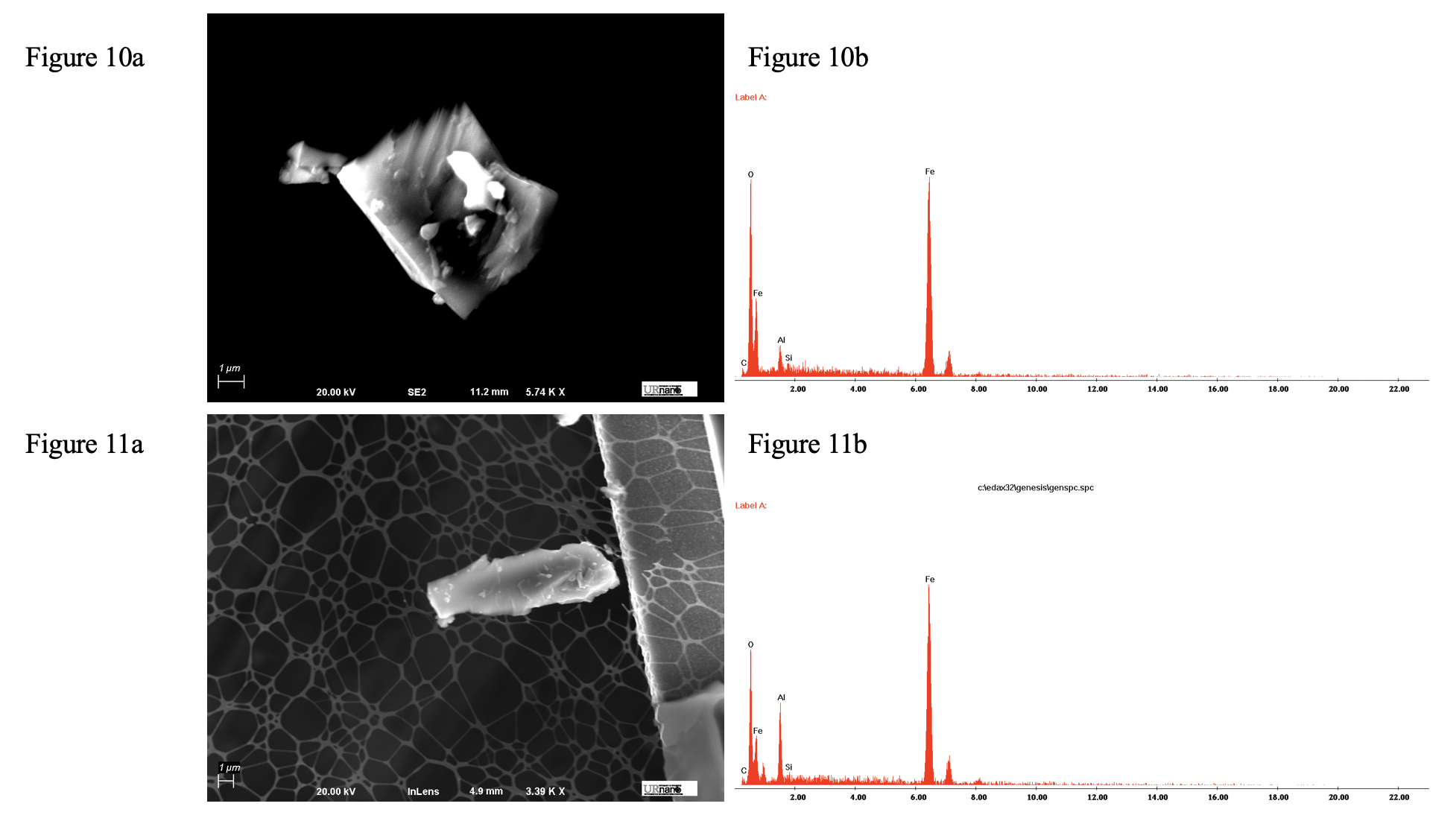
Figure 10a – 10b: a) SEM SE2 image of a magnetically separated grain from the plagioclase feldspar grain’s sample. b) EDAX elemental abundance data of the grain.
Figure 11a – 11b: a) SEM InLens image of a magnetically separated grain from the pyroxene grain’s sample. b) EDAX elemental abundance data of the grain, considerably more aluminum.
Although the very small magnetically separated grains were found using backscatter, the images of the grains are in SE2 and InLens. Backscatter was used to quickly locate the very small, dense grains against the much lighter carbon grid and empty space background. In backscatter, the images are very difficult to get in focus, since the nature of the backscatter detector requiring a larger working distance to accommodate the inserted detector causes it to receive less electrons than an InLens or Secondary Electron Detector. Imaging in SE2 and InLens yields much better results for structures this small, although since the dust was not coated, SE2 produced a blurrier picture. Both EDAX figures showed consistent results with previous data, supporting a composition of the inclusions of primarily iron and oxygen. However, there are aluminum and carbon peaks in each EDAX figure. This can be attributed to the carbon grid the inclusions were mounted on, and the aluminum SEM stage stub the carbon grid was stuck to. Figure 11b shows much more of an aluminum peak than Figure 10b. This could be due to the EDAX point of measurement being less centered completely on the inclusion, containing more of the aluminum stub in the area of analysis than Figures 10a – 10b.
There was a great variety of inclusion size between the two samples. In the pyroxene grain, the inclusions were mostly consistent in size, averaging about 50 microns long, as shown in Figures 12 and 13.
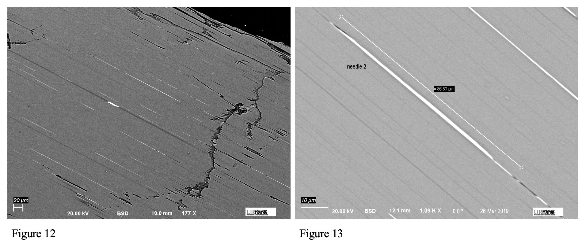
Figure 12: Low magnification backscattered image of the iron oxide needles within the pyroxene grain.
Figure 13: An example of the length of one of the longer needles, greater magnification.
These needles all have a similar shape and are similar lengths. Again, they are all oriented the same direction. All of this data supports the preliminary results we obtained in the laboratory, suggesting single domain behavior of the pyroxene grain.
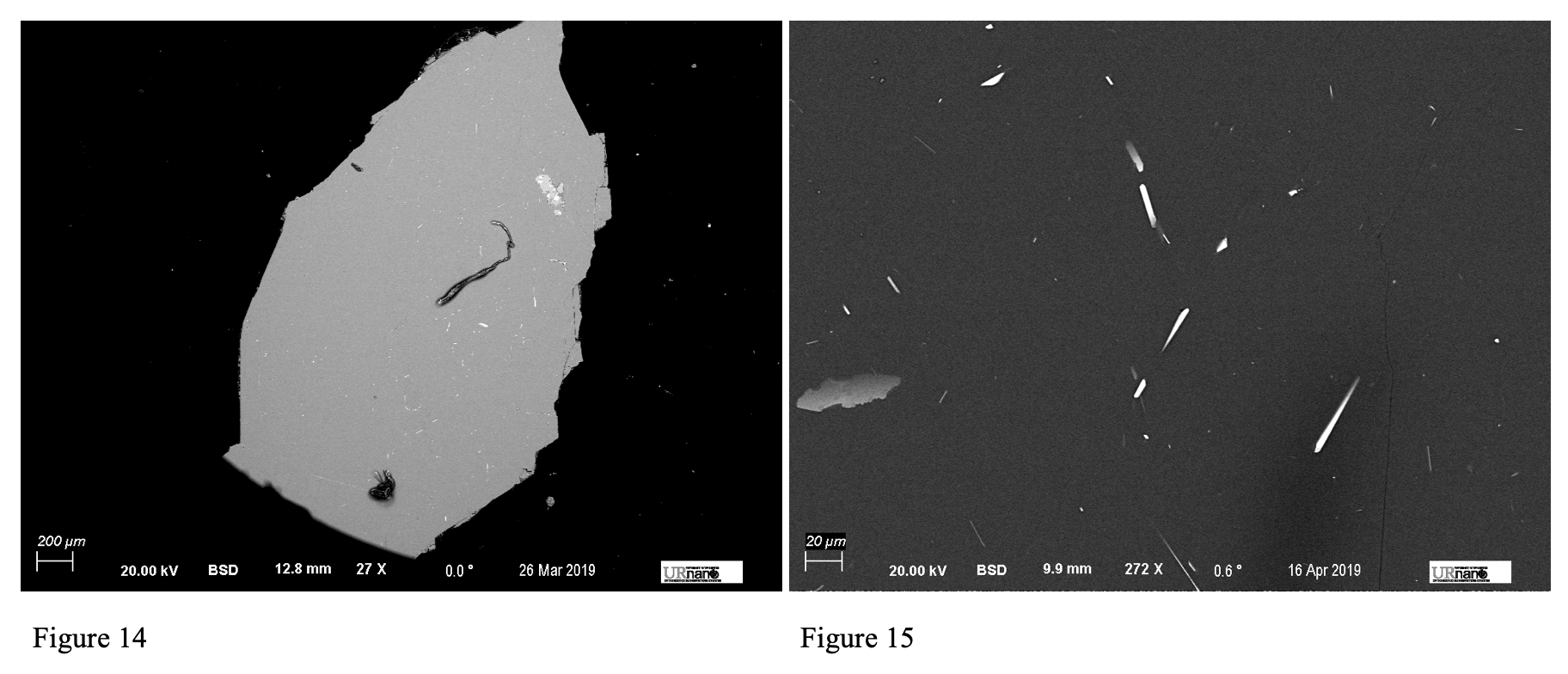
Figure 14: Low magnification backscattered image of the iron oxide inclusions within the plagioclase feldspar grain. Note the wider scale.
Figure 15: Higher magnification backscattered image of more iron oxide inclusions within the plagioclase feldspar grain. Note the much smaller scale than Figure 14.
In the plagioclase, the size and shapes of the needles vary much more, as seen in both Figure 14 and 15. These needles also appear to have much less of a preferred orientation when compared with the pyroxene grain, again coinciding with the previous multidomain behavior of this grain.
In order to confirm the presence of only clinopyroxene, as suggested by the Light Microscopy results, an elemental abundance analysis was done on the ground mass of the inclusions, the actual pyroxene grain itself.
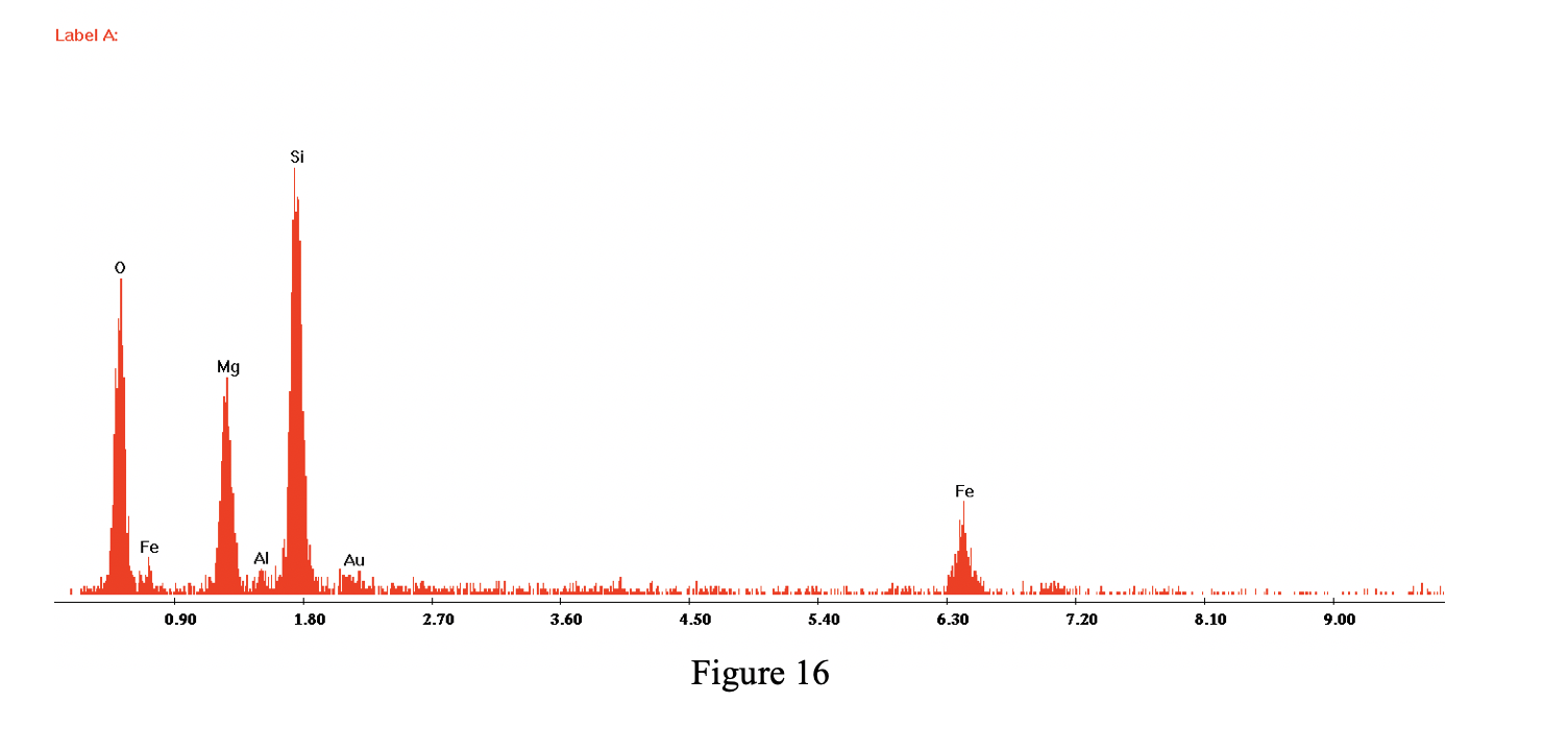
Figure 16: EDAX data of pyroxene grain. Note the absence of calcium.
Due to the absence of calcium in this elemental abundance analysis, the pyroxene could not be a clinopyroxene. Since all clinopyroxenes have calcium, we can conclude that this grain is indeed an orthopyroxene, thus the samples must contain both clinopyroxene and orthopyroxene.
C. TEM and Electron Diffraction:
In order to gain further insight as to what the actual mineralogical composition of the iron oxide inclusions is, TEM electron diffraction can be used. However, the grains that were prepared for the SEM were too thick to examine in the TEM, and the pyroxene and plagioclase feldspar groundmass would have made it impossible to image only the inclusions. To solve this issue, a magnetic separation was done twice of the samples the grains came from. The magnetic dust that was left was able to be spread across a carbon grid and imaged in the TEM to obtain electron diffraction data. The best diffraction data for the pyroxene grain sample’s rock is seen in Figure 17. Figure 18 is the diffraction pattern produced. The best diffraction data for the plagioclase feldspar grain sample’s rock is seen in Figure 19, and its respective diffraction pattern is shown in Figure 20.
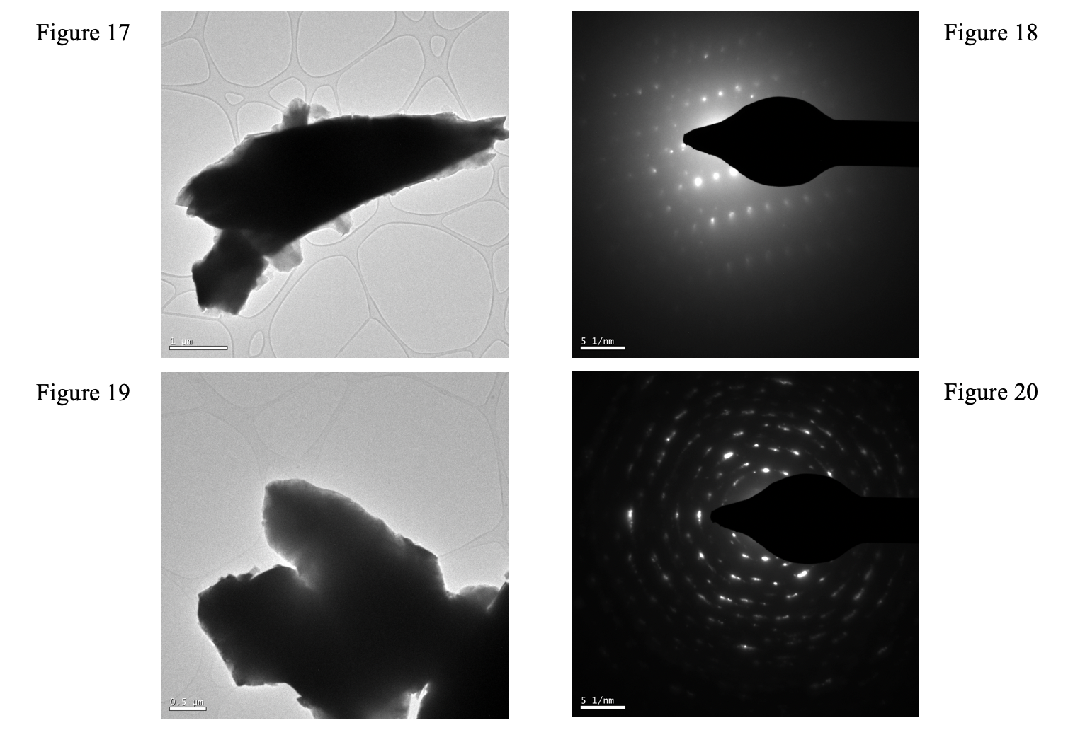
Figure 17: Iron oxide inclusion on carbon grid imaged by TEM (pyroxene sample’s rock)
Figure 18: Electron diffraction pattern produced by Figure 17.
Figure 19: Iron oxide inclusion on carbon grid imaged by TEM (plagioclase feldspar sample’s rock).
Figure 20: Electron diffraction pattern produced by Figure 19.
As seen on both diffraction patterns, the bright spots arrange themselves in a cubic, or isometric fashion. This means the crystal structure of the mineral being imaged is isometric which is very characteristic of magnetite. Figure 18 shows a single crystal, inferred by the absence of rings, while Figure 20 shows more of a polycrystalline structure, as the spots appear to be aligning themselves in rings. Either polycrystalline or monocrystalline, the diffraction patterns add more supporting evidence that the mineral these iron oxides are composed of is indeed magnetite, instead of hematite or titanomagnetite, which do not have isometric crystal structures.
4. Conclusion:
These rocks collected in Labrador, Canada, are indeed labradorites that contain both clinopyroxene and orthopyroxene. Their inclusions are made out of iron oxides, compositionally ranging from magnetite with exsolution of ilmenite to titanomagnetite, as supported with multiple techniques such as EDAX of inclusions in-situ and magnetically separated inclusions, and electron diffraction. In the pyroxene grain, it can easily be seen that the iron oxide needles are single domain, as the inclusions all measure similar lengths, and are very well-formed needles oriented in the same direction. On the other hand, the plagioclase feldspar grain is characteristic of a multidomain grain. The iron oxide inclusions have a wide range of sizes, shapes, and compositions, oriented every which way. Due to these properties of each grain, it is clear that out of the two, the pyroxene grain would be more useful in future paleomagnetic analysis, as it would better preserve magnetic data from when it formed.
5. Further Work:
Since many other pyroxenes tested in our laboratory showed similar numbers suggesting single domain properties while almost all other plagioclase feldspar grains showed multidomain data, for further Paleomagnetic analysis pyroxene grains would be the easiest and most useful grains to use. However, since there is clinopyroxene and orthopyroxene in the samples, it could be important to test each grain to see which it is, as clinopyroxene is inherently better to use while running paleomagnetic tests, since it more usually contains the iron oxide needles we are interested in. Also, further investigation into the odd aluminum peak section of the inclusion within the plagioclase feldspar grain is needed to expand our understanding of the processess the rock and more specifically, the grain, has undergone.
Acknowledgments
I would like to acknowledge Brian McIntyre for extensive help and patience for this project, advisor Dr. John Tarduno, TA Ralph Wiegandt, and lab partner Logan Bashford.