University of Rochester, Department of Physics and Astronomy
1. Introduction
Octopus is one of the most mysterious creatures of nature. It is the master of disguise; it can change the color and texture of the skin in the blink of an eye. It has eight flexible arms with numerous suckers which allow it to move from one place to another, hold onto substratum, catch its prey or fight with its predators etc. These suckers also grants them to the ability to smell or taste with their arms. These truly extraordinary abilities of octopus bestowed upon by millions of years of evolution are mesmerizing.
In this project, these amazing body parts and related mechanism were explored using different microscopic techniques. In particular, the structure and anatomy of the skin, the arms and the suckers of a Caribbean Octopus (Octopus briareus) were studied using light microscopy and scanning electron microscopy techniques.
2. Microscopic techniques:
Total six microscopic techniques were used in this project:
3. Structure of Octopus Skin
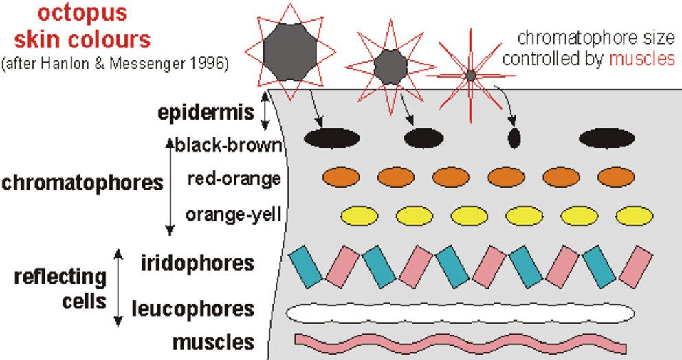
The skin of octopus consists of a transparent epidermis formed of columnar epithelial cells and interspersed mucous cells, and a dermal layer of varying thickness formed of connective tissue that includes a number of chromatophores, iridophores and reflecting cells[1]. A schematic diagram of the structure of octopus skin is shown in figure 1.1.
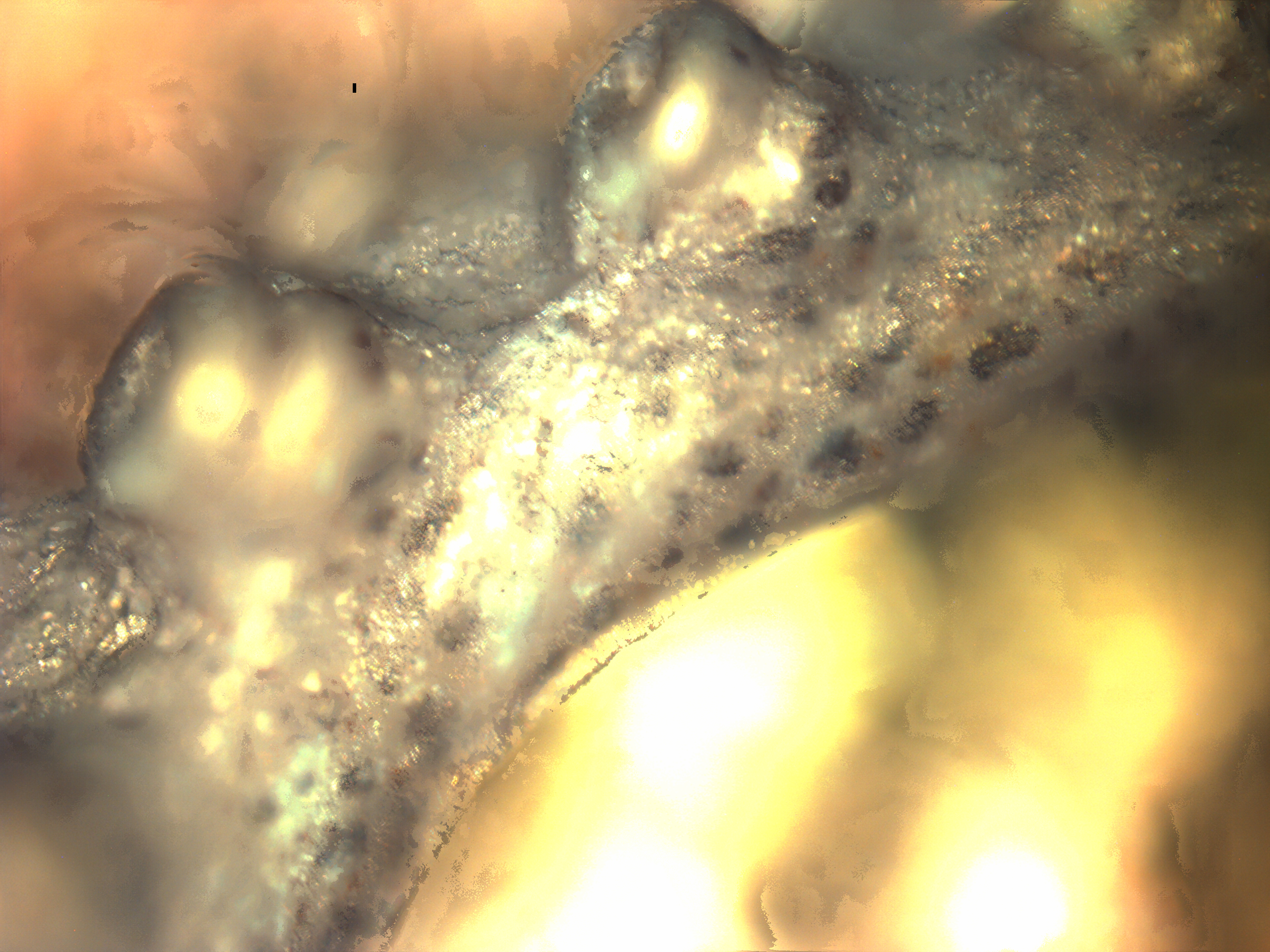
The organ which gives the octopus it’s signature coloring changing ability is called the Chromatophore. Chromatophores of different colors can be observed in figure 1.2. As the depth of field of the light microscope was limited, it was difficult to capture an image of the the specimen which is entirely in focus. To overcome this problem, ImageJ software along with a plugin called Extended Depth of Field was employed which integrated multiple images to create a single composite in-focus image of figure 2.
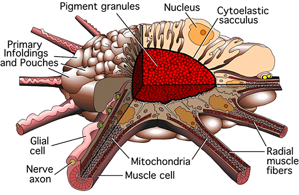
The chromatophore organ consists of five different kinds of cells: 1) the chromatophore proper: containing the pigment granules; 2) the radial muscle fibers attached to the margins of the chromatophore; 3) the neuronal processes (axons) associated with each muscle fiber; 4) the glial cells that accompany each axon; and 5) the sheath cells that surround and embrace the chromatophore, nerves and muscle fibers[2]. A schematic of the chromatophore organ is given in figure 3.
Using these specialized muscles in their skin, octopus can alter these chromatophores from punctate to expanded states or vice-versa. It enables them to expand or shrink the pigment containing chromatophore proper at will, thus allowing it to display any combination of colors by expanding or shrinking chromatophores of different colors (Figure 1.1).
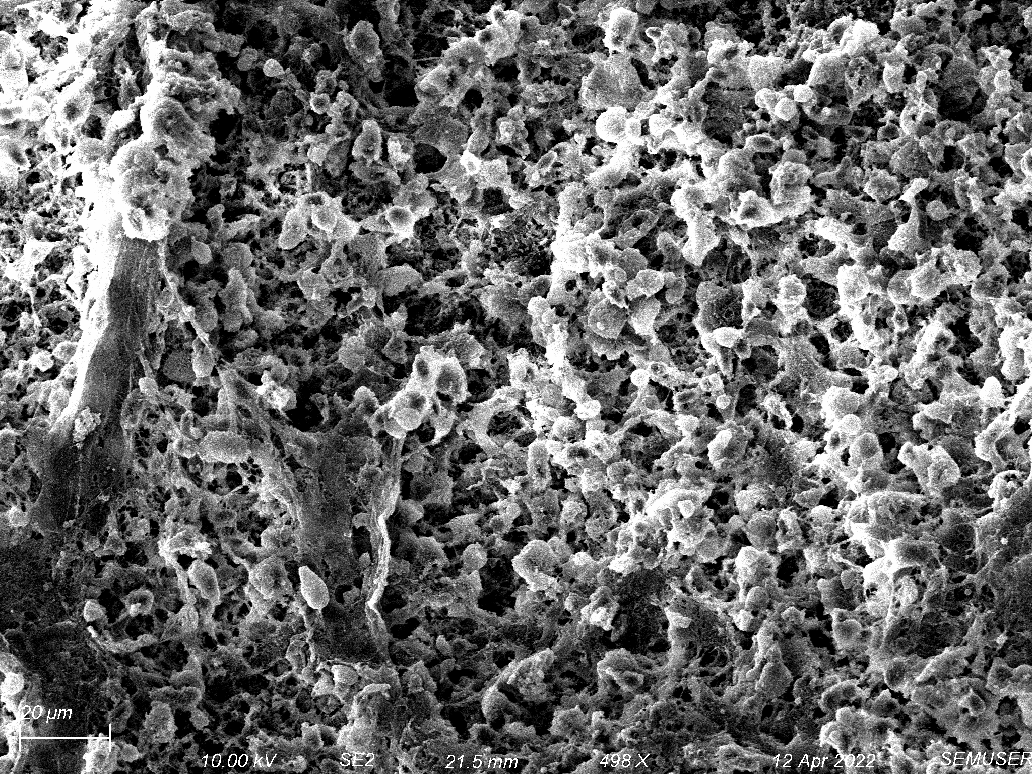
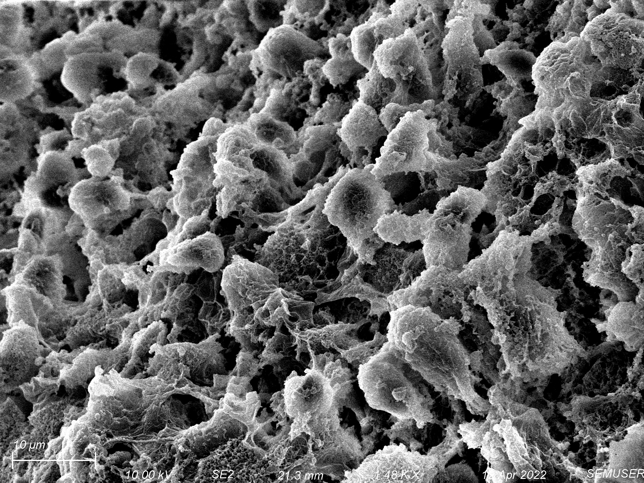
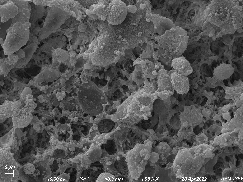
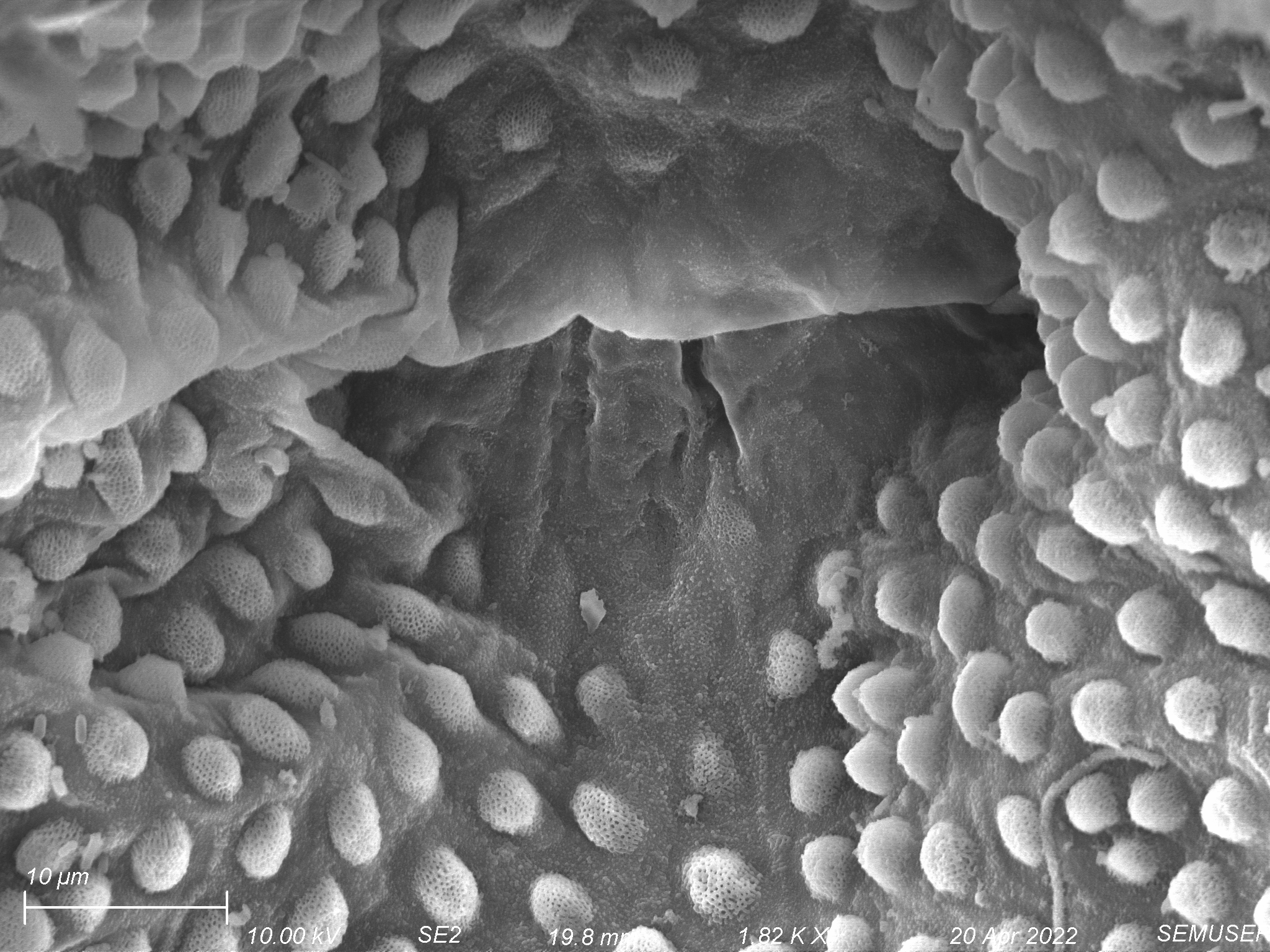
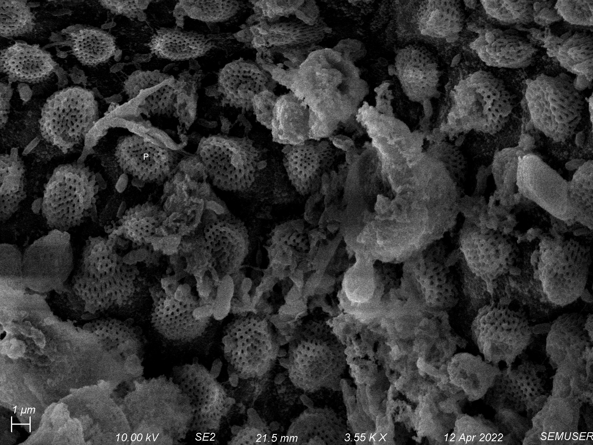
When the specimen was examined using the light microscope, it showed little detail of the epithelial cells as they were transparent to light but when they were examined under electron microscope revealed complex interdigitation of lateral cell surfaces as well as the presence of an apical microvillous layer. This microvillous layer forms an anastomose meshwork which can be seen below.
3. Structure of Octopus Arm
Octopus has eight arms which work as muscular hydrostat and contain longitudinal, transverse and circular muscles around a central axial nerve. They can extend and contract, twist to left or right, bend at any place in any direction or be held rigid.
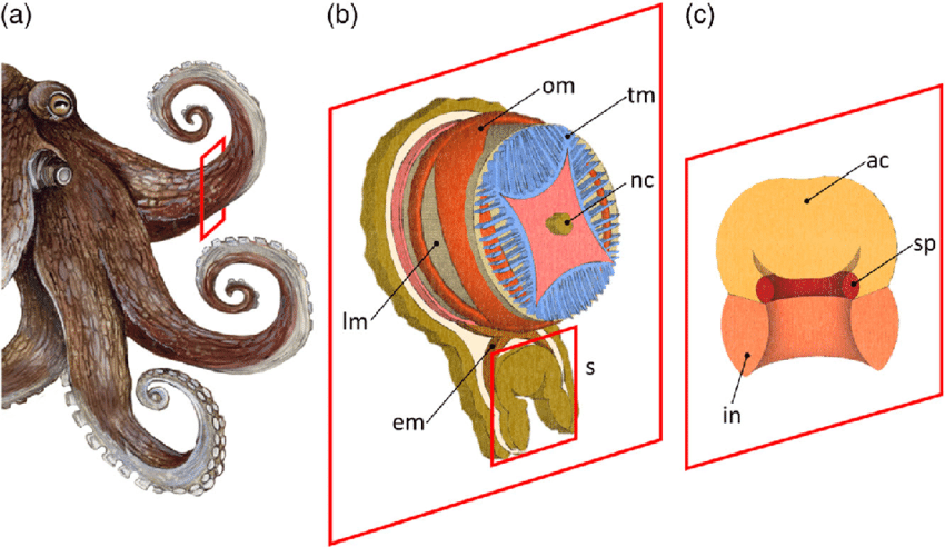
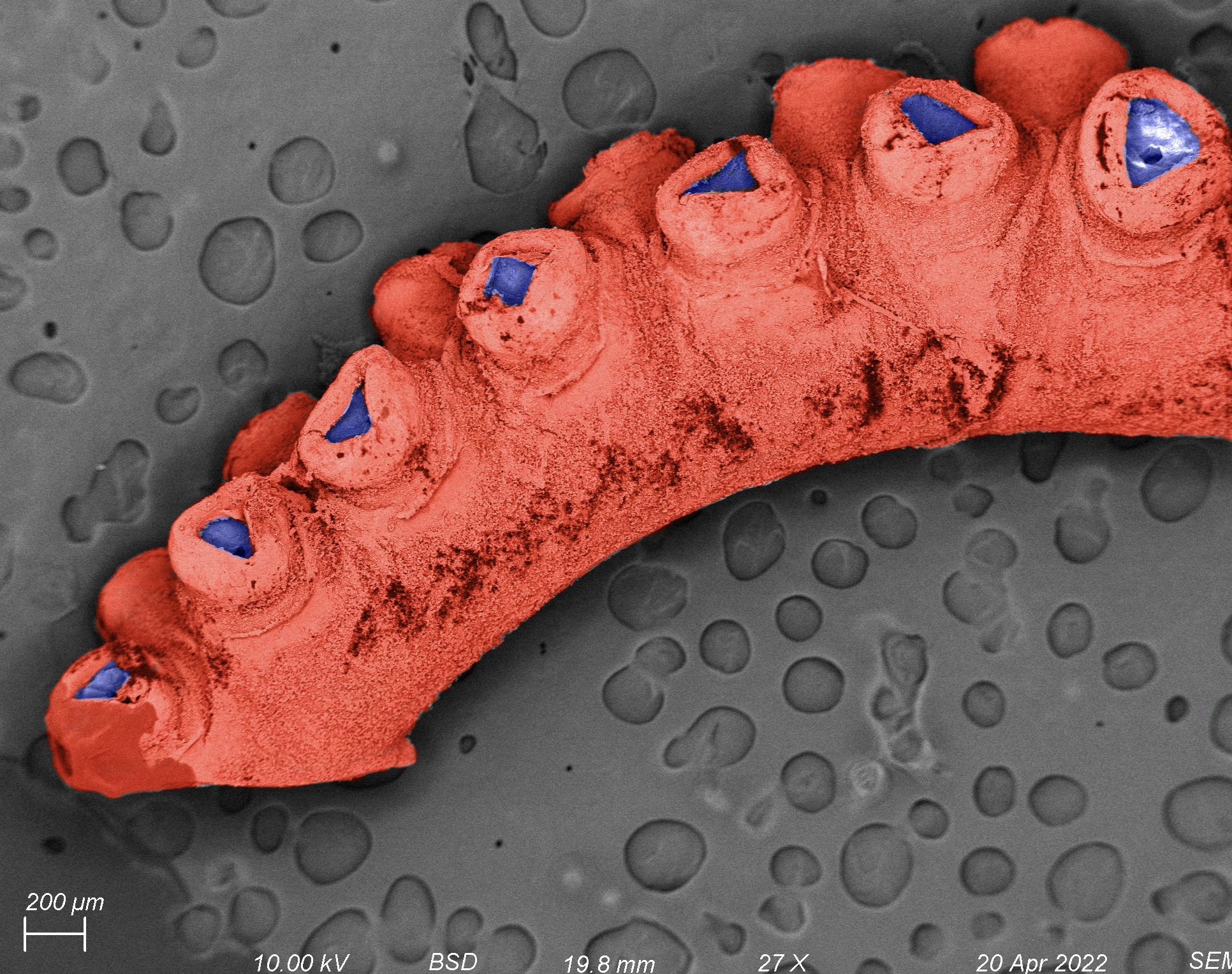
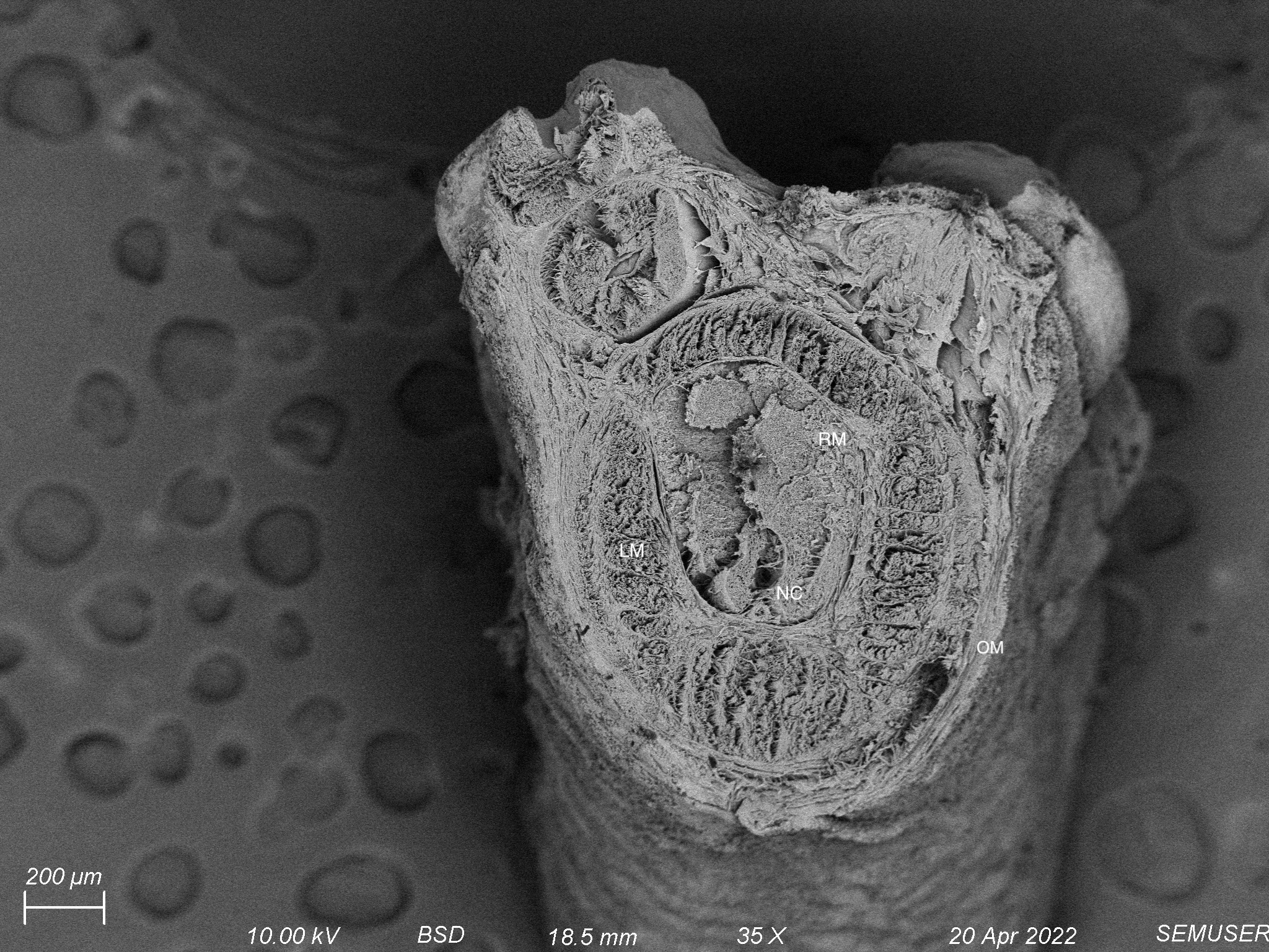

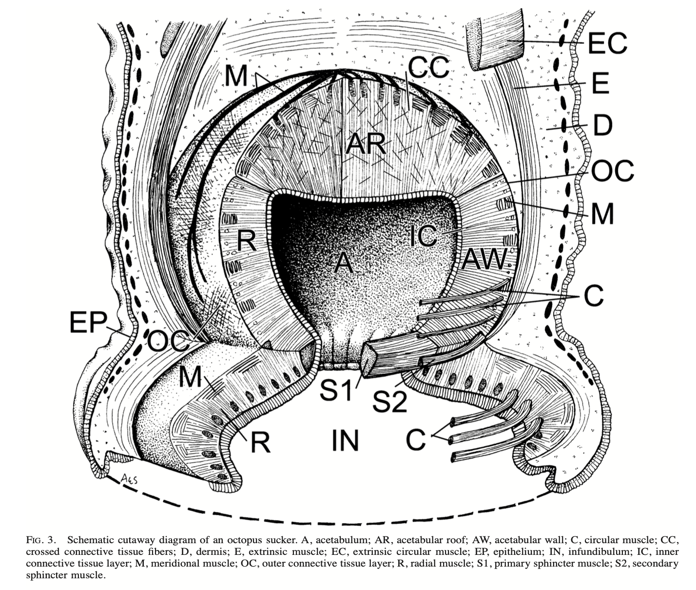
4. Structure of Octopus Sucker
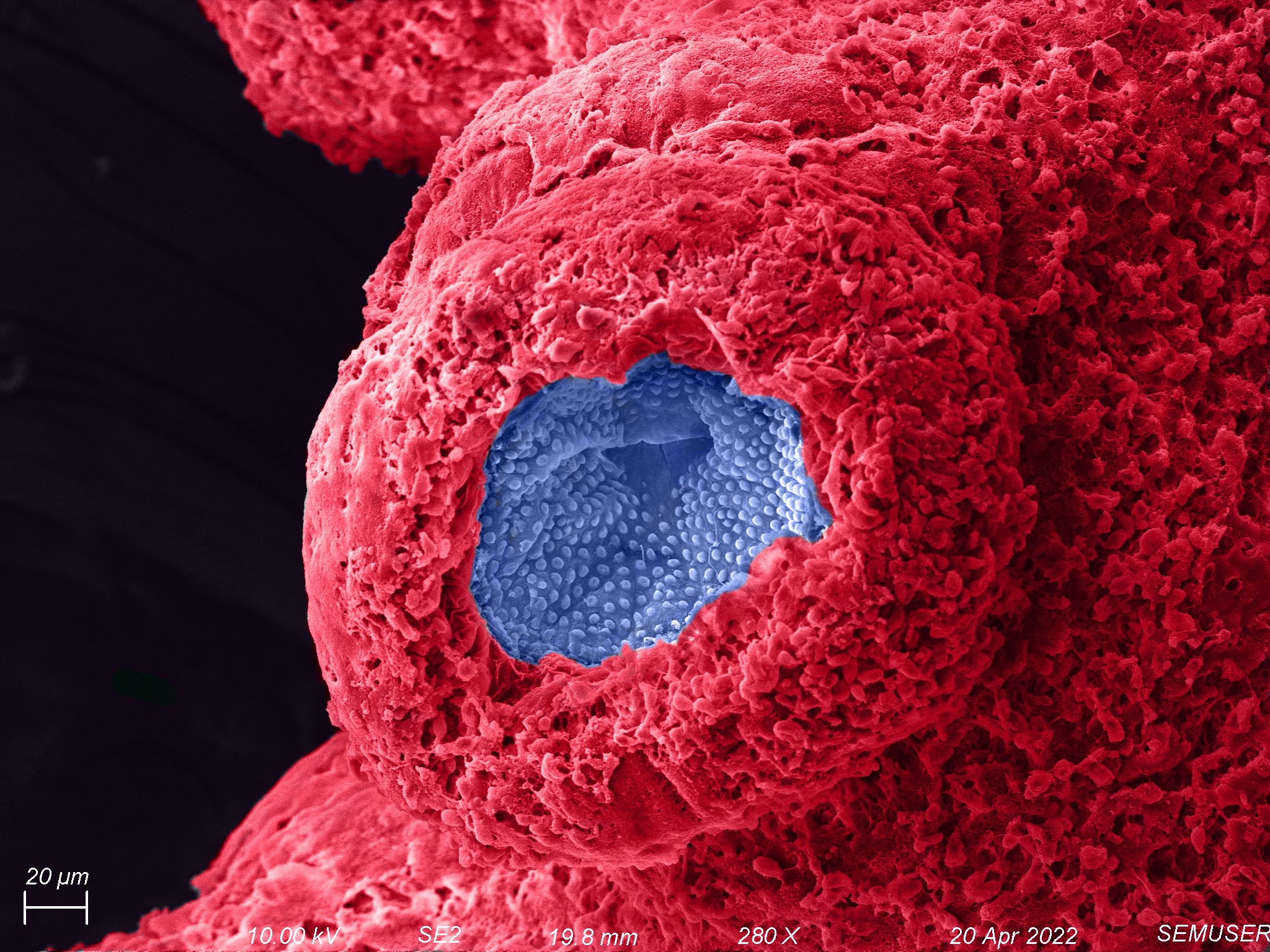
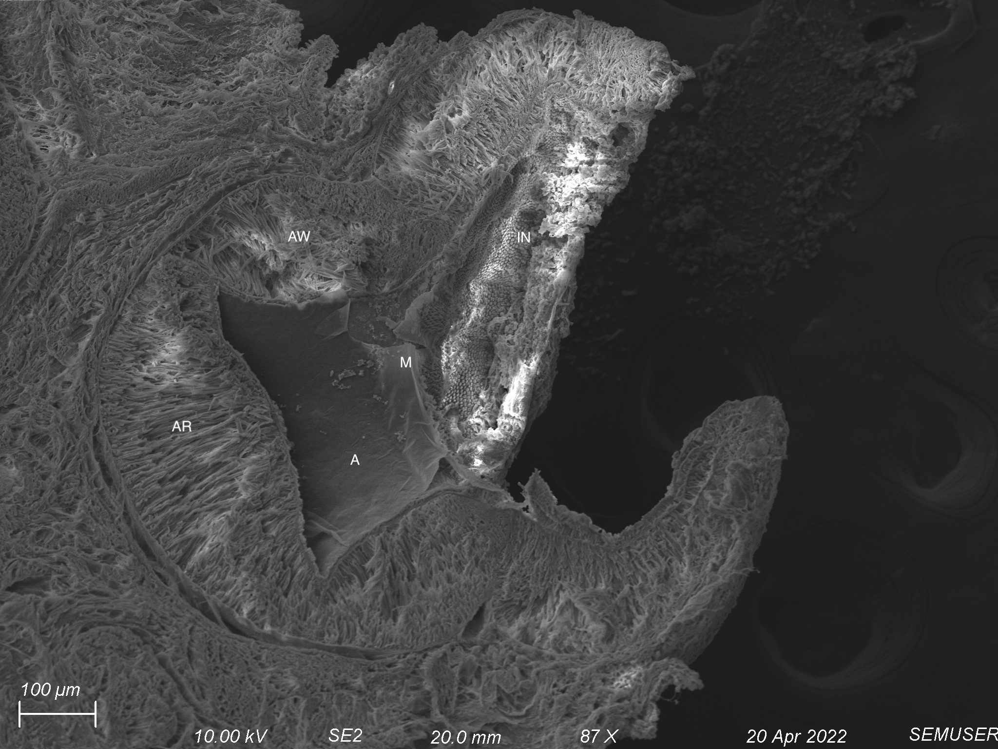
The interior surfaces of the arms are covered with circular, adhesive suckers (Figure 3.1 and 3.2). Each sucker is usually circular and bowl-like and has two distinct parts: an outer shallow cavity called an infundibulum and a central hollow cavity called an acetabulum. A cross-section of a whole sucker is presented in figure 3.3 in which different components of the suckers are identified. Figure 3.4 reveals the internal structure of the acetabulum. The outer wall of the infundibulum was found to contain numerous pegs upon closer inspection (Figure 3.5). These pegs are endowed with minute pores.
When a sucker attaches to a surface, the orifice between the two structures is sealed. The infundibulum provides adhesion while the acetabulum remains free, and muscle contractions allow for attachment and detachment.
Acknowledgments
I would like to thank our beloved instructor Brian McIntyre for his invaluable guidance and support throughout my project. I am grateful to him for introducing me to this incredible world of microscopy.
References
[1] Olóriz, F., & Rodríguez-Tovar, F. J. (1999). Advancing Research on Living and Fossil Cephalopods. Springer Publishing.
[2] Cloney, R. A., & Florey, E. (1968). Ultrastructure of cephalopod chromatophore organs. Zeitschrift Für Zellforschung Und Mikroskopische Anatomie, 89(2), 250–280. https://doi.org/10.1007/bf00347297
[3] Mazzolai, B., Mondini, A., Tramacere, F., Riccomi, G., Sadeghi, A., Giordano, G., del Dottore, E., Scaccia, M., Zampato, M., & Carminati, S. (2019). Octopus‐Inspired Soft Arm with Suction Cups for Enhanced Grasping Tasks in Confined Environments. Advanced Intelligent Systems, 1(6), 1900041. https://doi.org/10.1002/aisy.201900041
[4] Kier, W. M. (2002). The Structure and Adhesive Mechanism of Octopus Suckers. Integrative and Comparative Biology, 42(6), 1146–1153. https://doi.org/10.1093/icb/42.6.1146