A Comparison of Gun Shot Residue Through the Eras
Paul Bloom
University of Rochester, Institute of Optics
1. Introduction
Every firearm discharge produces microscopic gun shot residue particles (GSR) that can be collected and analyzed in the SEM to determine particle size, morphology, and chemical composition. Particles consistent with GSR are typically between half a micron and 10 microns and resemble partially melted spheres. These particles are the minority of GSR particles, but are easily classifiable and suggest that other chemically similar, structurally irregular particles may also be discharge residues.
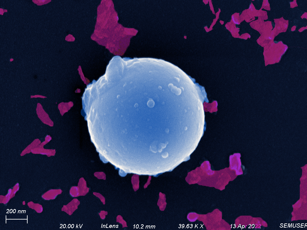
Figure 1: A particle of rifle residue consistent with the expected morphology (colorized).
GSR consists mainly of heavy elements (most commonly lead) and other taggants (synthetic markers manufactured into explosives such as gun powder) that appear bright under Backscattered electron detection. Energy Dispersive Spectroscopy (EDS) must also be performed to verify that the elements found in all GSR are present in our sample and determine which taggants were deposited during discharge.
This project sought to compare the morphologies and elemental makeup of residues from a flintlock musket replica and a modern 22 caliber rifle.
2. Gun Shot Residue Collection and Sample PreparationTo collect GSR from the desired firearms, Silicon was placed on sticky dots and the guns were fired at a proximity to ensure that some of the residue would settle on the silicon.
Figure 2: Pieces of silicon were placed on sticky dots and placed within proximity of the gun's discharge.
Experimental Note
Initially, we aimed to have samples from a rifle, musket, and a 10 gauge shotgun. However, either due to the diffusion of the shotgun residue, impact force of the shotgun pellets, or improper sample handling, GSR could not be imaged on the silicon that was supposed to collect the particles.
Interestingly, the tweezers initially used to transport the shotgun residue silicon left an imprint of stainless steel on the shot gun's designated silicon chip that was either not present or not easily visible on the other samples. The lack of shotgun residue in addition to the visibility of tweezer marks suggests that a combination of few, extremely forceful collisions between the silicon and the rapidly diffusing cone of shotgun residue, weak adhesion of the desired residues, and poor sample manipulation (or placement) prevented us from including comparisons on the morphology and chemical makeup of shotgun residue.
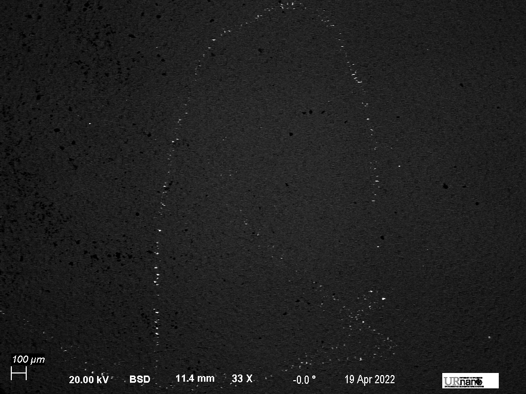
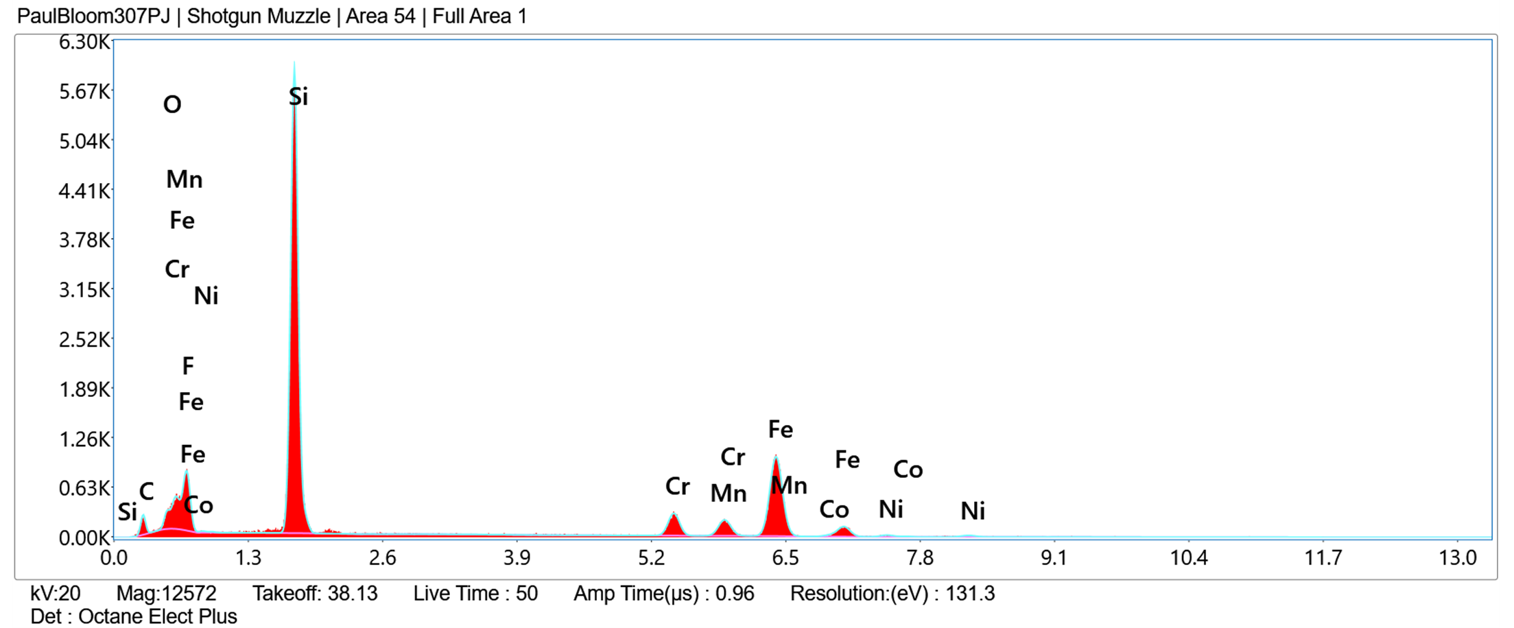
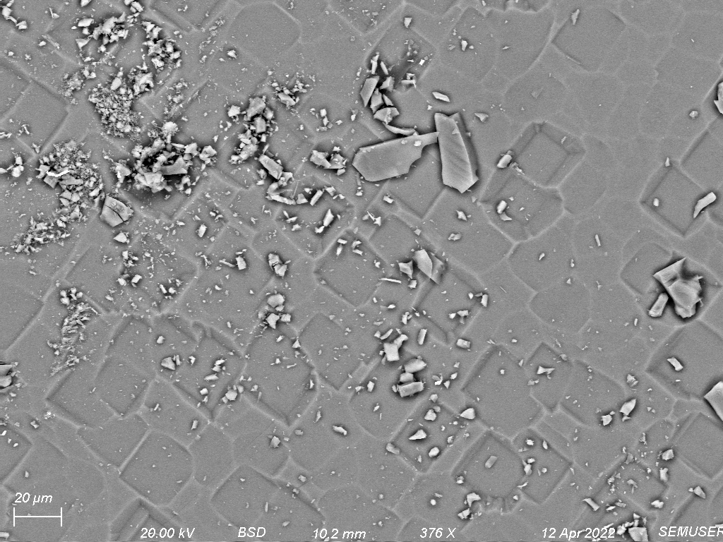
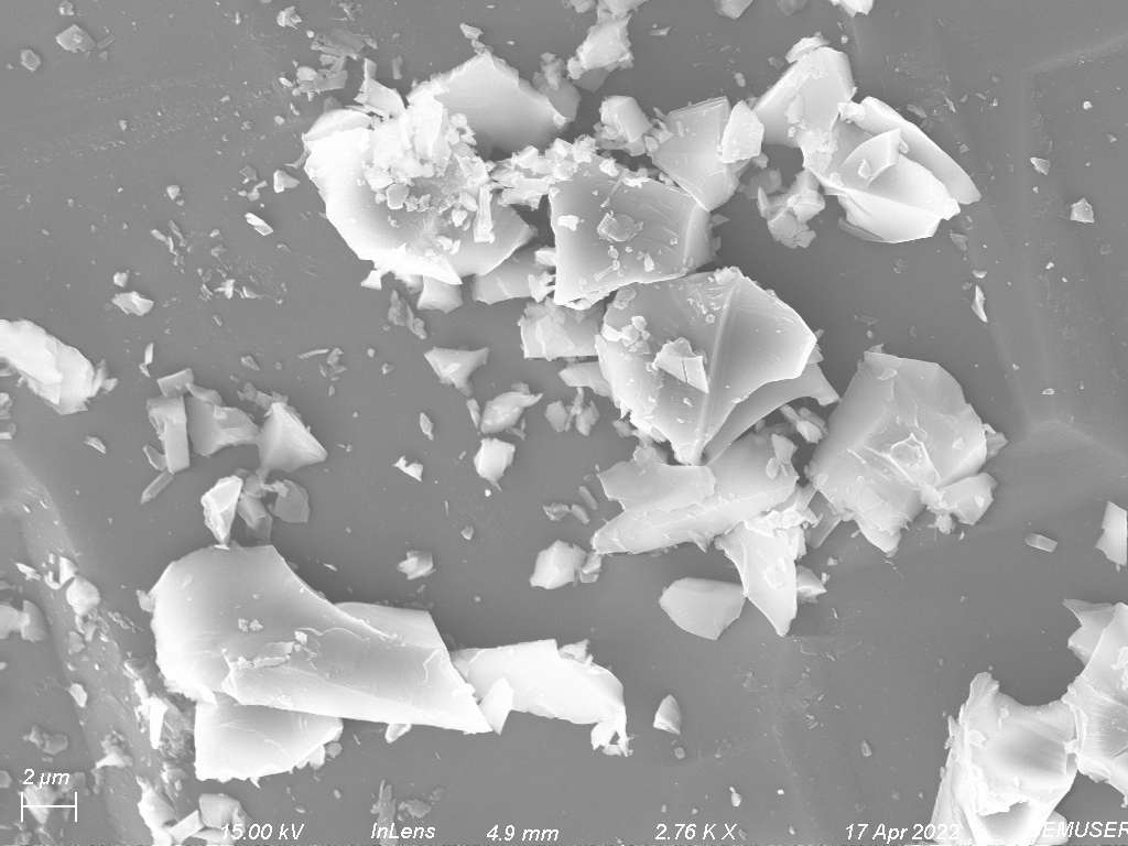
Figure 3: Top: tweezer imprint on silicon chip meant to hold shotgun residue and its x-ray spectra. Bottom: Images show stainless steel contamination.
Ample rifle and flintlock residues were collected on the designated silicon chips.
All samples were then sputter-coated with platinum to decrease charging and allow easier sample imaging without introducing artifacts.
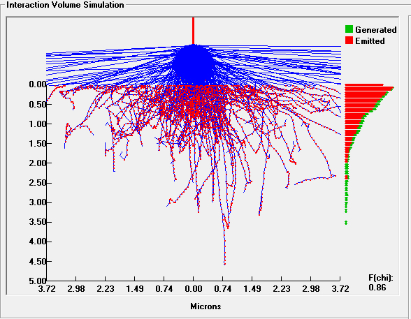
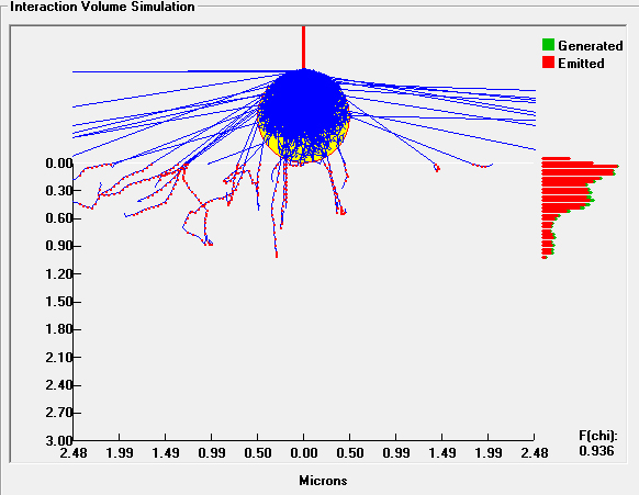
Using the EDAX EDS software and x-ray detector in the SEM, the elemental spectra and maps of our regions of interest in our samples were collected and, with backscattered and secondary electron images, we could verify that the particles collected on the silicon chips were, in fact, GSR and compare the residues between the rifle and flintlock. Interaction volumes and escape depths were then modeled via Monte Carlo simulation for electron collisions with lead particles on a bulk silicon substrate at different accelerating voltages.
Figure one was artificially colorized for viewing convenience.
3. Results
I. Morphology
To determine the plausibility of GSR being present on the samples we imaged, the first step was to look for spherical particles (figure 4) that could indicate that other, irregular particles on our silicon chip were GSR as well.
Most GSR particles will not resemble the burnt spheres that we see here, but a good way to be sure that we are imaging GSR (and not outside contamination) is to find that some are present on the samples we image.
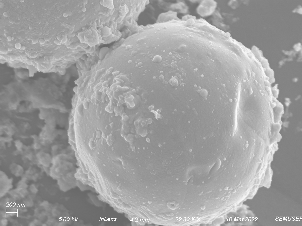
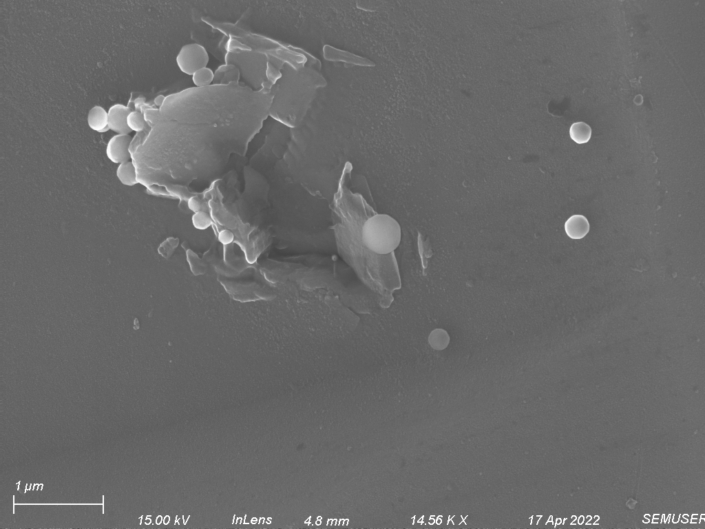
Figure 4: Spherical rifle (left) and flintlock (right) residue allows us to assume that other particles on the sample are also GSR.
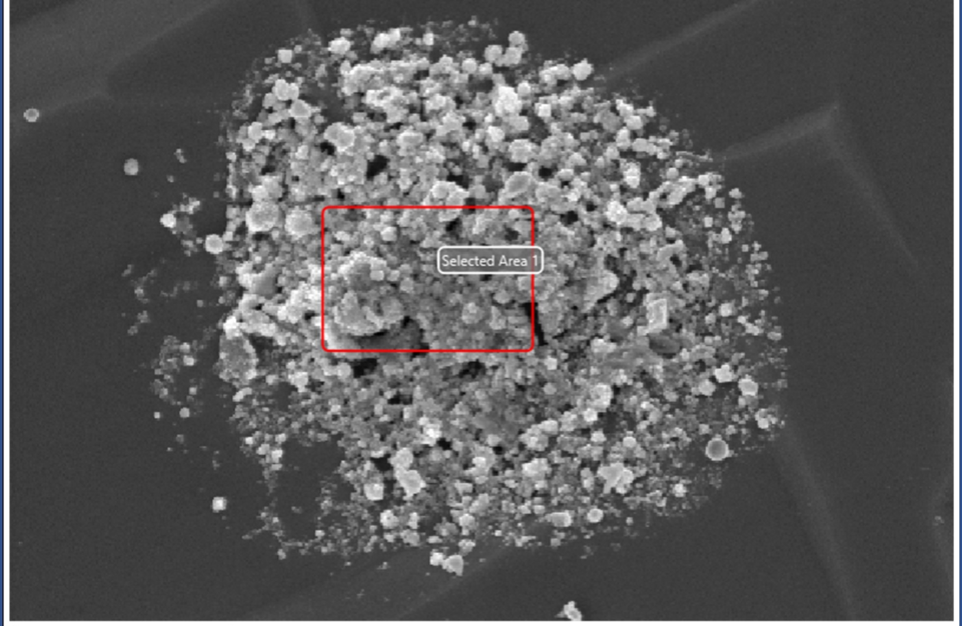
Particles on silicon chip designated for the rifle residue had a characteristic shape of particle, but it also had a common particle distribution.
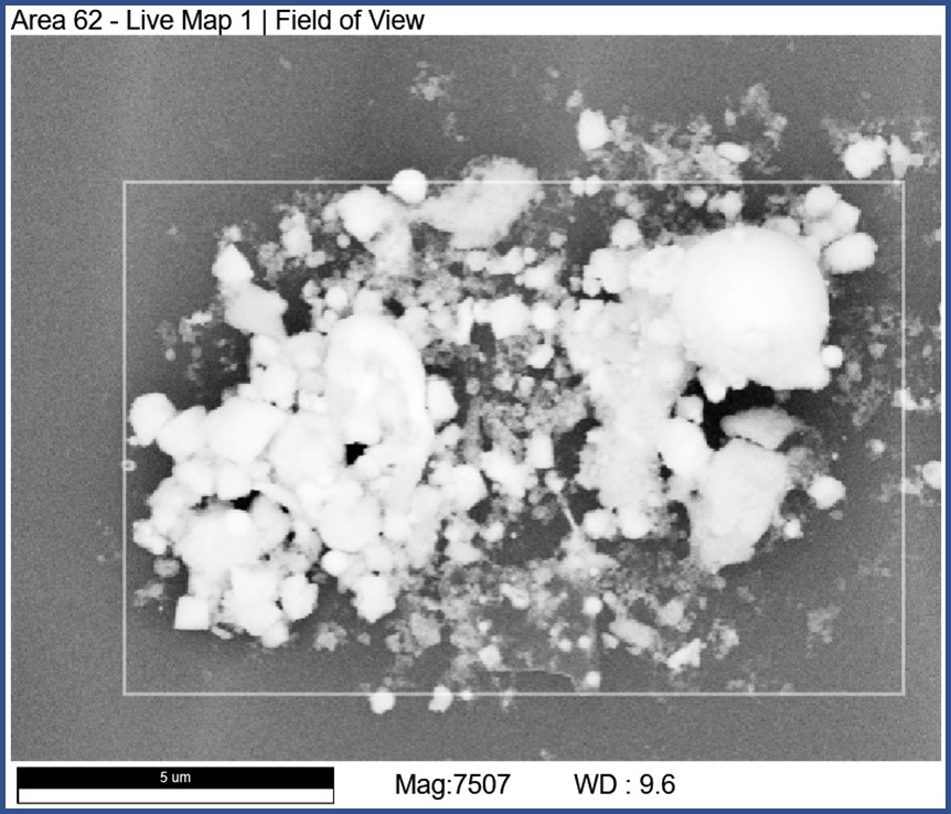
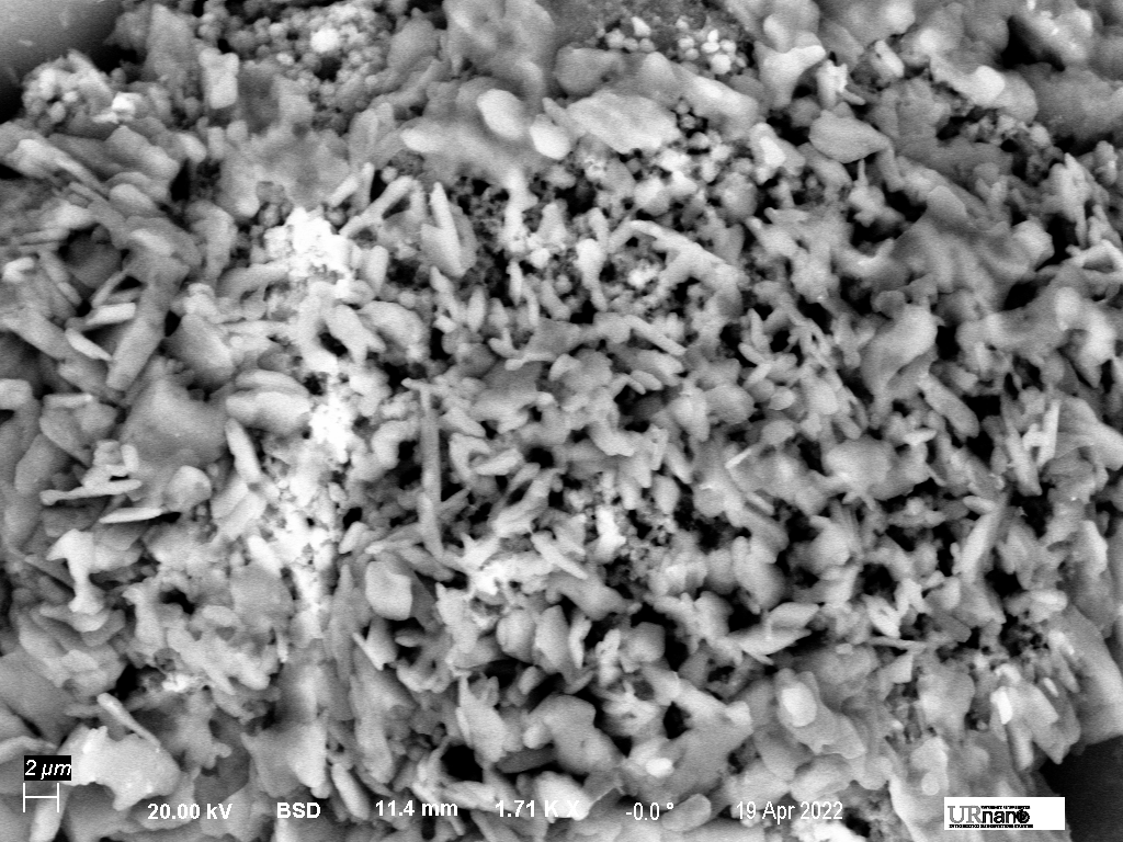
As we can see in figure 5, rifle residue was typically found in dense collections of particles ranging in sizes appropriate for GSR particles (.5-10um) and although they do not make up a majority of particles, many spheres are visible in the conglomerates at every level of magnification.
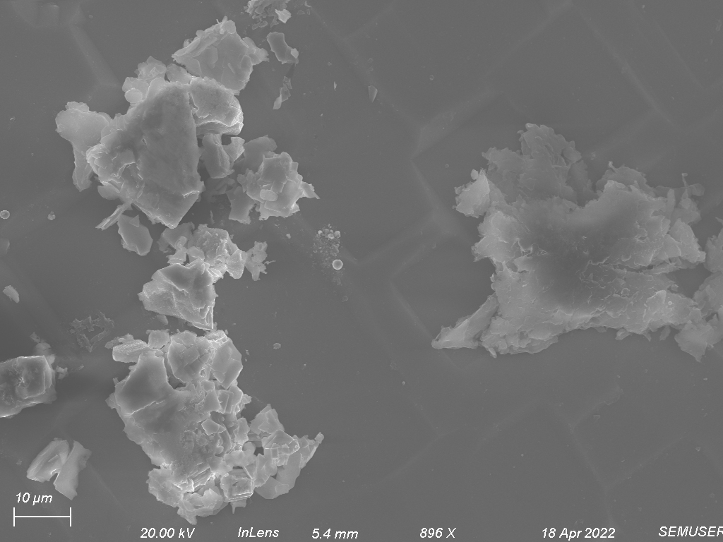
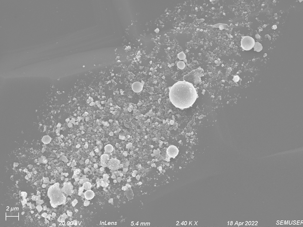
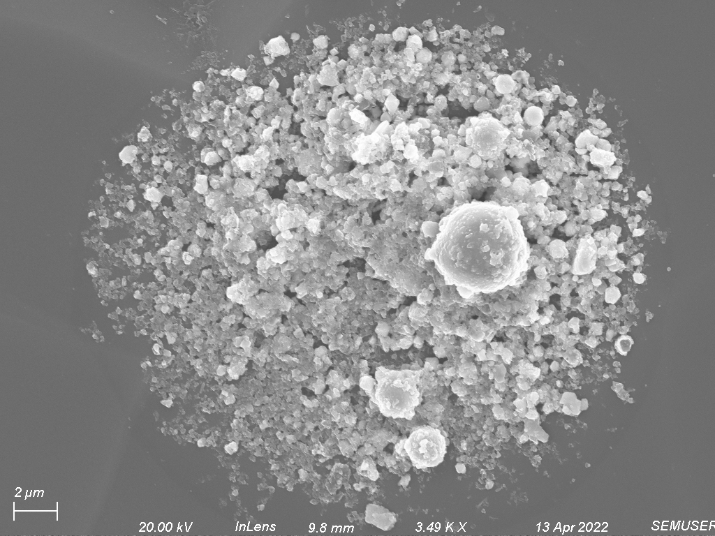
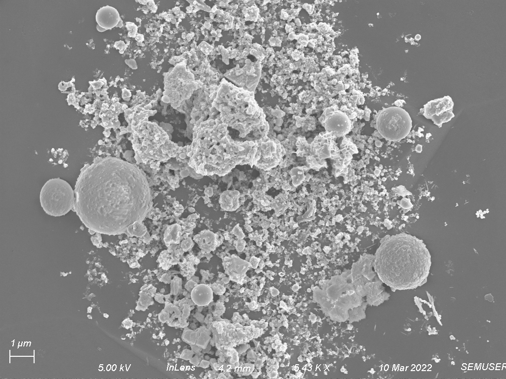
Figure 5: Clusters of rifle residue increasing in magnification.
These porous structures are frequent throughout the sample and are likely a result of extreme heat upon gun powder ignition combined with heavy metal residue that melts and solidifies after firing.
The flintlock residues have some similar characteristics, but many notable differences. Structures in figure 6 are the most common features in flintlock residue and using x-ray analysis turn out to be gunpowder. Heavier elements are also present in these gun powder deposits, as can be seen through this backscattered electron image.
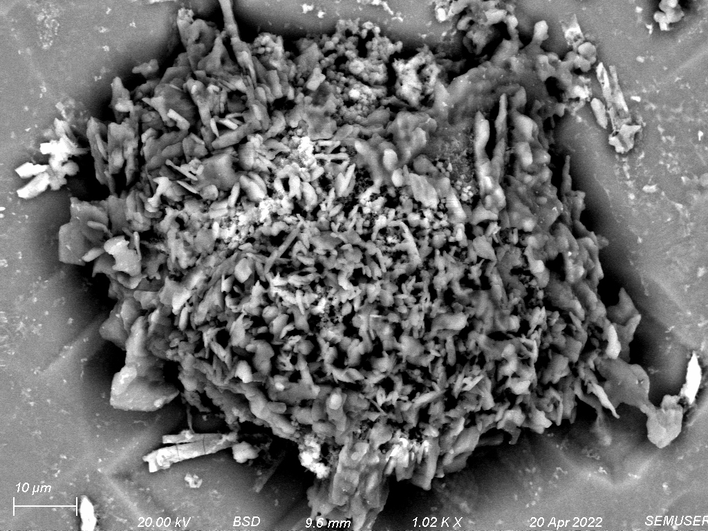
Figure 6: A gun powder deposit collected on the silicon chip designated for flintlock residue
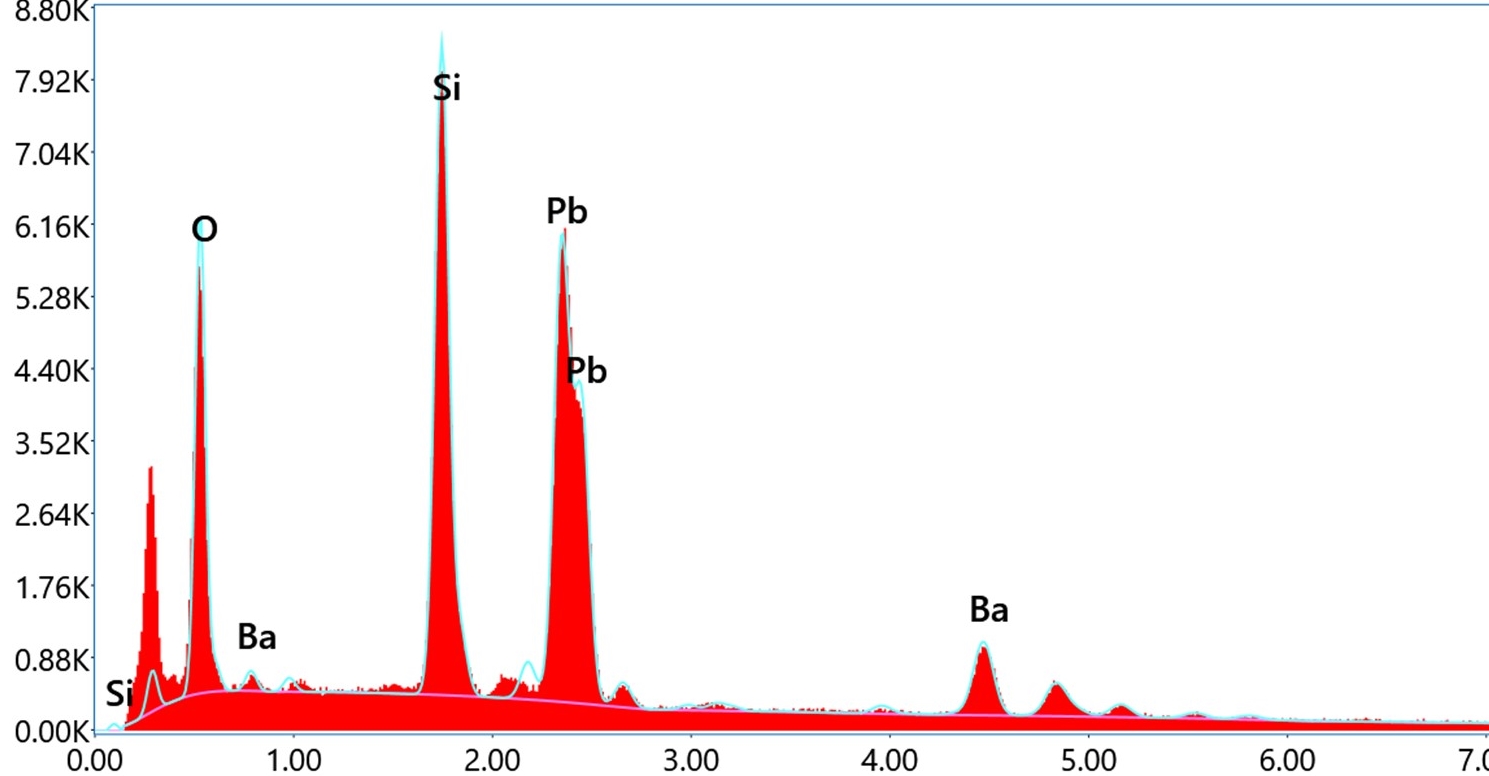
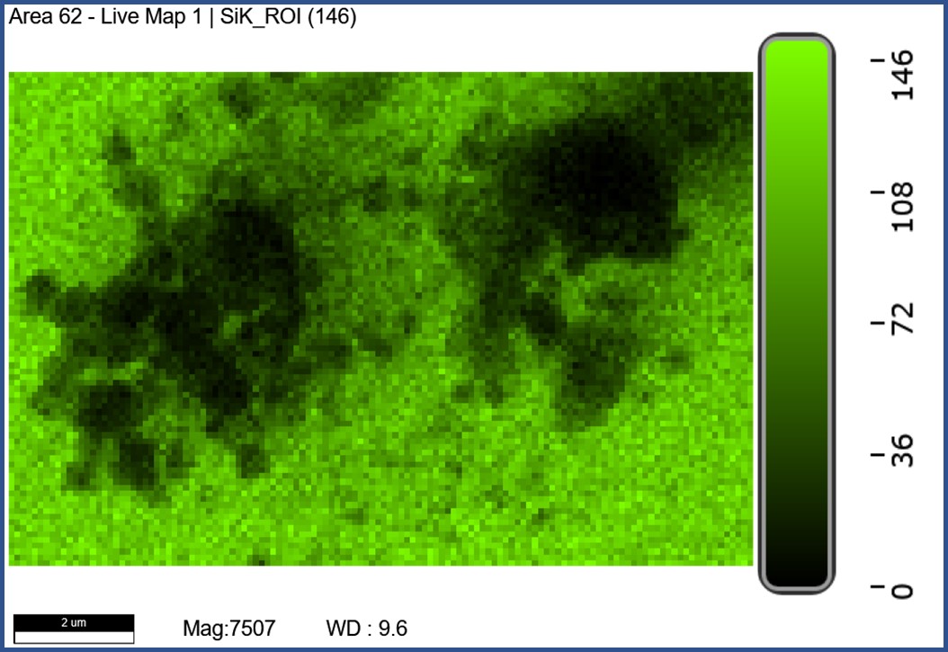
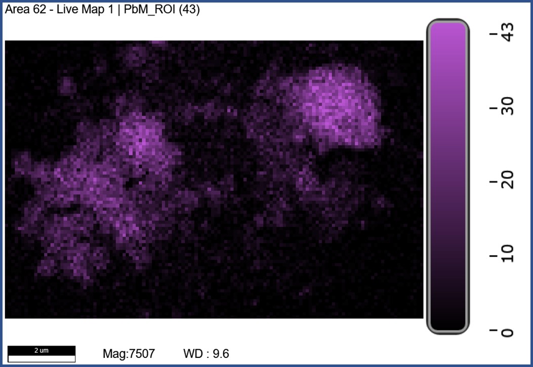
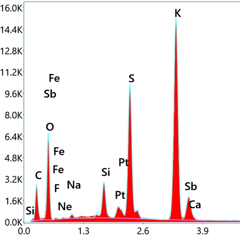
II. Elemental AnalysisX-ray and Backscattered electron detection are also crucial steps in any forensic analysis. Morphological analyses allow us to assume that the particles under investigation are GSR, but EDS must also be carried out to ensure that the particles of interest consisted of the elements we know to be common to all GSR. Surveying the particles using BSE is an easy way to identify high atomic number elements because they appear far brighter than the silicon background. As we can see in the rifle BSE images, many of the particles we identified as GSR using secondary electrons possess regions of dense elements.
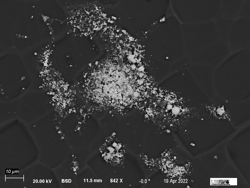
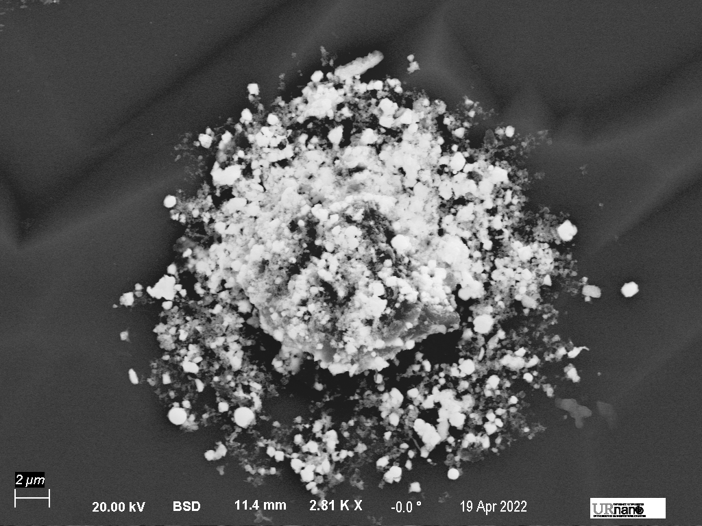
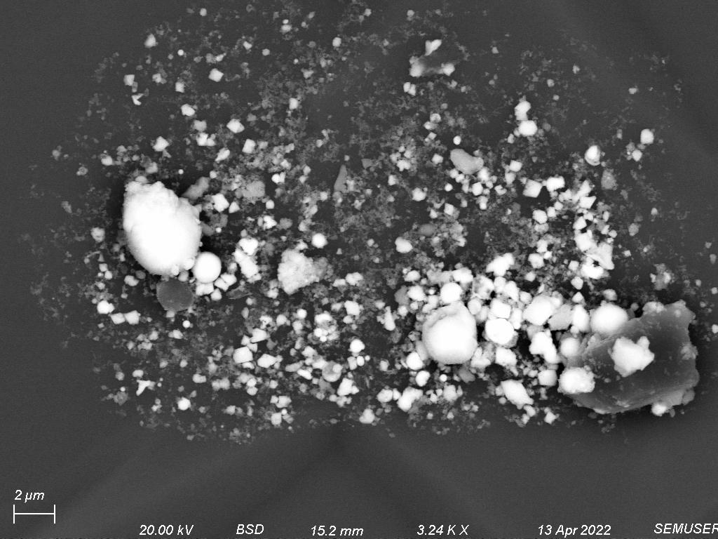
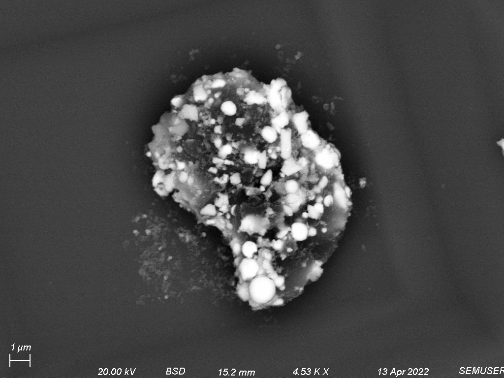
Figure 7: BSE images of rifle GSR. BSE contrast is indicative of atomic number differences on the sample. Images are organizing by increasing magnification.
Taking x-ray spectra of these regions, we find large amounts of lead and silicon signals. Because EDS was performed with a 15 or 20 kV electron beam, the penetration and escape depths of x-rays would be well into the silicon chip. This interaction volume is modeled below and shows the different x-ray counts that can escape the silicon substrate from beneath a particle of lead at a these accelerating voltages.