
Image Gallery
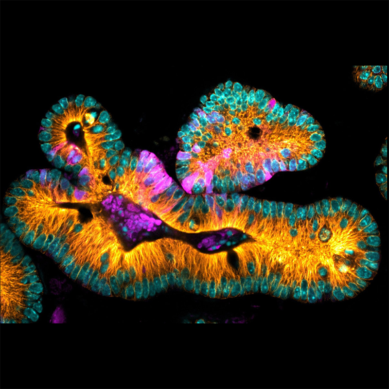
Mouse Intestinal Organoid expressing LGR5-GFP
Mouse Intestinal Organoid expressing LGR5-GFP to visualize stem cells (magenta) microtubules (orange) and DNA (cyan). Image courtesy of Nicole Dawney from the Bergstralh Lab and High Content Imaging Core, University of Rochester.
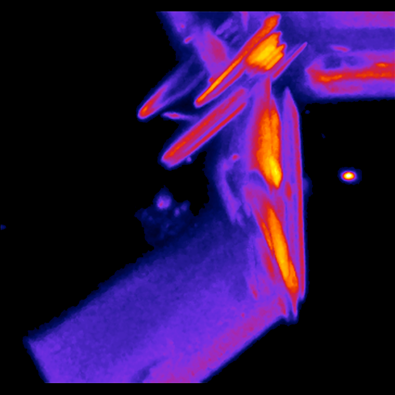
Nucleoid-associated proteins from E.coli combined with DNA
Nucleoid-associated proteins from E.coli combined with DNA to form a gel, which can be mechanically proved and characterized using optical tweezers. Imaged with laser scanning confocal on Lumicks C-trap. Image courtesy of Elio Abbondanzieri (Meyer Lab).
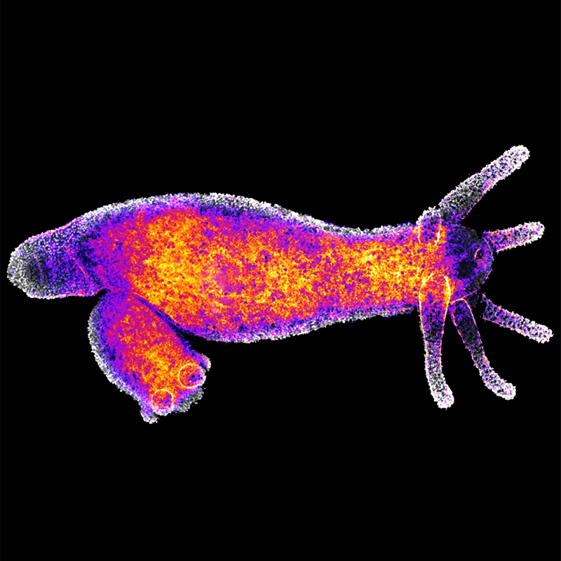
Hydra vulgaris adult with bud
Nuclei (white) and actin (fire LUT) highlighted. Imaged on Andor Dragonfly spinning disk confocal microscope. Image courtesy of Lindsay Rathbun (Bergstralh Lab).
iPSCs (induced pluripotent stem cells) with ZO-1, Claudin-5, and nuclei highlighted
Imaged on the Andor Dragonfly spinning disk confocal microscope. Image courtesy of Molly McCloskey and the McGrath Lab.
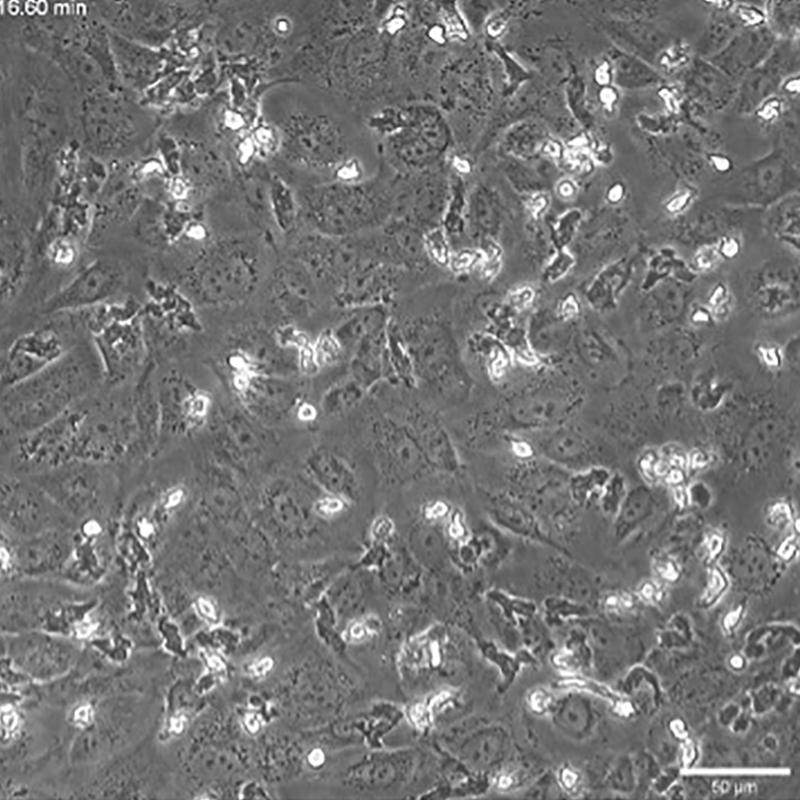
Neutrophil migration across a silicon nano-membrane
Phase contrast image on Etaluma Lumascope courtesy of McGrath Lab.
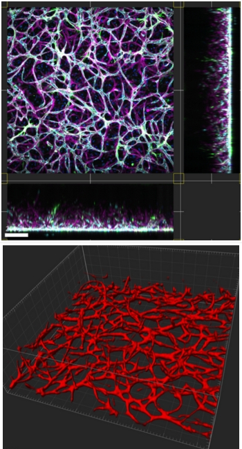
HUVECs and hMSCs forming 3D microvasculature inside µSIM devices. VE-cadherin (teal), collagen IV (magenta), alpha-SMA (green), Hoescht (blue), and CD31 (red) highlighted
Spinning disk confocal (top) and 3D surface rendering (bottom) images courtesy of Kevin Ling (McGrath Lab).
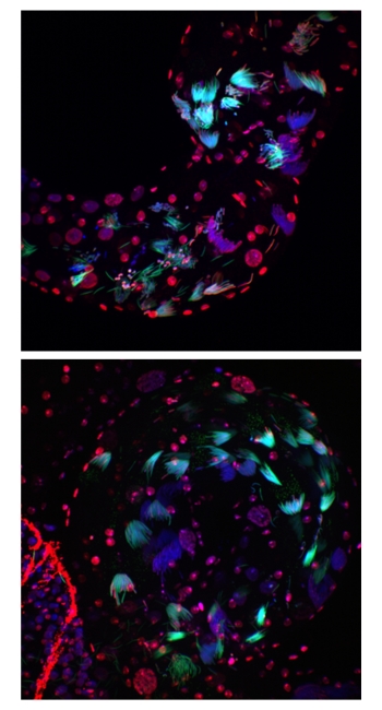
Spermatogenesis in Drosophila melanogaster. Protamine B (green), DNA (DAPI, blue), and dsDNA (red) shown
Andor Dragonfly spinning disk confocal images courtesy of Logan Edvalson (Larracuente Lab).
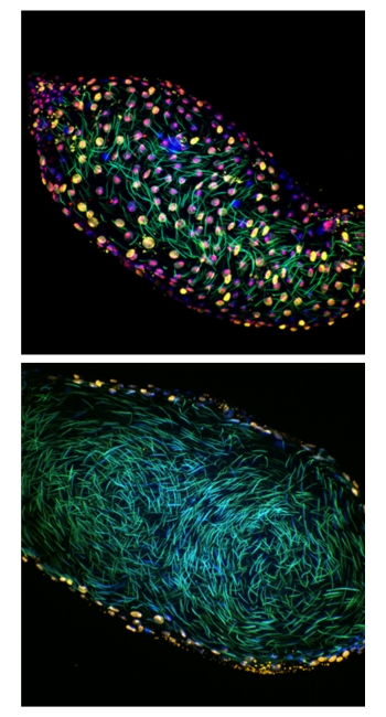
Spermatogenesis in Drosophila melanogaster. Protamine B (green), DNA (DAPI, blue), H3K9me3 (red), and dsDNA (yellow) shown
Andor Dragonfly spinning disk confocal images courtesy of Logan Edvalson (Larracuente Lab).