Setting and Background
Hansen Point BasaltsLate Cretaceous to Paleogene volcanic rocks exposed in the High Canadian Arctic region provide an opportunity to examine the field close to (or within) the ancient tangent cylinder. Recent work has focused on a phase of magmatism at ~77 Ma, postdating eruption of flood basalts of the Strand Fiord Formation (~95 Ma). The younger volcanics are exposed in a series of down-faulted basins stretching more than 150 kilometers along the northwestern coast of Ellesmere Island.
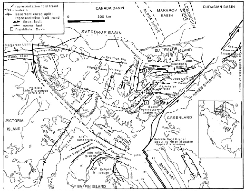
The series of volcanic and magmatic emplacements that compose the informally titled "Hansen Point volcanics" unconformably overly Paleozoic Nansen Formation rocks in the sampled regions of northwestern Ellesmere Island. In general, this region's volcanism is categorized by Embry & Osadetz (1988) as members of Late Cenomanian-Maastrichtian cycle, which is the fourth and final cycle of emplacement for Cretaceous age volcanics. This unnamed region between Emma Fiord and Audhild Bay was described as "Audhild Peninsula" (AP) by Daniel Sinnett in his senior thesis, which will be adopted for the remainder of this paper to describe the area, is considered by Embry and Osadetz (1988) to be the most litholigically diverse region of the Hansen Point volcanics.
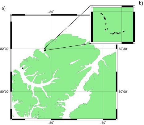
In 2008, the Paleomagnetic Research Group led by Dr. John Tarduno returned to Ellesmere Island to continue study on the Late Cretaceous volcanics. Freshly melted snow allowed for sampling within the “Hansen Point” region (82.5° N, 82° W) of a series of basaltic, plagioclase phryic dikes cutting through rhyolite. This series of dikes is believed to post-date previously studied Hansen Point and Audhild Bay volcanics. A total of 21 sites were sampled, collecting over 140 various cores and hand samples for study.
Rocks containing titanomagnetite, when exposed to water, can undergo maghemitization, in which oxidation results in a loss of iron and an introduction of oxygen transforming titanomagnetites into titanomaghemites. The potential for a reversal in the recorded moment during this oxidation exists since the dominant B sublattice magnetic moment in titanomagnetite is replaced by the A sublattice as the dominant magnetic moment within titanomaghemite, resulting in a change in polarity of the recorded moment (Doubrovine and Tarduno, 2003). This oxidation results in an exsolution of titanomagnetites into ulvospinel-rich and magnetite-rich end members; the mechanism of inversion is most likely spinodal decomposition (Price, 1980), which requires low temperature environments. This method of inversion requires low temperature environments, consistent with observed temperatures of inversion in oceanic basalts (~250-500 °C depending on amount of oxidation) (Doubrovine and Tarduno, 2003).
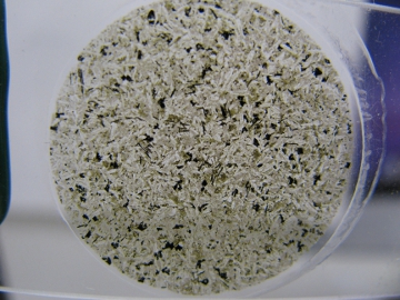
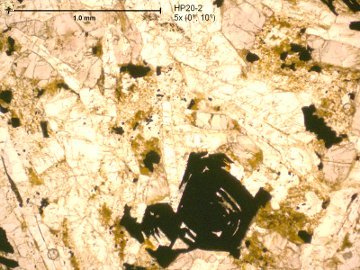
Samples collected from Hansen Point are basaltic in composition and have been exposed to an aqueous enough environment in which self-reversal is a potential occurrence. Preliminary lines of evidence suggest that some of the titanomagnetite within the rocks have undergone inversion in titanomaghemite. Scanning electron microscopy of thin sections (prepared by Gerry Kloc at the University of Rochester) shows lamellar planes of light and dark material found within grains of iron-rich oxides. Energy dispersive x-ray spectroscopy and elemental mapping revealed inversely correlated concentrations of titanium and iron within the thin section. This evidence strongly suggests that exsolution (and resulting self-reversals) is a significant concern with basalts sampled from Hansen Point.
Sierra Nevada Granites
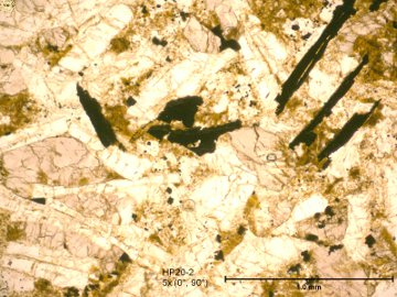
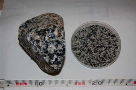
The Sierra Nevada mountain range in California and Nevada ranges 640 km north to south and is ~110 km across. Magmatism took place in the Sierra Nevada between 220 Ma and 80 Ma (Saleeby et al. 1987), although most of the exposed plutons yield ages of 125-85 Ma (Saleeby et al. 1990). The Sierra Nevada batholith consists of several plutons with surface outcrops ranging in size from less than 1 km2 to over 100 km2. The compositions of plutons are mainly tonalitic to granodioritic and granitic (Ducea et al., 1998). The batholith covers ~90% of the central and southern Sierra Nevada range, with only ~65% coverage of the northern part. No significant metamorphic event has affected the Sierra Nevada batholith after its formation (Ducea et al., 1998). Due to the lack of significant metamorphism, much of the igneous textures are preserved. Sampling from the Yosemite granodiorites have allowed for unusually large (2-5 mm) plagioclase and quartz crystals to be isolated, future work will constrain the rock magnetic characteristics. This project focused on collecting high depth-of-field micrographs of the individual plagioclases.
Methods Used:
- Thin section light microscopy conduted on a Nikon light microscope with camera (before project)
- Sputter coating (not shown on site): Thin sections of the Hansen Point volcanics were cut and sputter coated with about 60-120 Angstroms of gold to produce a conductive surface using the Denton Vacuum metal sputtering system
- Secondary Electron Detection (SE2 detector) using the Zweiss-Auriga FIB/SEM.
- Backscattered Electron Detection using the Zweiss-Auriga scanning electron microscope with backscattered electron detector insert
- Energy Dispersive Spectroscopy using the EDAX x-ray spectrometer
- Elemental Mapping using the EDAX in scanning mode
- 3D Anaglyphs using SE2 micrographs, compiled together using GIMP 2.0