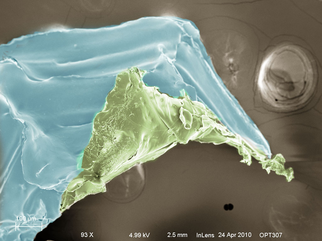
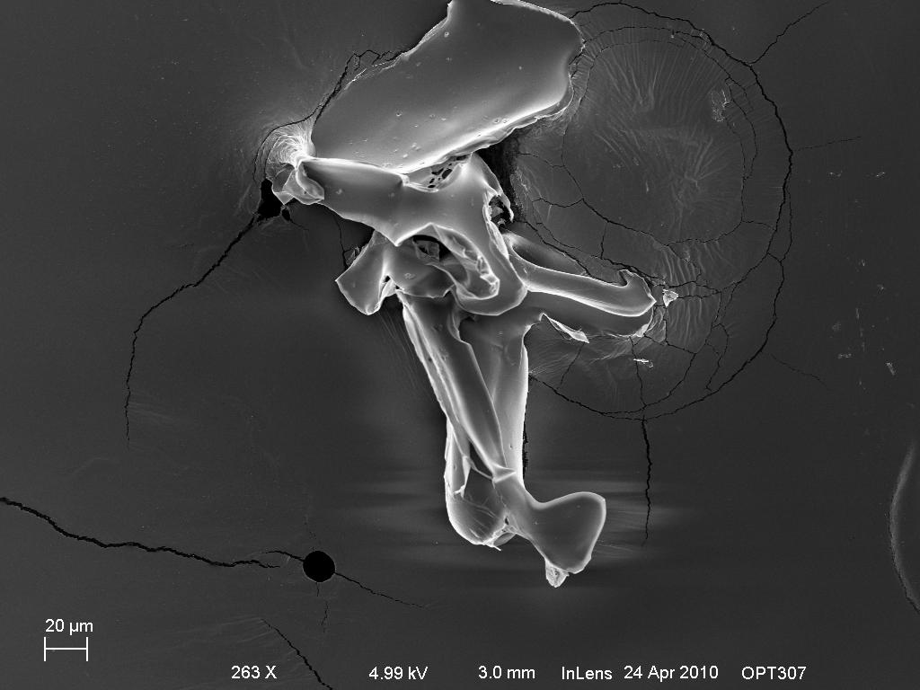
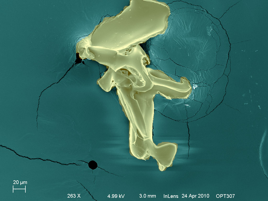
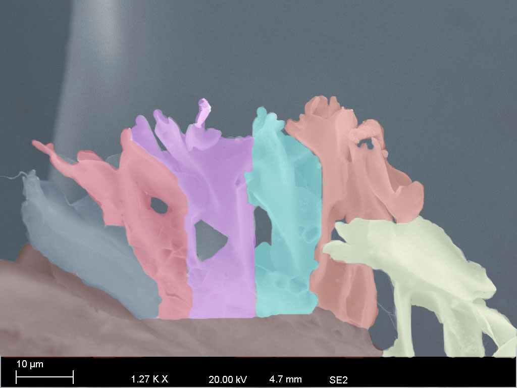
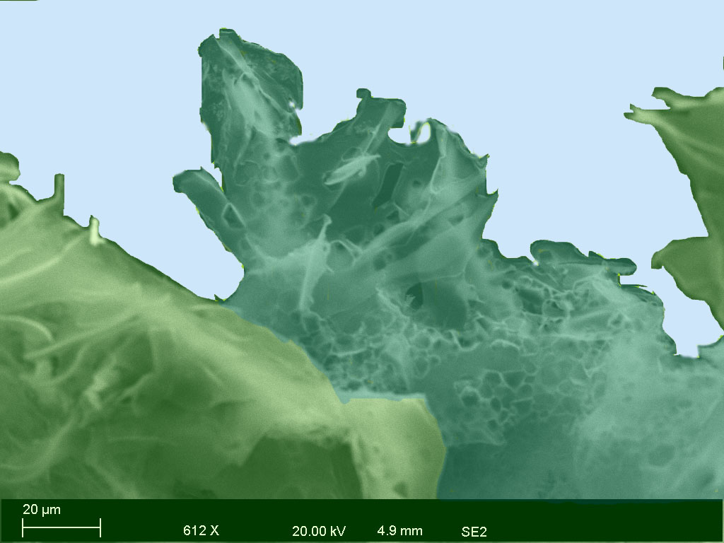
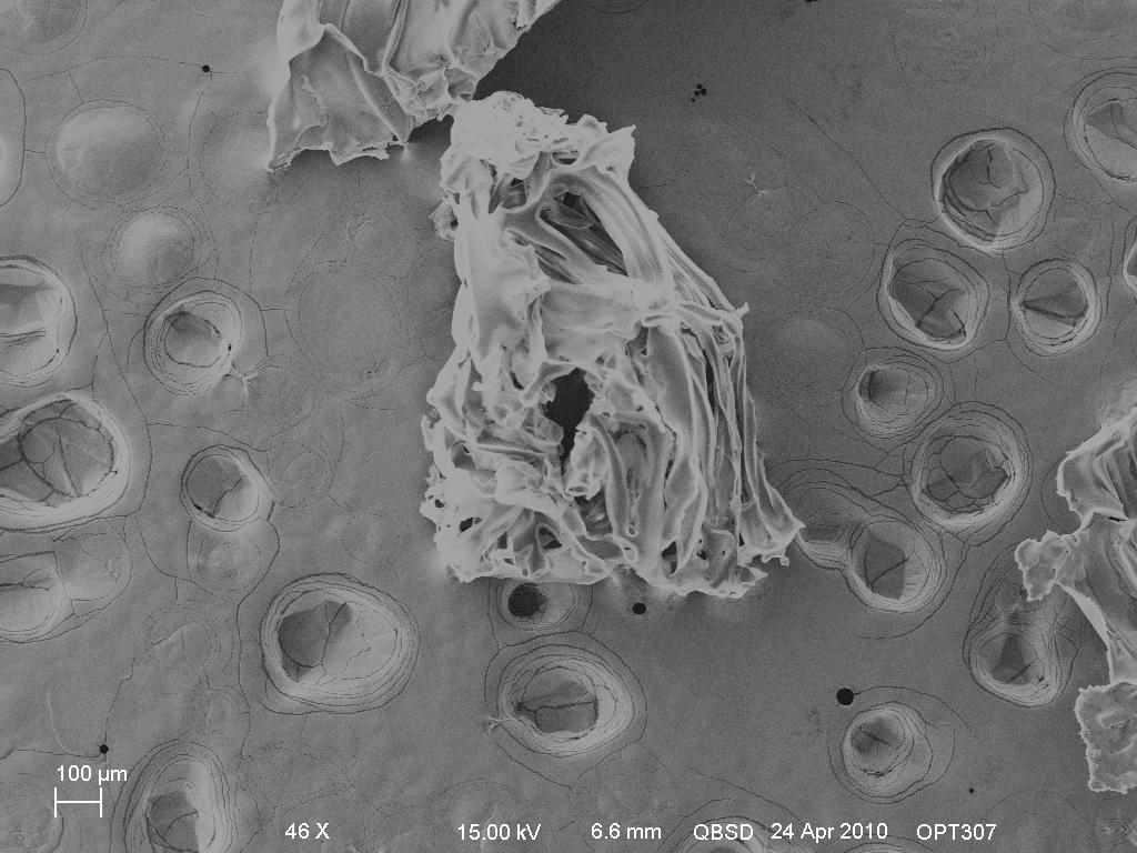
The following images are all micrographs taken using
the secondary electron imaging technique (SE2,and inlens detector).
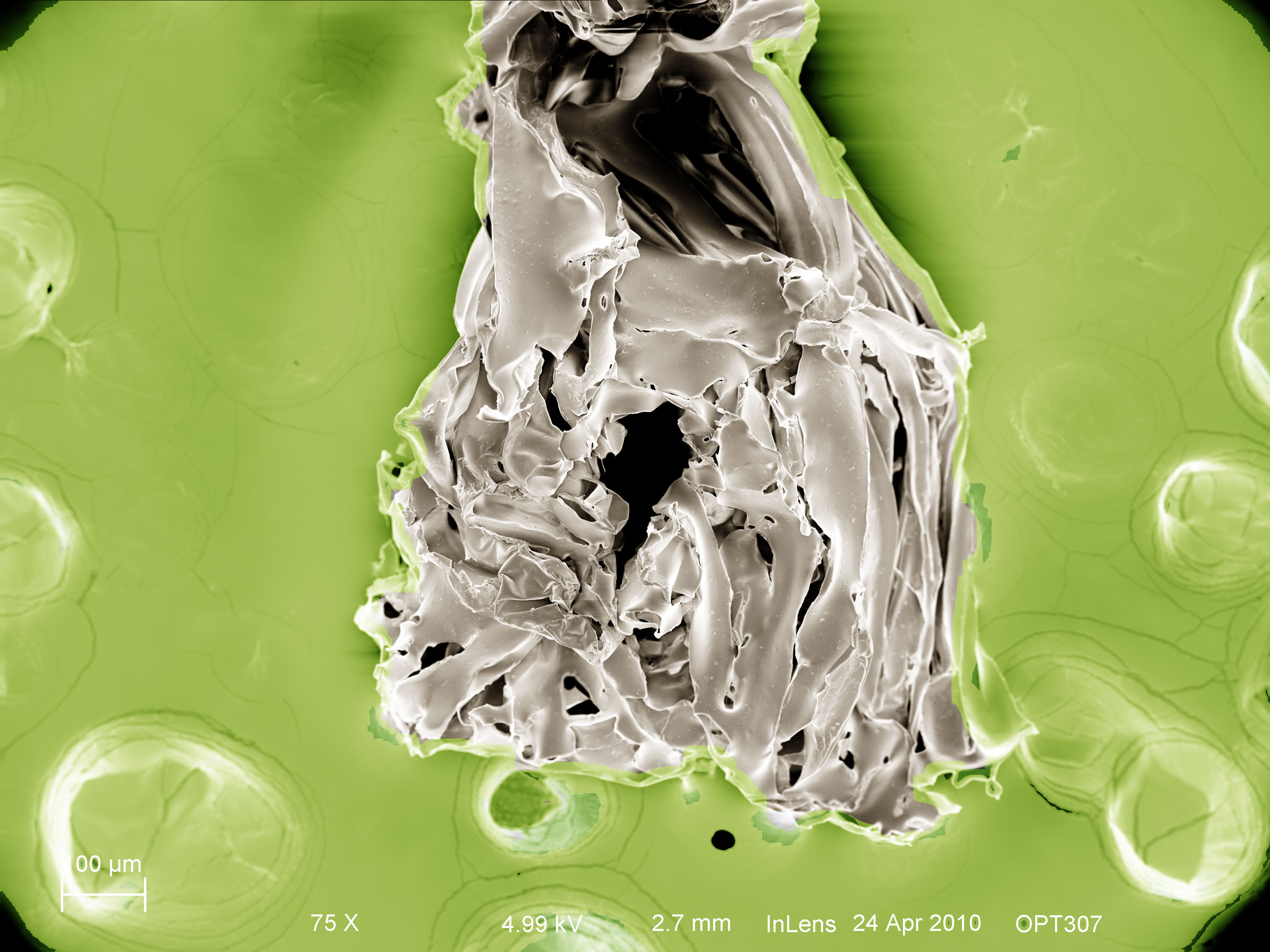
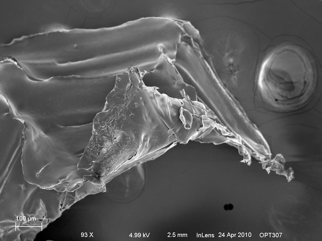
Because the ice replicas are relatively large, in order to achieve,
the depth of field is not optimal. As we can see, the freezer
ice replica does not possess the same hexagonal
structure
as the snowflakes. This confirms our earlier assessment that
perfect
hexagonal snowflake structure can only be formed with proper
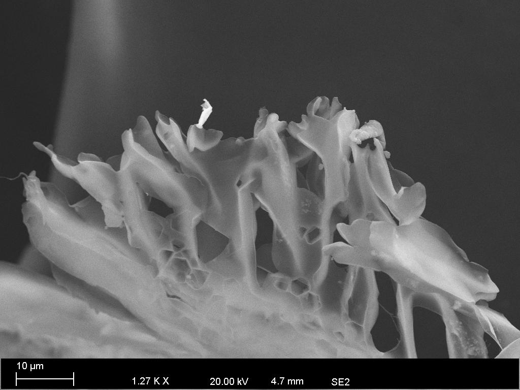
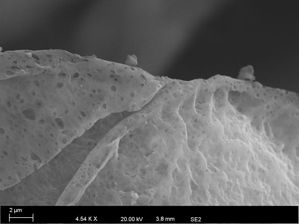
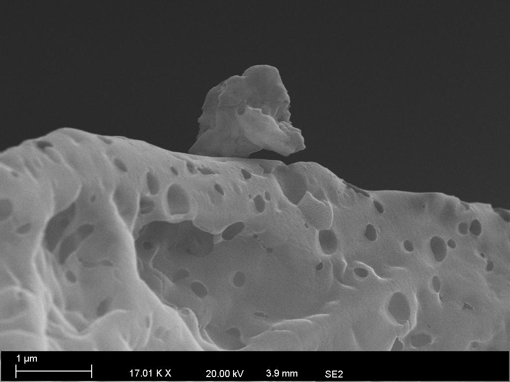
temperature and humidity.The last two images are previously
collected snowflakes samples which showed dramatic difference with
the freezer ice sample.
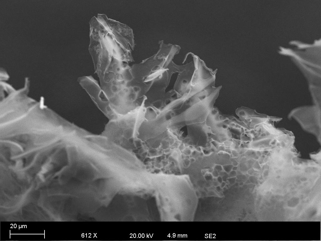
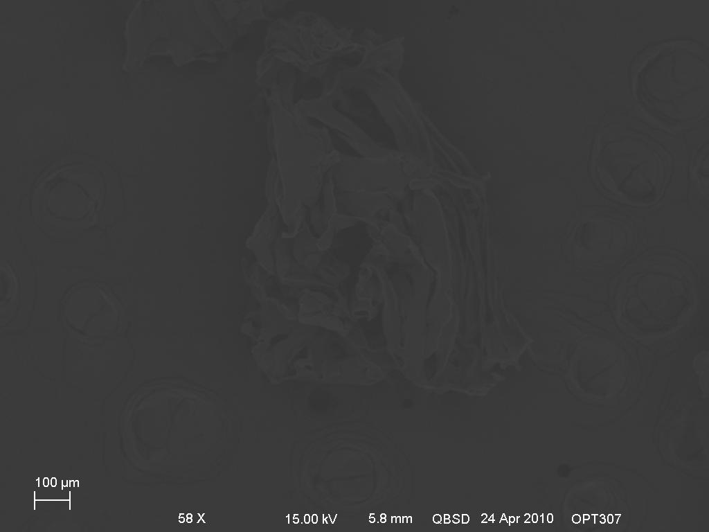
the following micrographs are taken with backscatter electron imaging technique, however, due to
there are no high atomic number element present in my sample, we can't really see much with this technique.
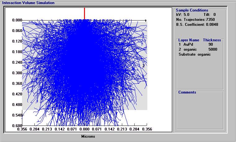
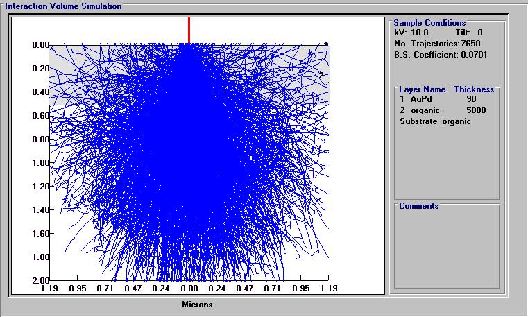
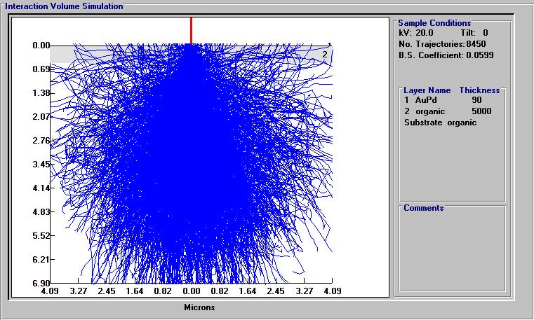
I used electron fly simulation technique to simulate the electron interaction volume. I used three different accelerating voltage, 5KV, 10KV, and 20KV.
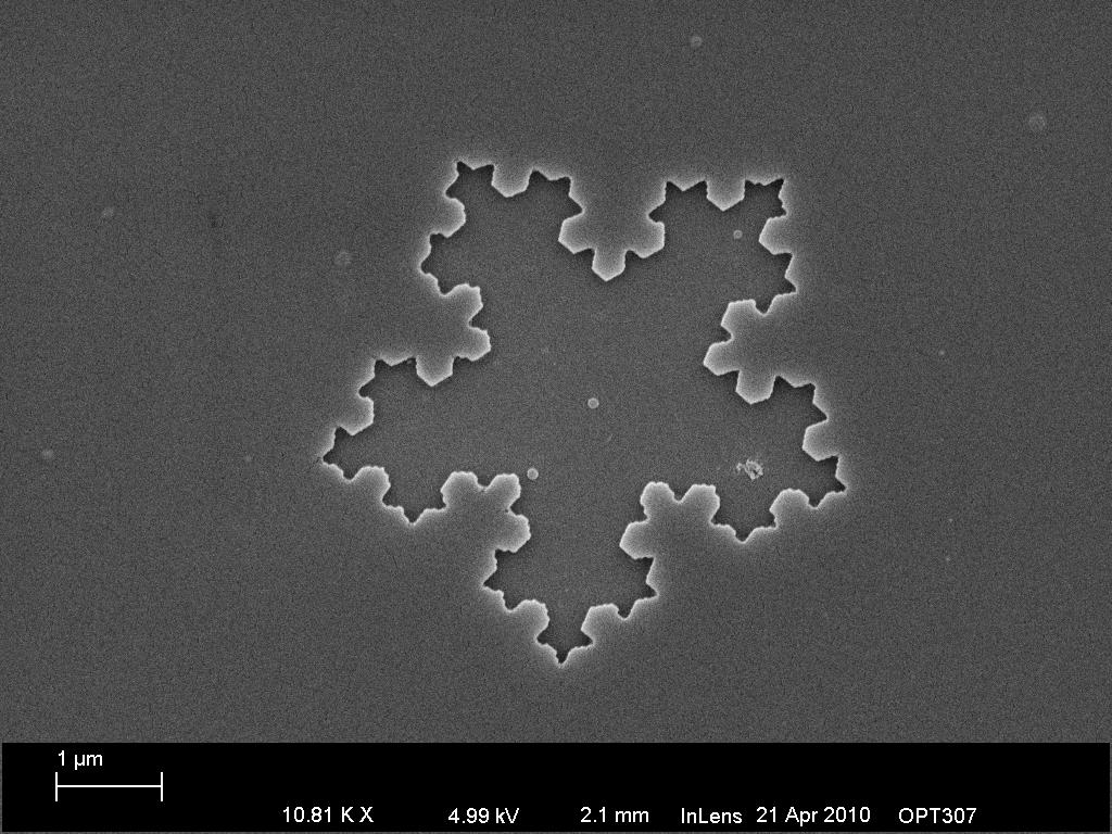
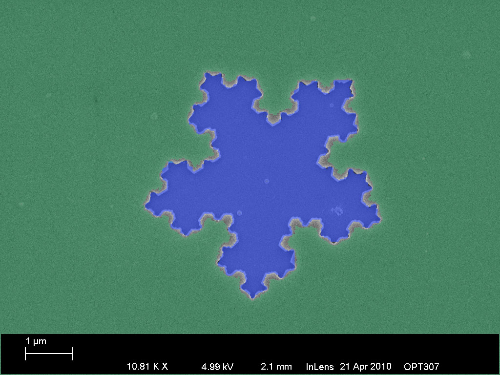
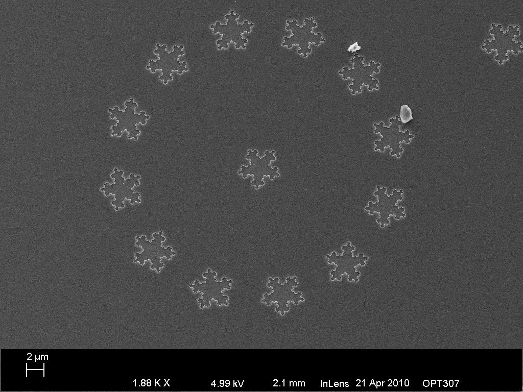
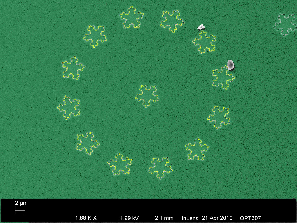
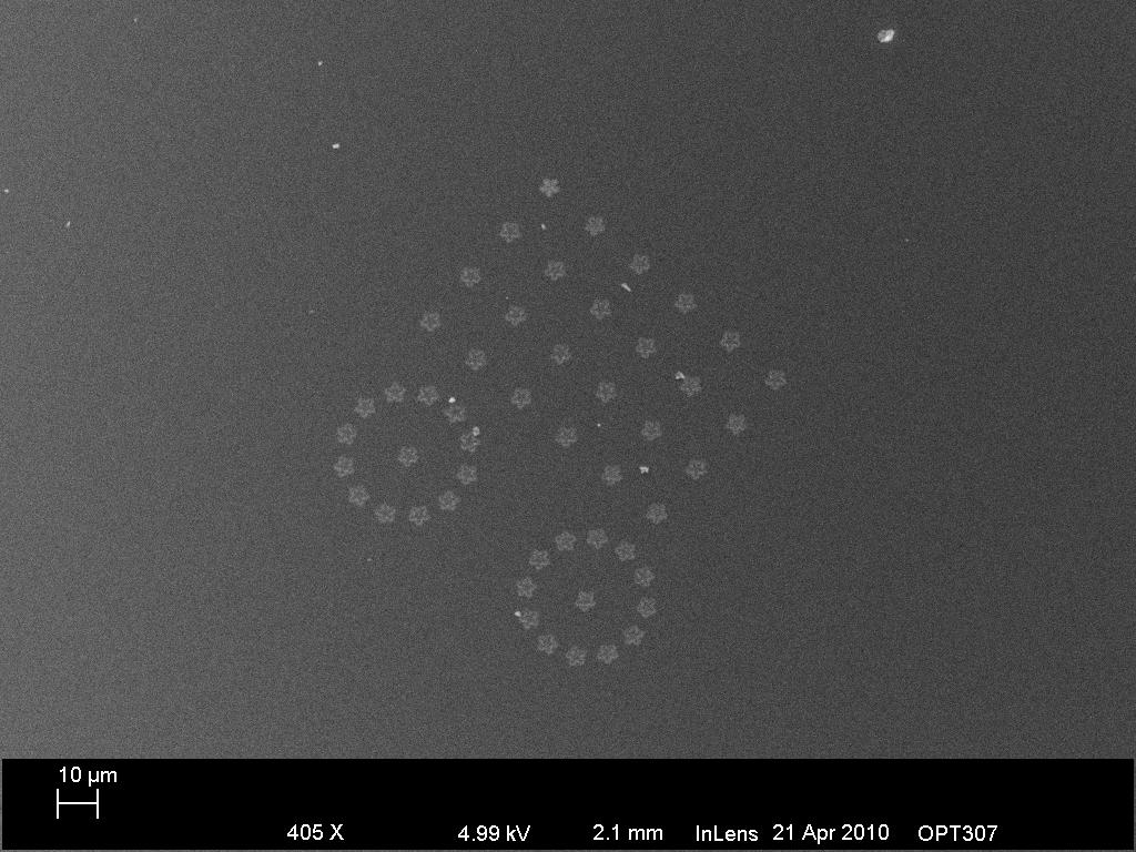
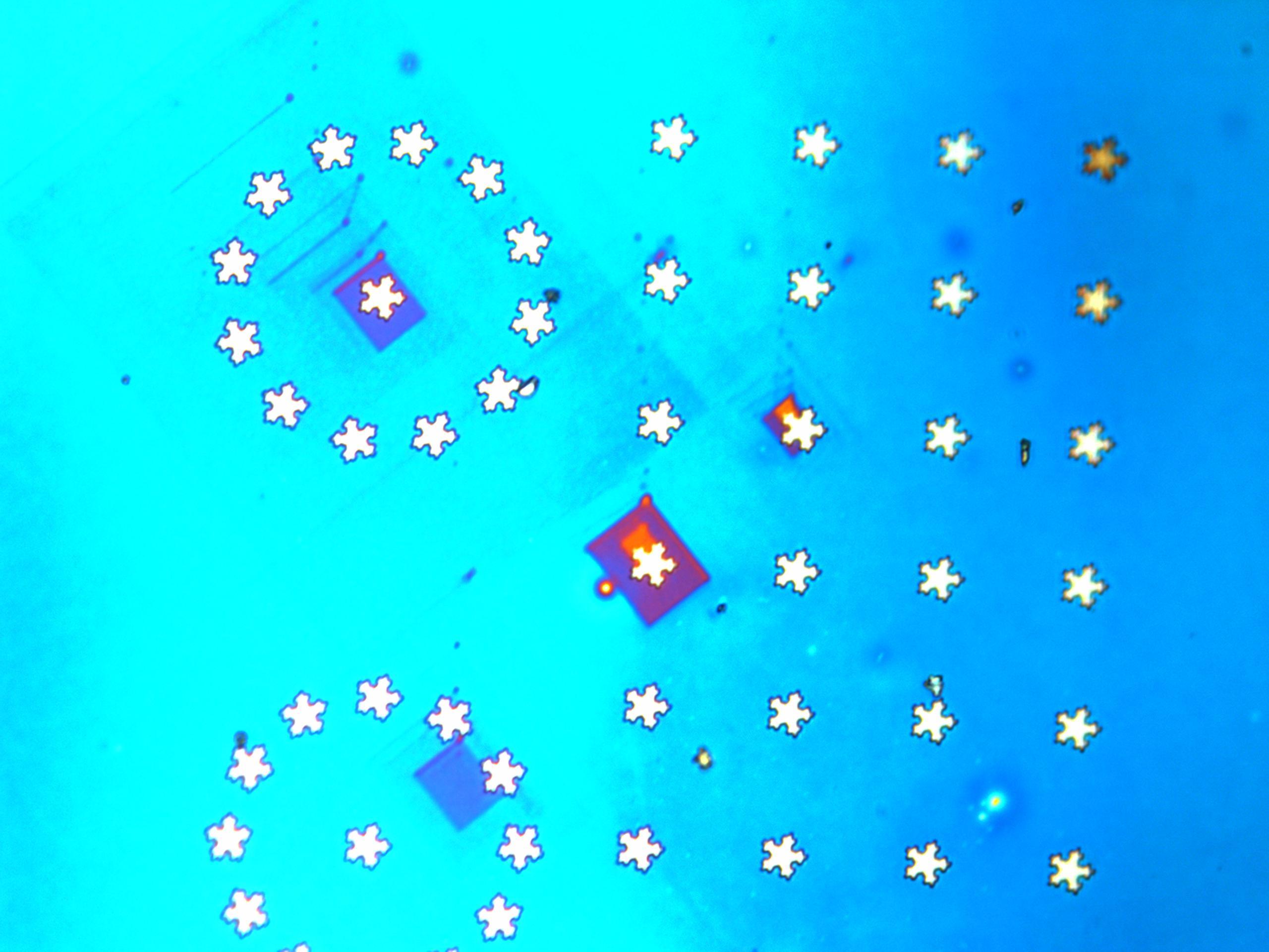
I also made snowflake patterns using E-beam lithography, it's all positive resist with line dose.