building room 116, the other one is located at the SEM preparation lab in the wilmot building of
the University of Rochester.
The ice crystals were collected via filter papers, then a couple droplets of 2% Formvar in
chloroform solution were dropped around the edge of the crystals.
To ensure that the temperature of the solution is lower than the temperature of the ice, the
solution was previously cooled by liquid nitrogen for three to four seconds.
After which, the filter papers were stored in room temperature so that the solution would
evaporate and only the plastic cover could remain.
Then, the portion of a filter paper was mounted on a SEM sample stub. The stub is
then sputter coated with gold for 90 seconds with 15mA current.
Finally, the preparation of the PMMA on silicon substrate was done by the teaching assistant
of opt 407 class- Andreas Liapis.
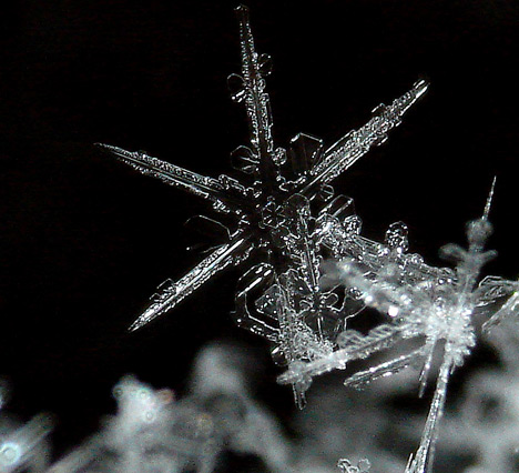
Figure.1 A SEM micrography of snowflake.