
Methods
The purpose of this project is to image the Marcellus Shale and any deformation features.
For this project we viewed the Marcellus shale with three tools, the light microscope, the atomic force microscope, and the scanning electron microscope. Each tool provides unique information and their combined use allows us to make significant progress in understanding the microscopic structures of the Marcellus shale. More detailed methods are discussed below.
Light Microscopy
Thin sections were prepared by Gerry Kloc at the University of Rochester EES department. Calcite Veins and bulk Marcellus shale were imaged using a petrographic microscope in plain and cross polarized light. Light microscopy is excellent at showing the different crystals within veins, and highlighting different minerals.
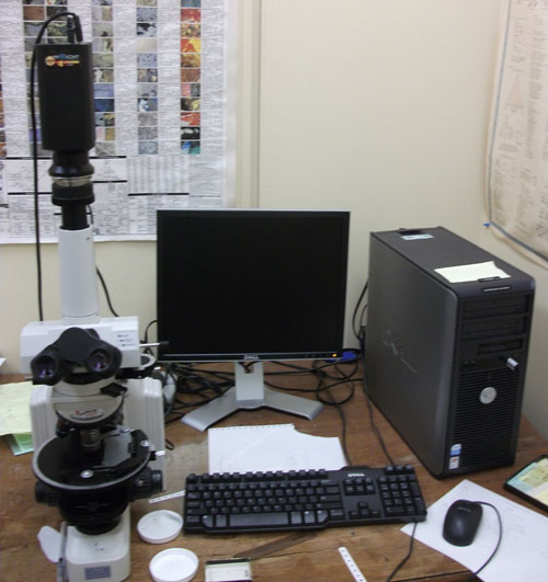
Petrographic Microscope and Camera
(University of Rochester, Structural Geology)
Atomic Force Microscopy (AFM)
With the help of Sergey Korjenevski, Atomic Force Microscopy was used to image shale grains which often have a submicron grain size. This technique moves a probe across the sample which is deflected by the atomic force of the sample surface. This probe uses a cantilever and tip system to interact with the sample and determine its shape without the use electrons or light. It can provide detailed 3-D mapping of the sample's surface topography.
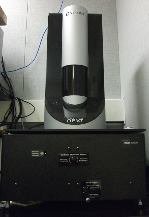
Atomic Force Microscope
(University of Rochester, Optics)
Sputter Coating
Sputter coating is a process which covers samples with a thin layer of gold. Metal atoms are ejected from a target that is bombarded by heavy gas ions (Ar), and coat all surfaces. All samples for this project were coated with 60 angstroms of gold. This process makes the surface of samples electrically conductive, allowing them to be imaged with a SEM.
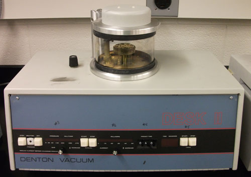
Sputter Coater
(University of Rochester, Optics)
Scanning Electron Microscopy (SEM/FIB with EDAX)
The Scanning Electron Microscope (SEM) is a tool can image the sample by scanning it with a beam of electrons. The backscattered electrons and secondary electrons which are emitted through a sample-beam interaction are imaged using an electron detector. Backscattered electrons show small changes in sample chemistry, while secondary electrons are excellent at providing high resolution imagery of the sample's surface. The SEM used for this project also included an EDAX detector for Energy Dispersive X-RAY Spectroscopy. This tool allows us to make chemical maps of the sample.
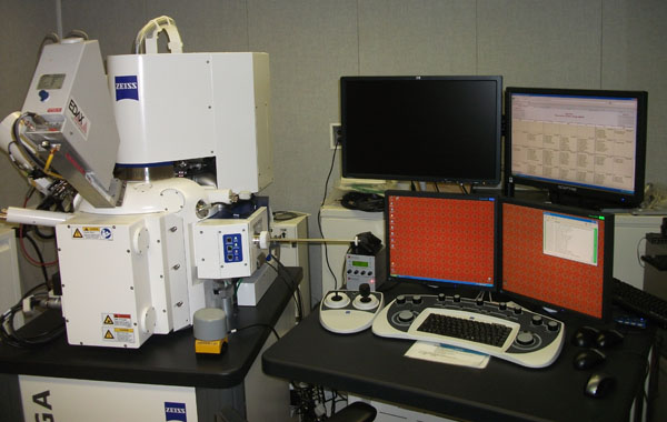
Zeiss Auriga SEM/FIB with EDAX
(University of Rochester, Optics)