Materials and Methods
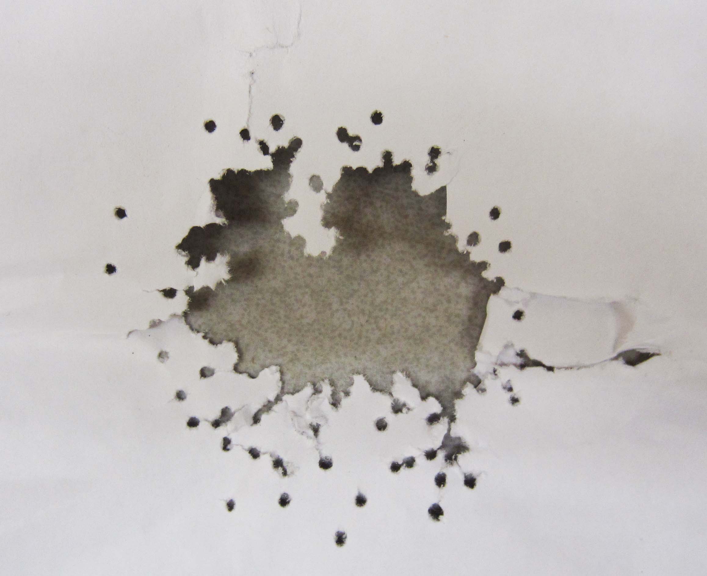
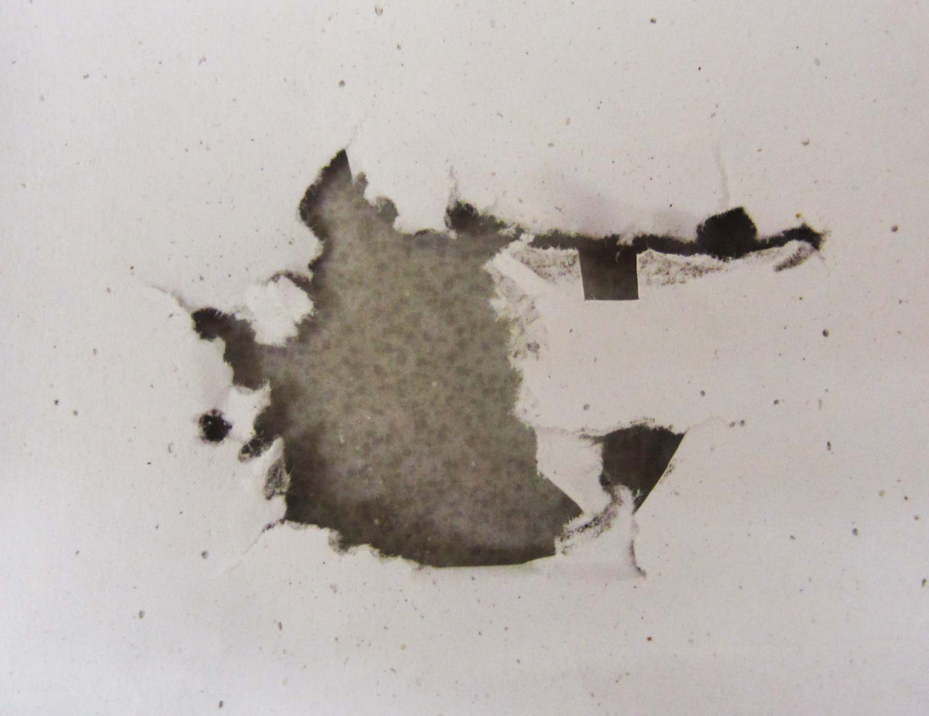
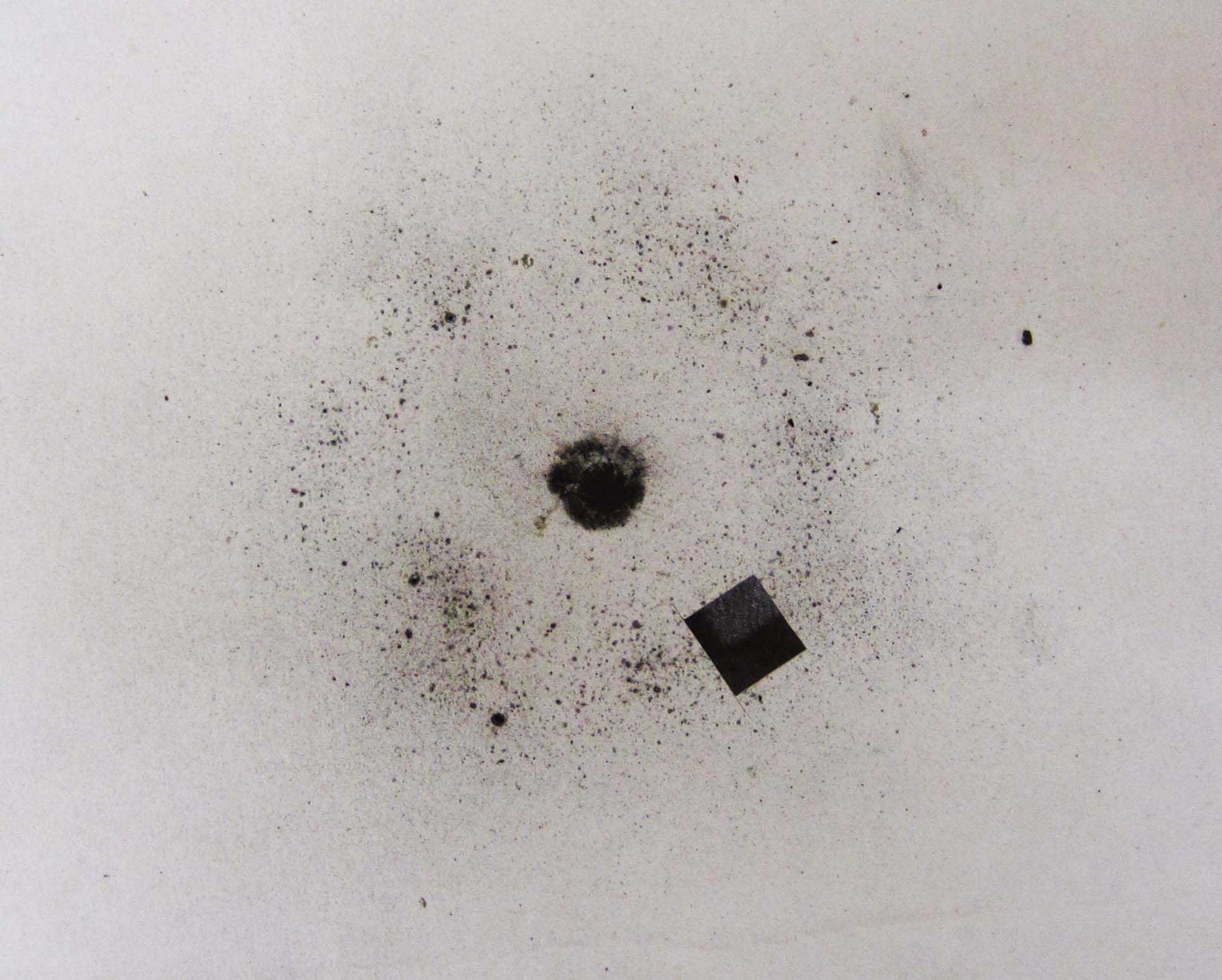
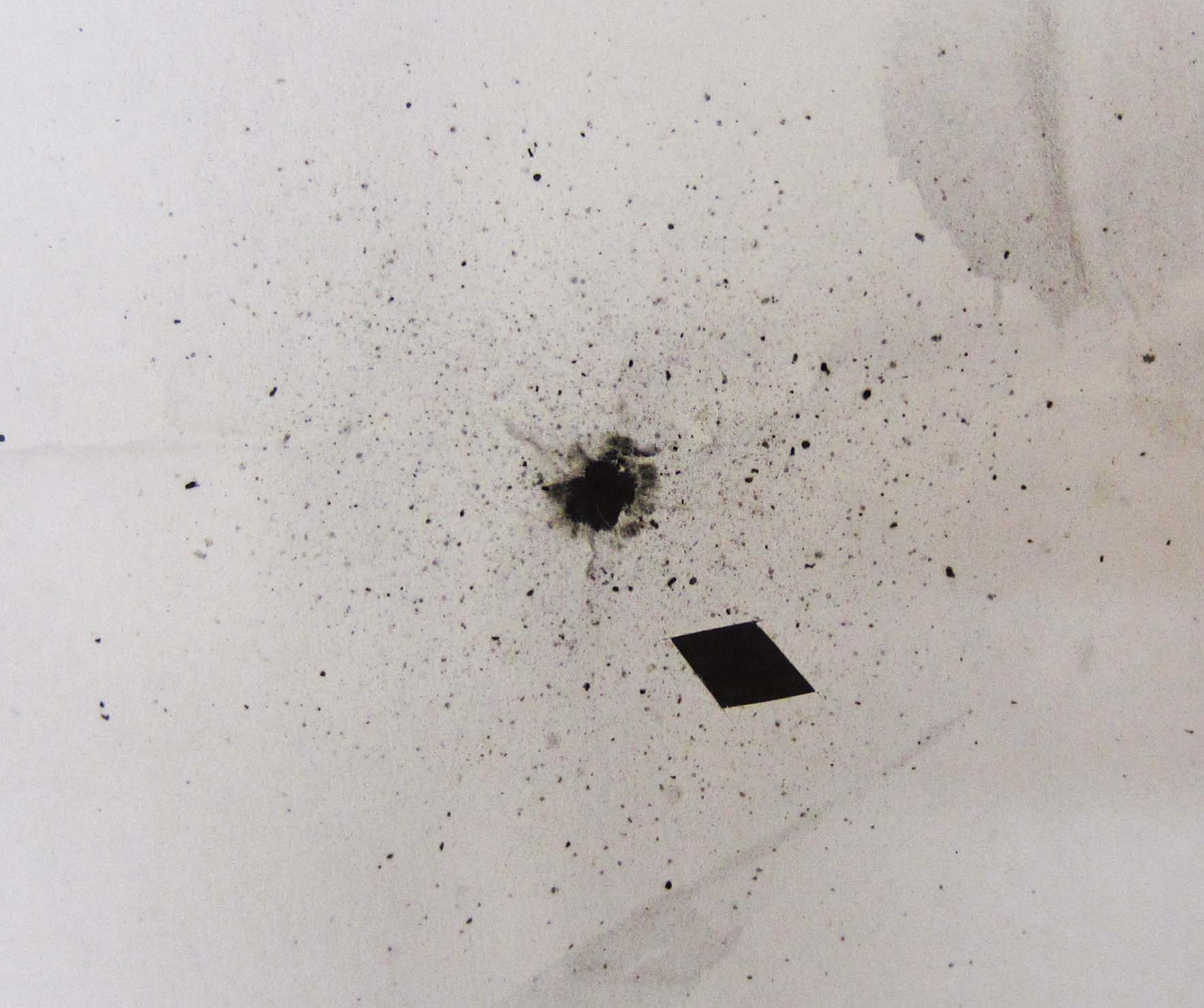
Four samples of GSR on white printer paper were collected by Brian McIntyre. The ammunition types used in the four samples were a 12-gauge Federal shotgun shell, a .410-gauge Winchester shotgun shell, a .22 caliber Remington rim-fire rifle round, and a .22 Federal rim-fire rifle round. In each case, the paper 'target' was placed on the ground and the firearm discharged from approximately 12 inches away. See Figure 1 below for images of the paper targets. A small piece was cut out of each sample for EM imaging and EDAX.
Figure 1 - Images of paper target samples collected for GSR analysis. Samples came from 12-gauge Federal (top left), .410-gauge Winchester (top right), .22 caliber Remington rim-fire (bottom left) and .22 caliber Federal rim-fire (bottom right) ammunition.
The two shotgun shell samples did not have significant GSR around the entrance 'wound.' I expect that because the lead shot spread out as it left the barrel of the gun, that the area of paper that may have collected significant residue was torn away instead. Despite this, I was able to find some torn edges that contained residue for analysis. Each sample was sputter coated with gold prior to EM imaging.
For each of the four samples, representative areas were selected for particle analysis. Three high-contrast micrograph images were captured from each sample using the back-scattered electron detector and 1000x magnification. These images were analyzed using ImageJ software obtained from the National Institutes of Health website. A 'watershed' particle separation process was employed, and particles on the edges were neglected.
Additionally, several representative x-ray spectra were collected from various particles in each sample. During each x-ray collection the raster was positioned such that the electron beam interacted exclusively with the particle of interest (neglecting electron interaction volume below the particle, which will be discussed later).