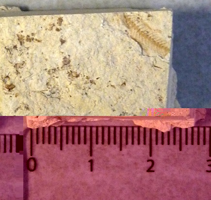
Sample
The sample is from a laminated oragnic rich shale. It was collected from the Cerro Pucara section near Yauri (14.79°S, 71.39°W). Fish and leaf fossils were visible with naked eyes on samples collected from this location. The fish sample was chosen for further analysis.
Methods Used
1. Light Microscopy:
The samples were first viewed under the reflected light microscope. Since the fish bones created uneven topography on the sample surface, it was hard to achieve focus. This technique did not provide good details of the structures of the bones.
2. Sputter Coating:
Since the sample was a non-conductive (rock) sample, so sputter coating was necessary to view it under the Scanning Electron Microscope. Initially 100Å gold coating was done but the sample started charging so gold coat of 200Å more were applied later. It was not necessary to view the sample under extremely high magnification, so creation of extra surface ornamentation due to the gold coating was not a problem.
3. SEM Imaging:
The sample was imaged using the SE2 andthe BSD detectors and also by the mixed detector signal from In-lens and BSD. Imaging of the sample under the SEM showed presence of diatoms, and these are also imaged in detail for identification. Much better images were collected by the BSD and the mixed detector signals comapred to the SE2 detector as With theSE2 detector, charging became an issue.
4. X-Ray Microanalysis:
The fossils were analyzed with the X-Ray to look at the elemental composition. It was used to check that fossils were not altered. X-Ray maps were also collected to look at elemental distribution .
5. Electron Flight Simulator:
This process was used to find the interaction volumes of both the fish and a diatom sample.
6. Colorization:
Most of the images were colorized using Adobe Photoshop for better visual representation.