Introduction-Samples-Morphology-3D Gallery-Composition-Acknowledgement
Before SEM, all the samples were coated by Au or Ag sputtering to remove the charging effect, which means to get good images.
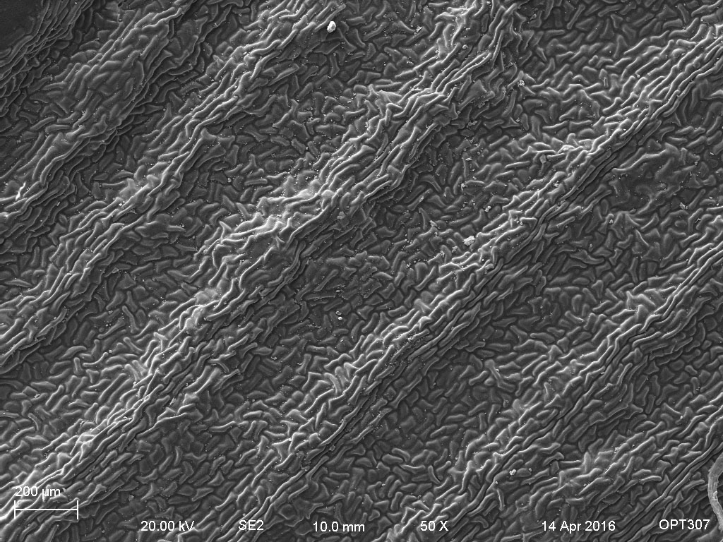
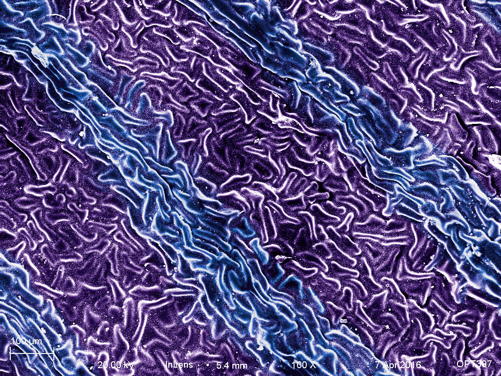
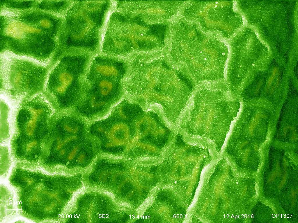
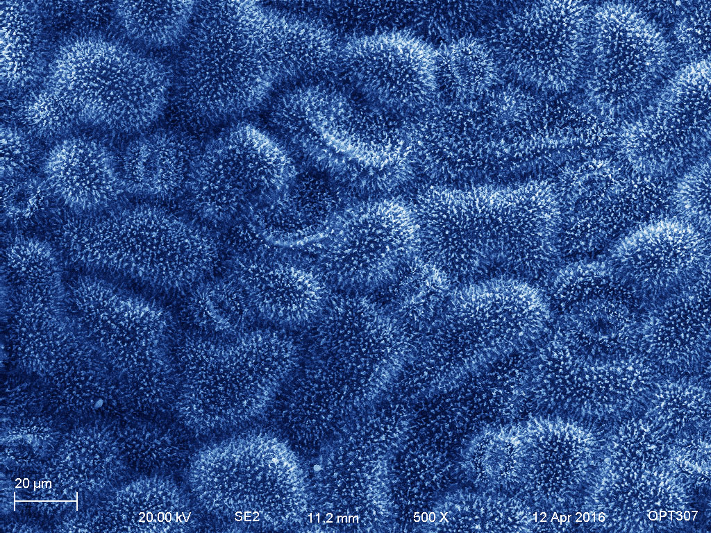
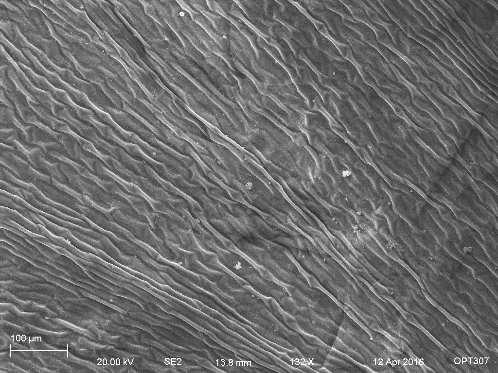
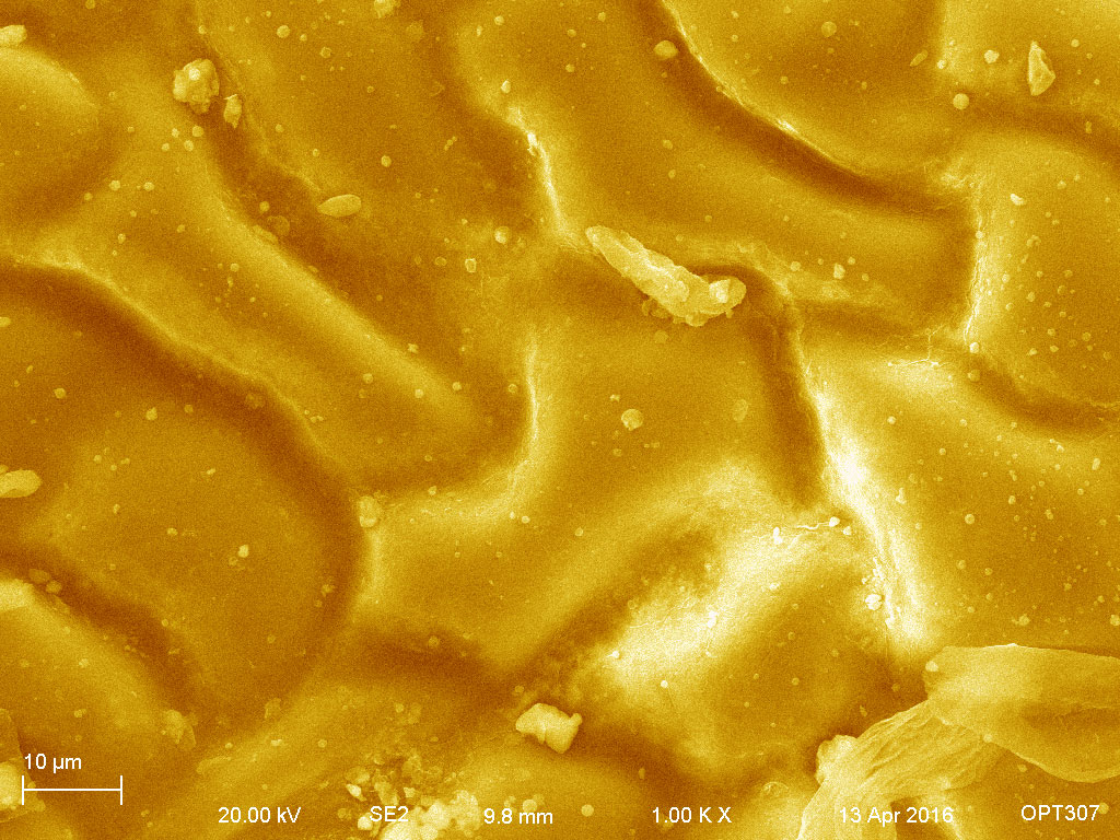
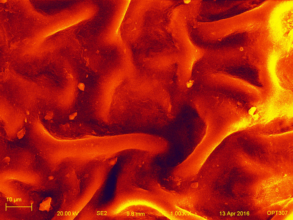
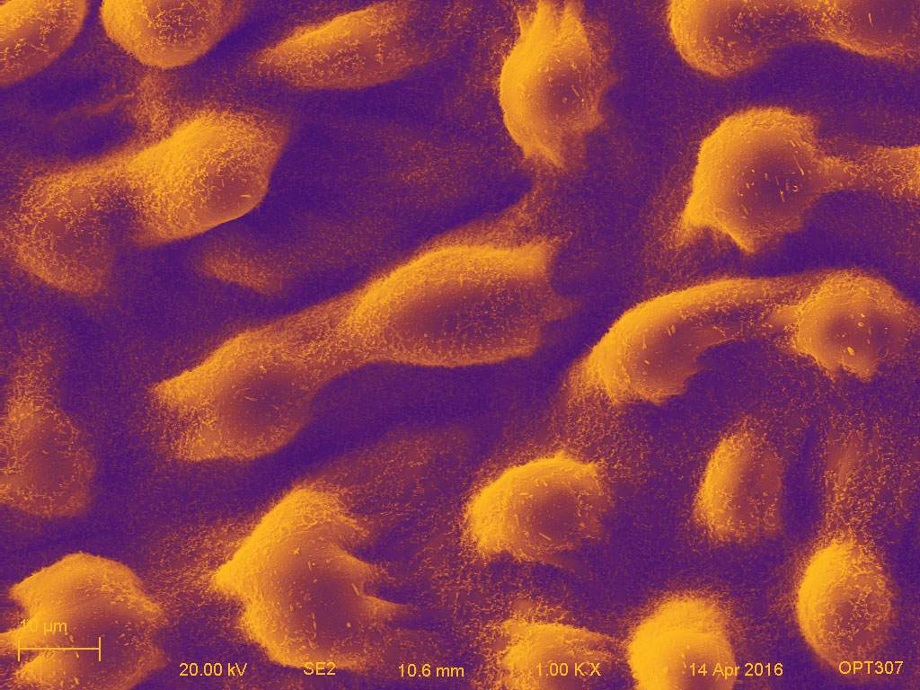
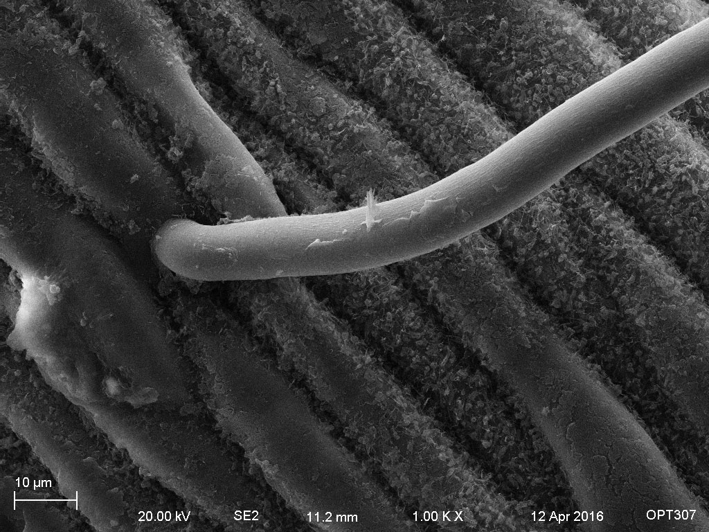
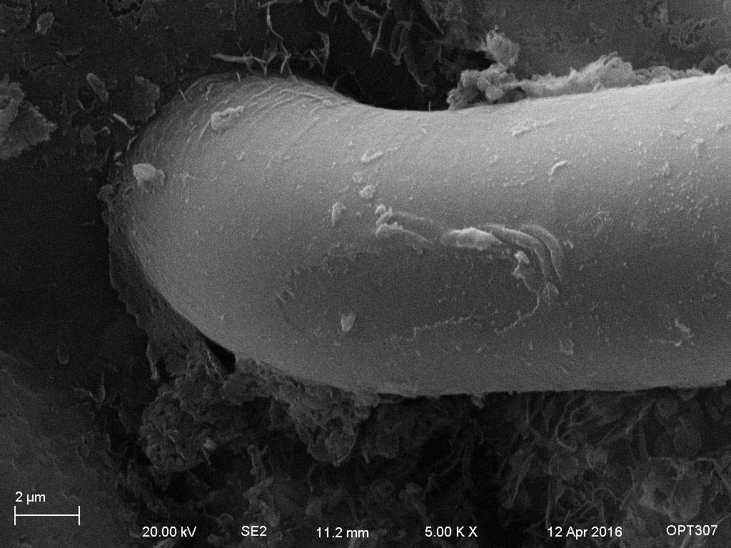
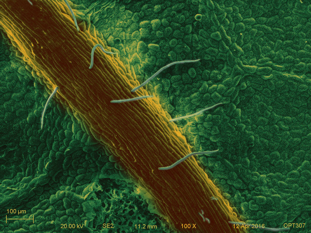
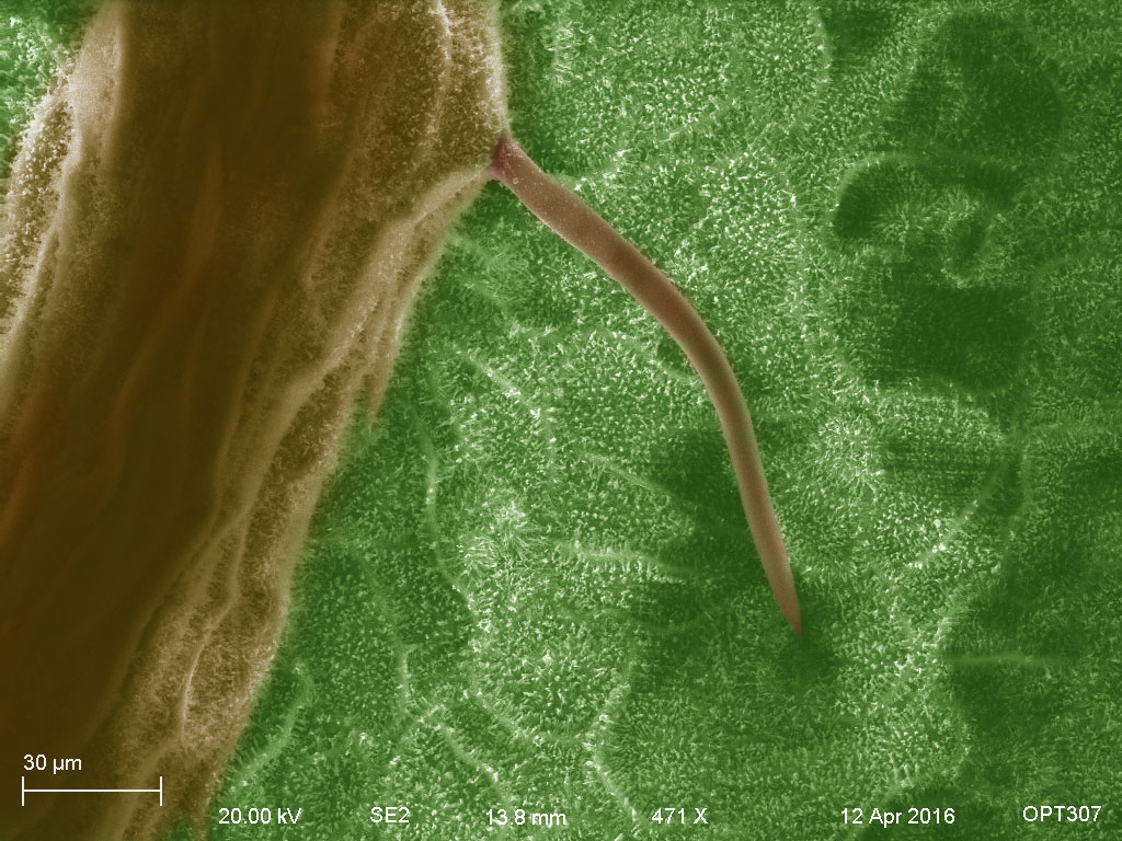
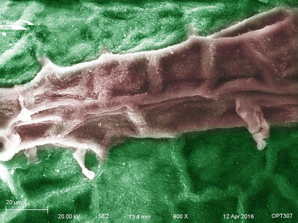
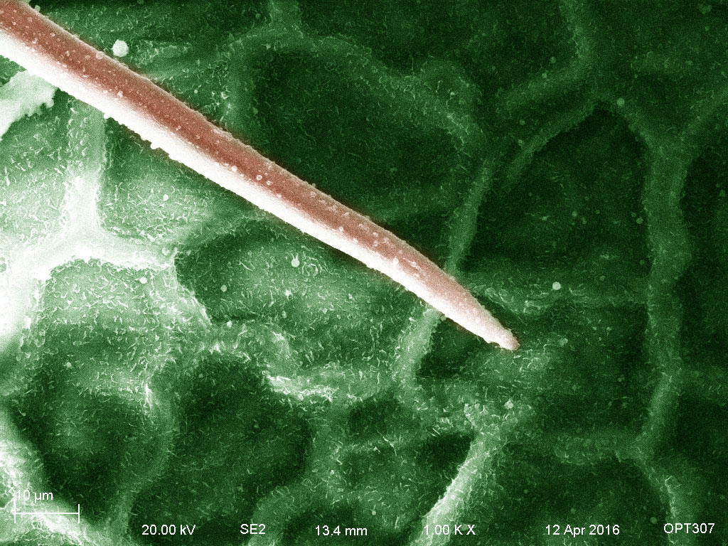
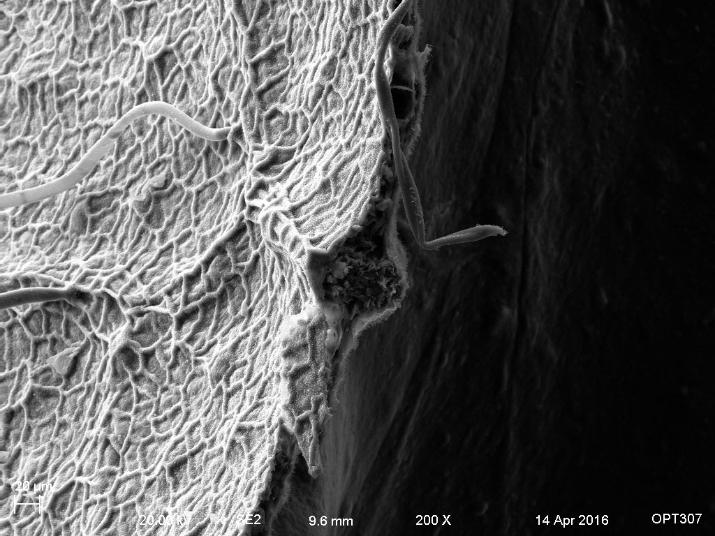
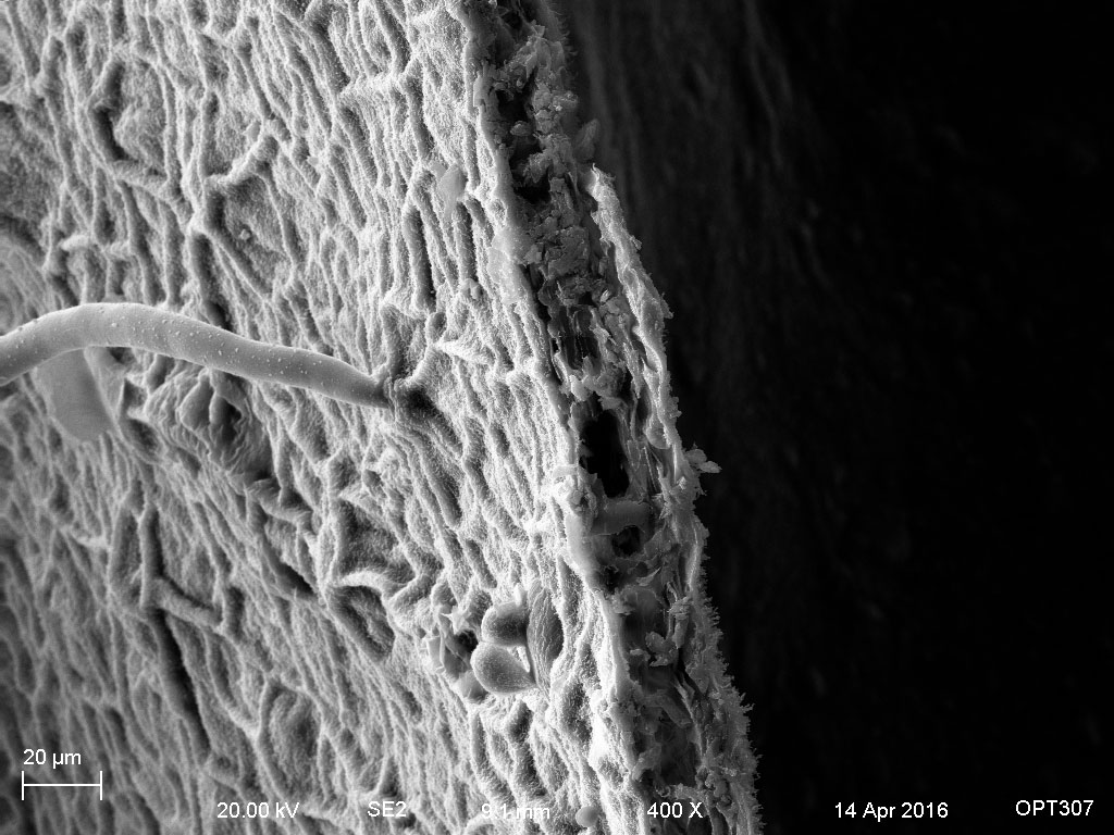
Although the leaves are different, but they share the similar features, such as epidermis, vein, trichome, stoma and small tubes to help transfer the food, oxygen, water, etc.
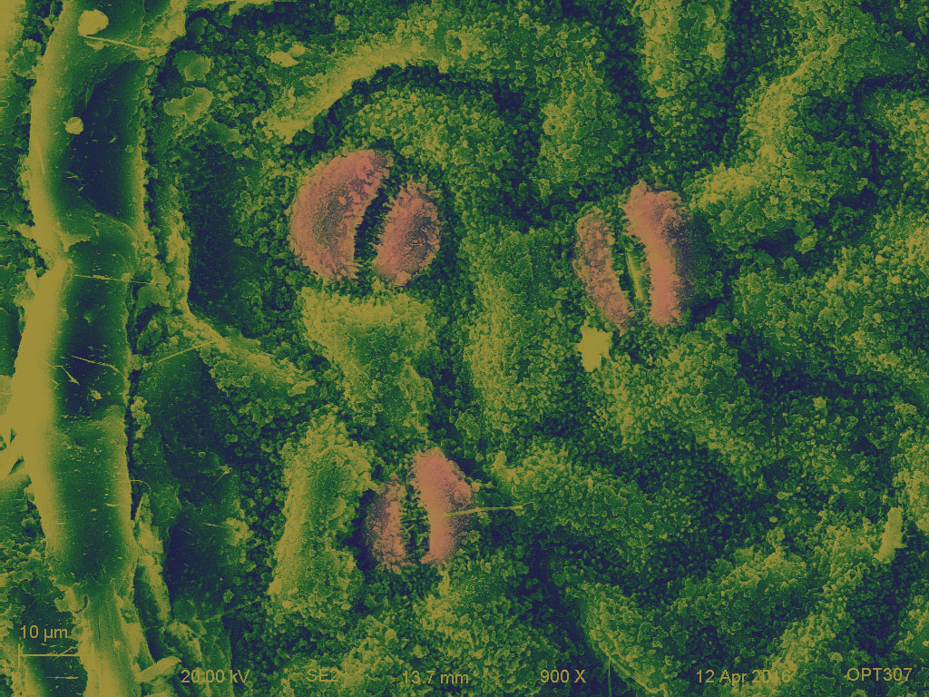
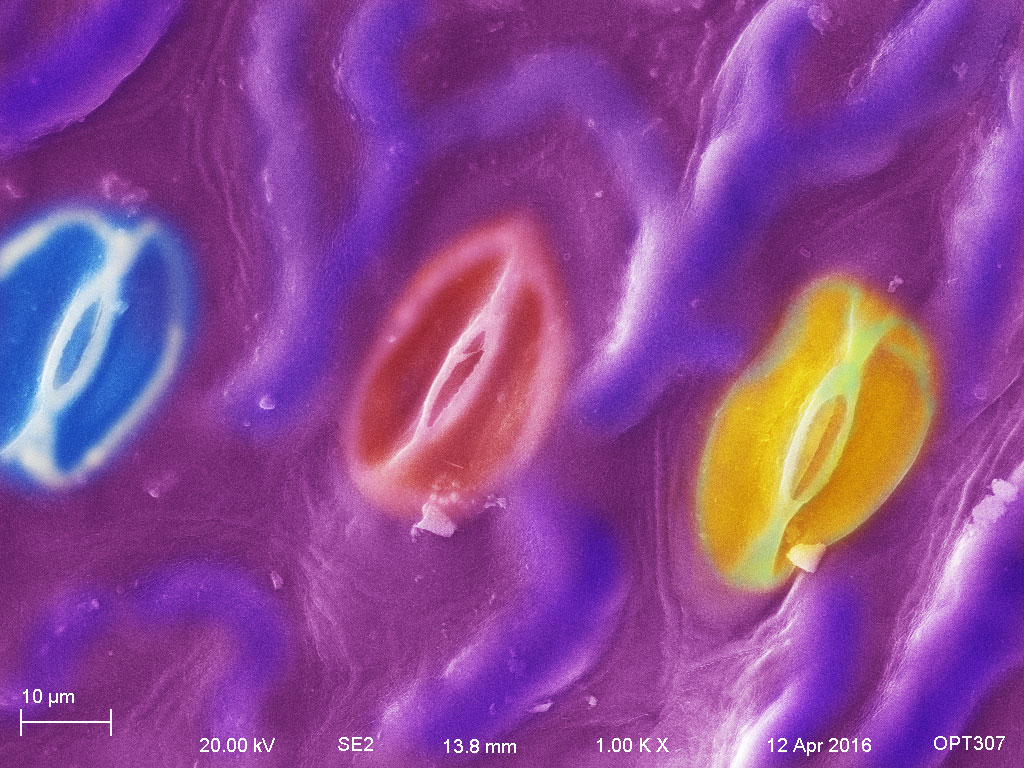
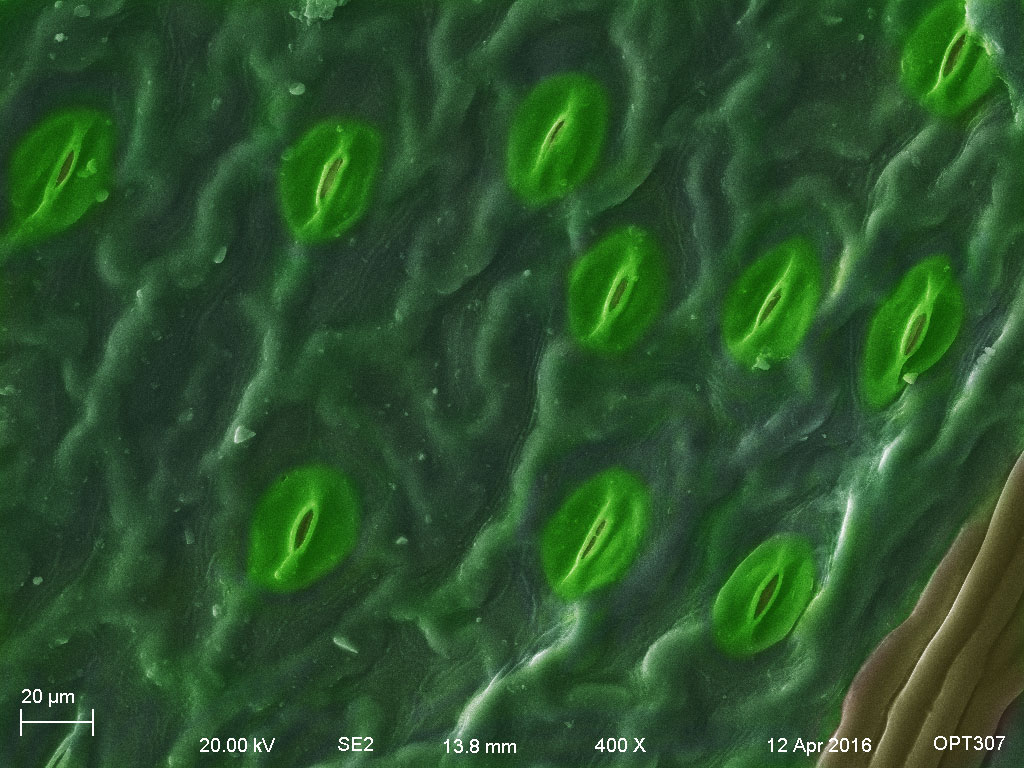
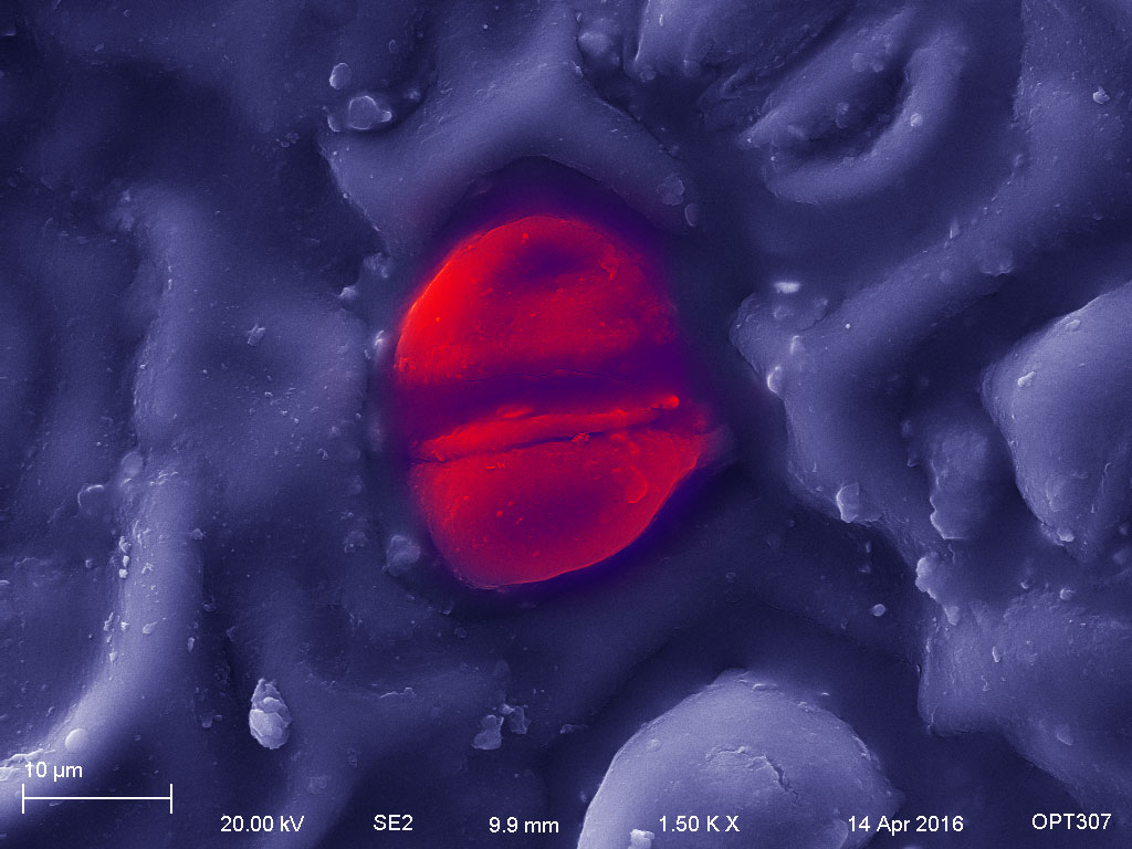
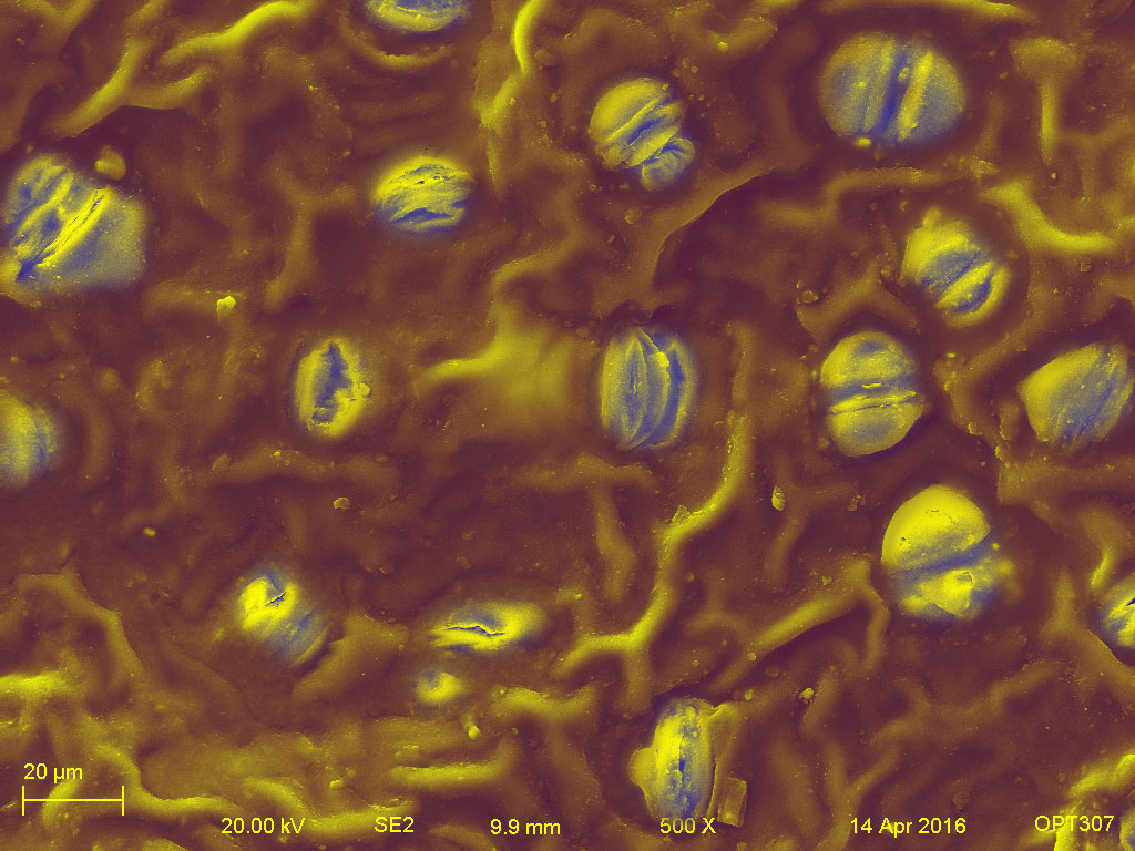
The upper and lower epidermises are both covered by cuticle, but the lower side usually has more stoma.
The leaf tissues include several cell types: epidermal cells, guard cells, subsidiary cells, and trichomes.
The epidermis cells is a single-layer of cells that covers the leaves. They separate the plant from the external environment to protect against the water loss, regulate gas exchange, secrete metabolic compounds, and absorbs water and mineral nutrients.
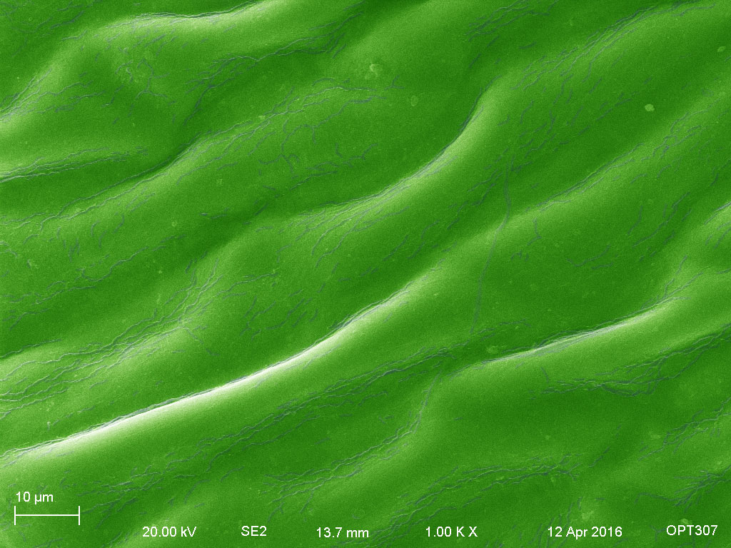
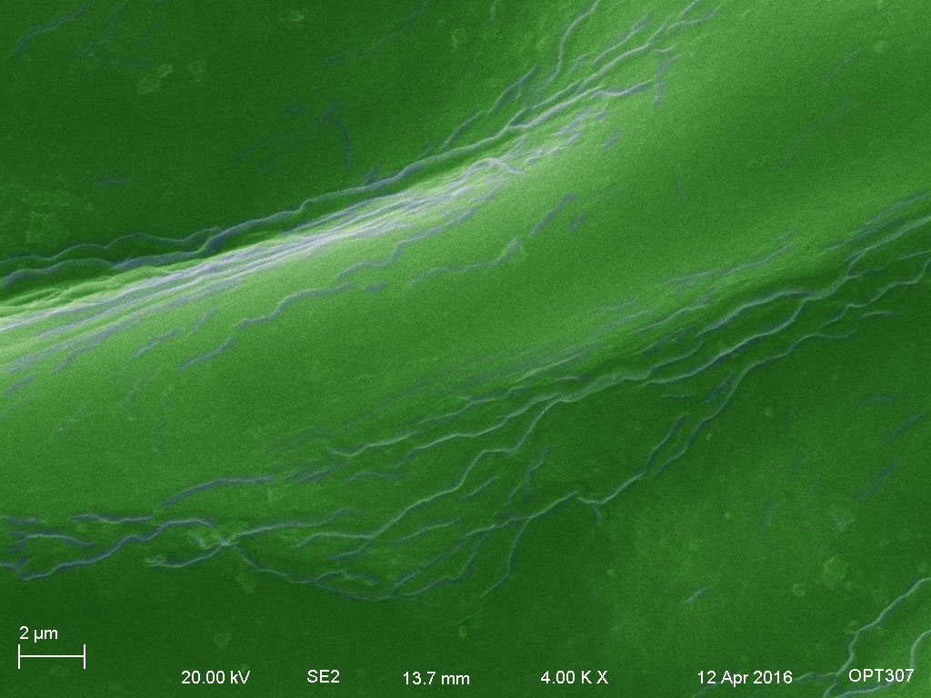
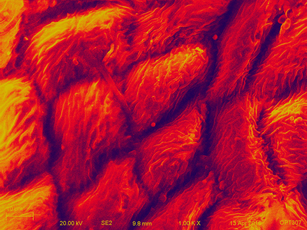
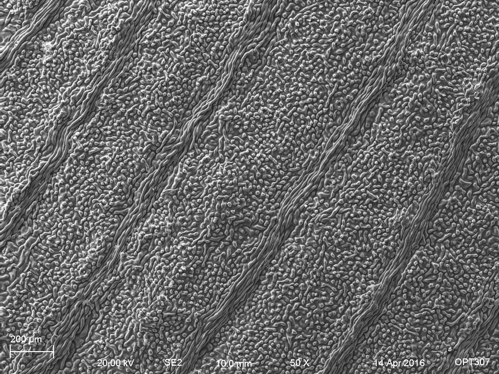
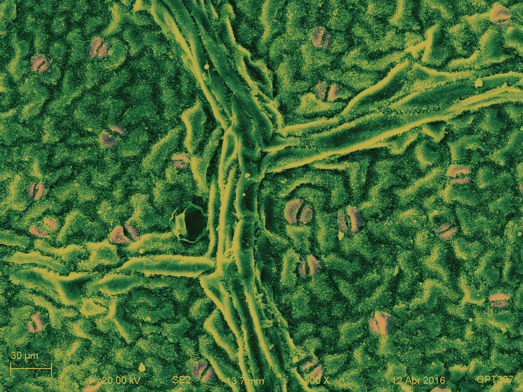
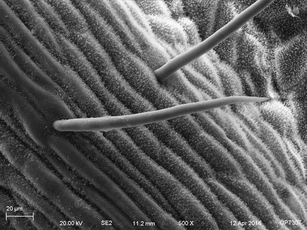
The veins are the vascular tissue of the leaf and are located in the spongy layer of the mesophyll. Trichomes are fine outgrowths or appendages on plants, algae, lichens, and certain protists.

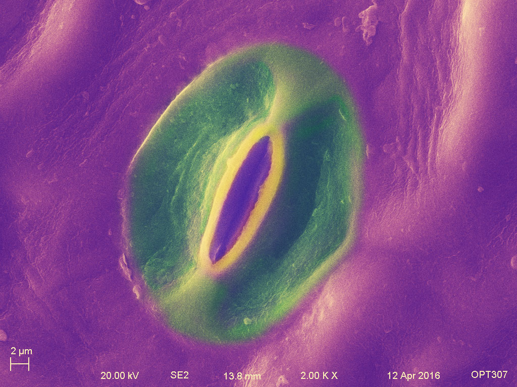
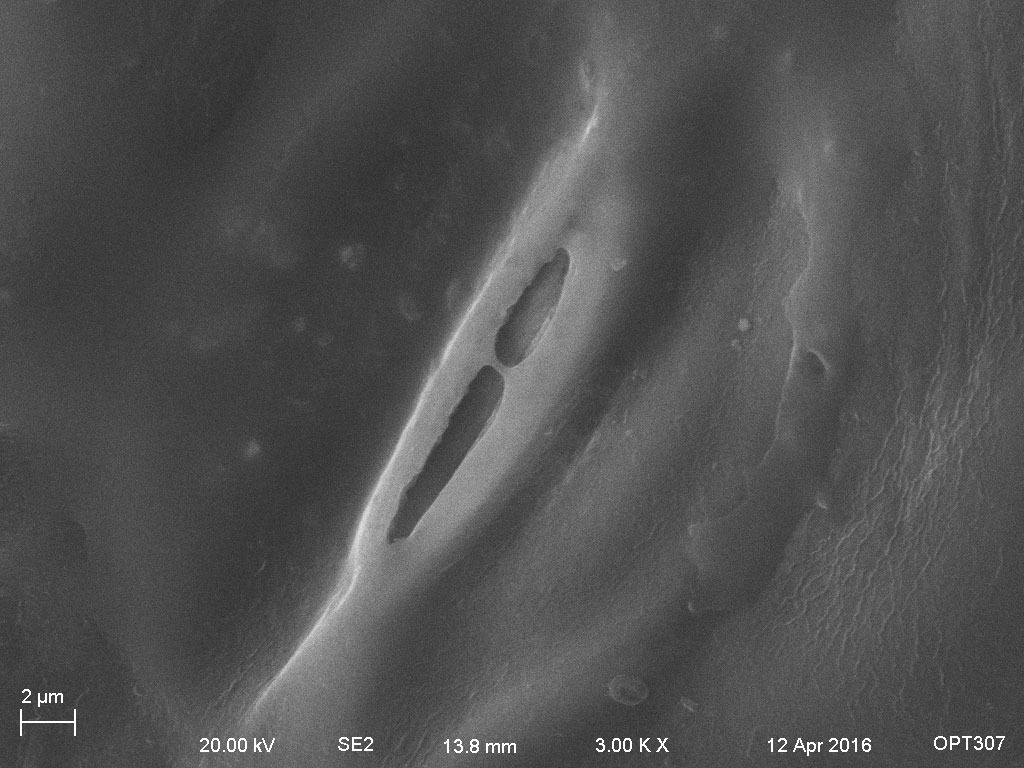
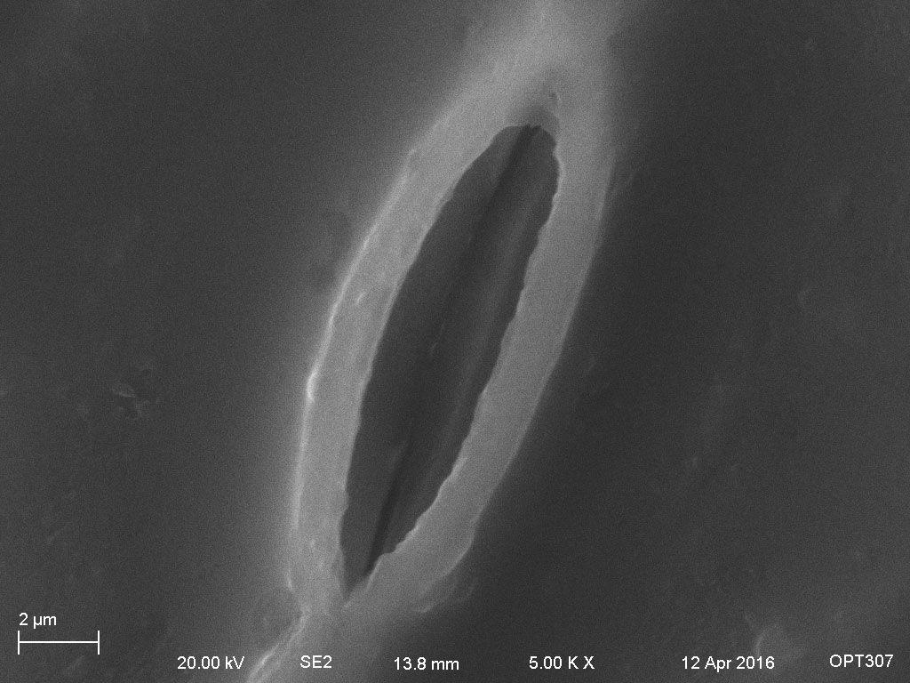
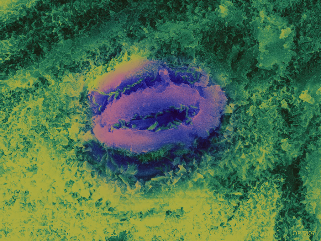
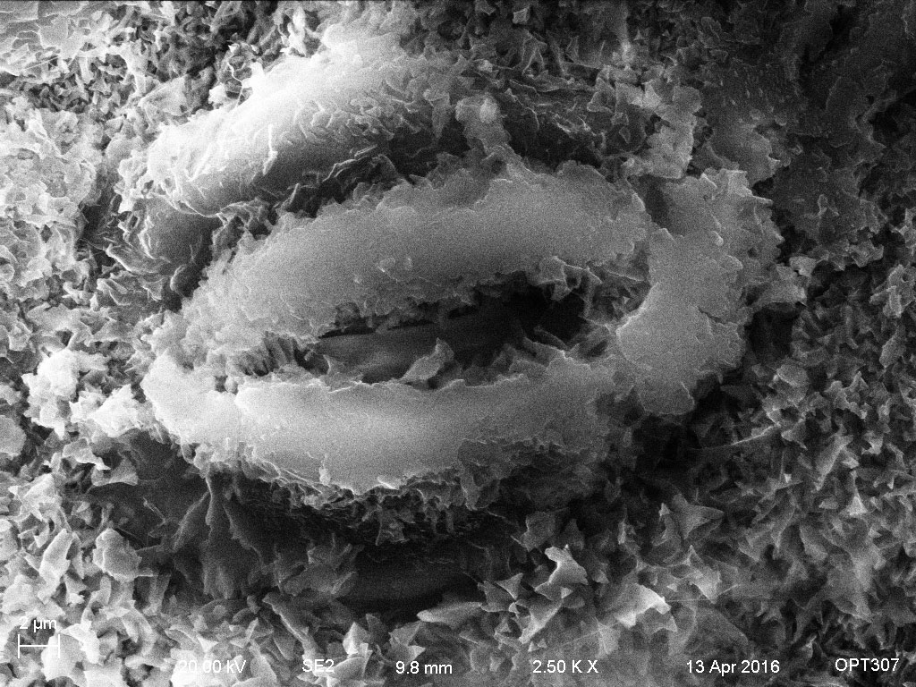
A stoma is a pore that is used to control gas exchange. The pore is bordered by a pair of guard cells that could regulate the size of the opening.
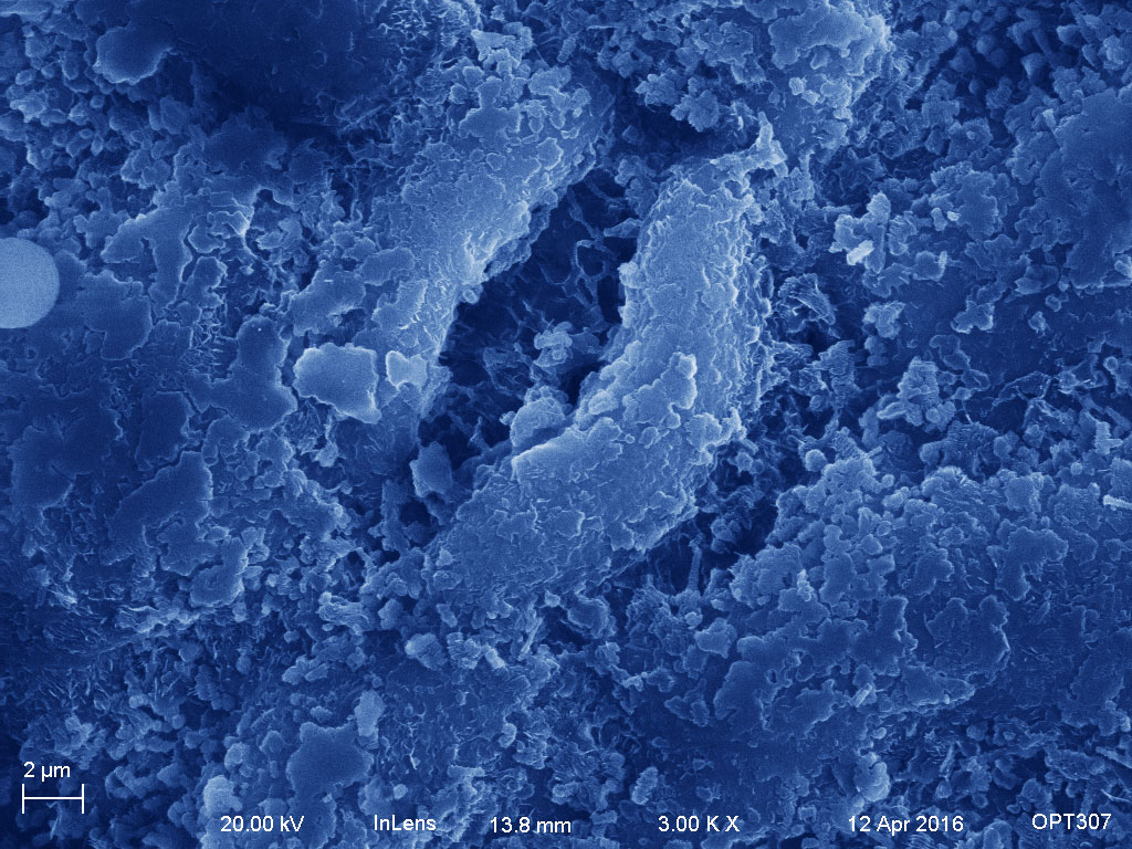
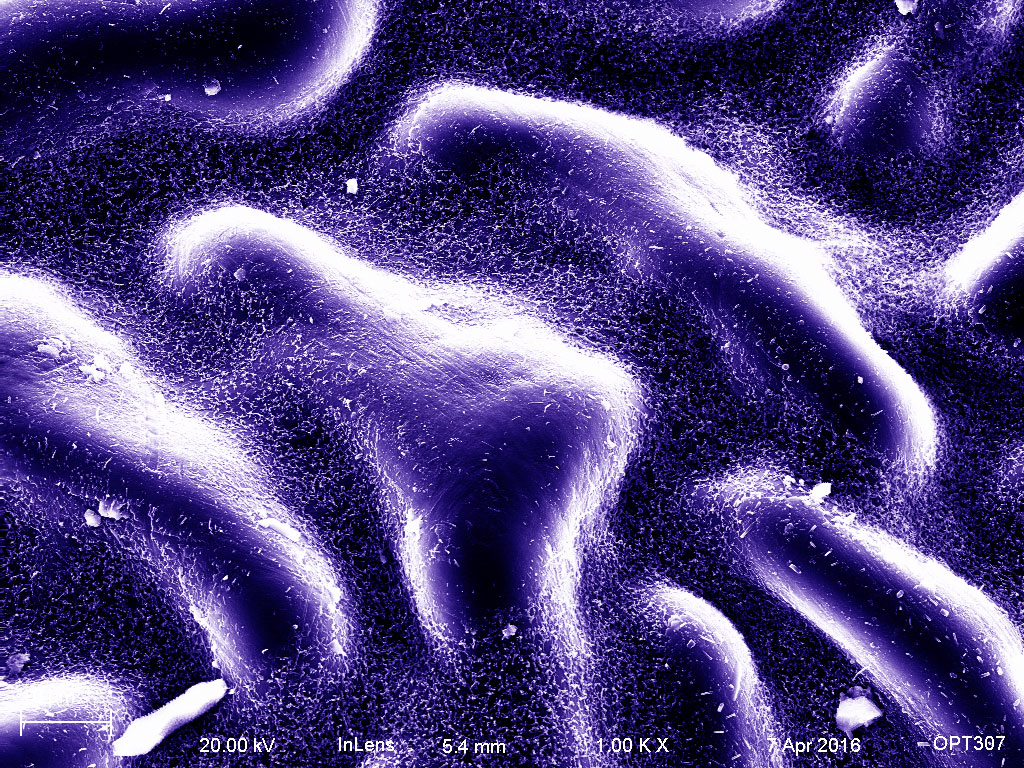
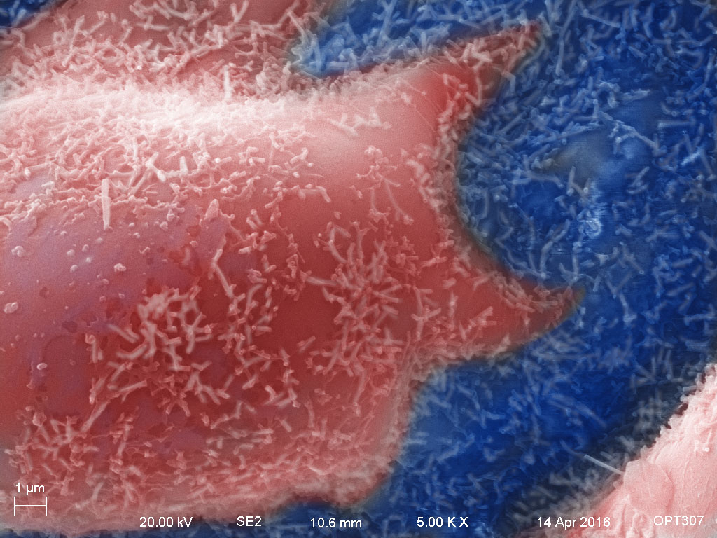
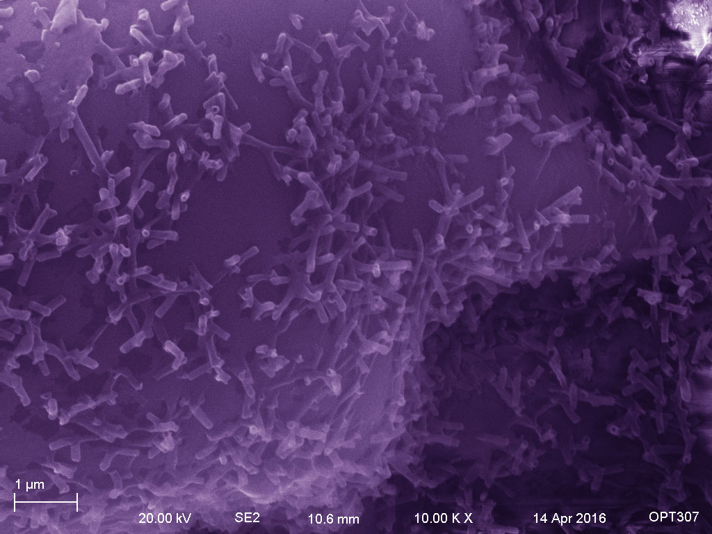
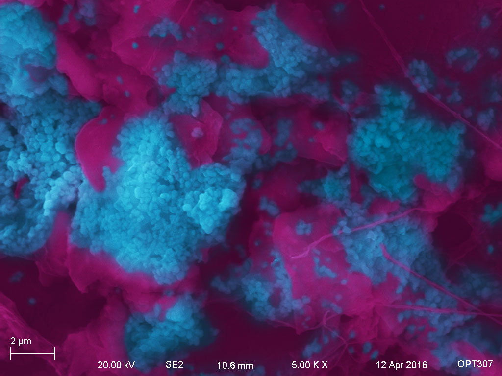
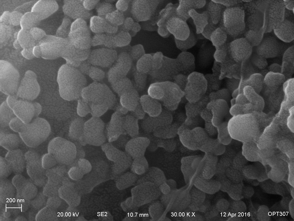
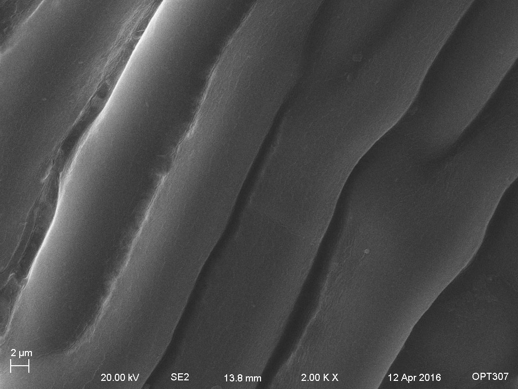
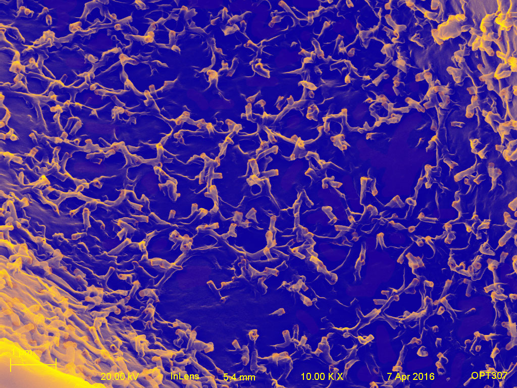
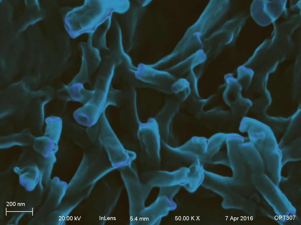
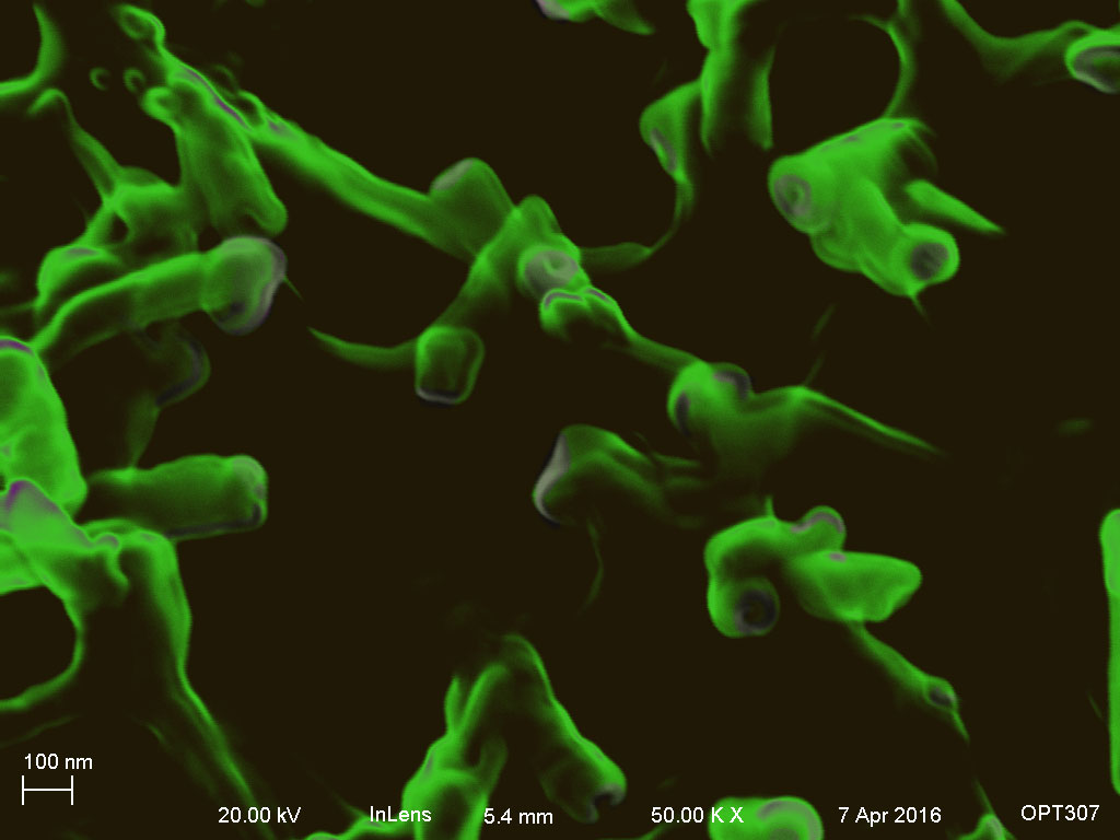
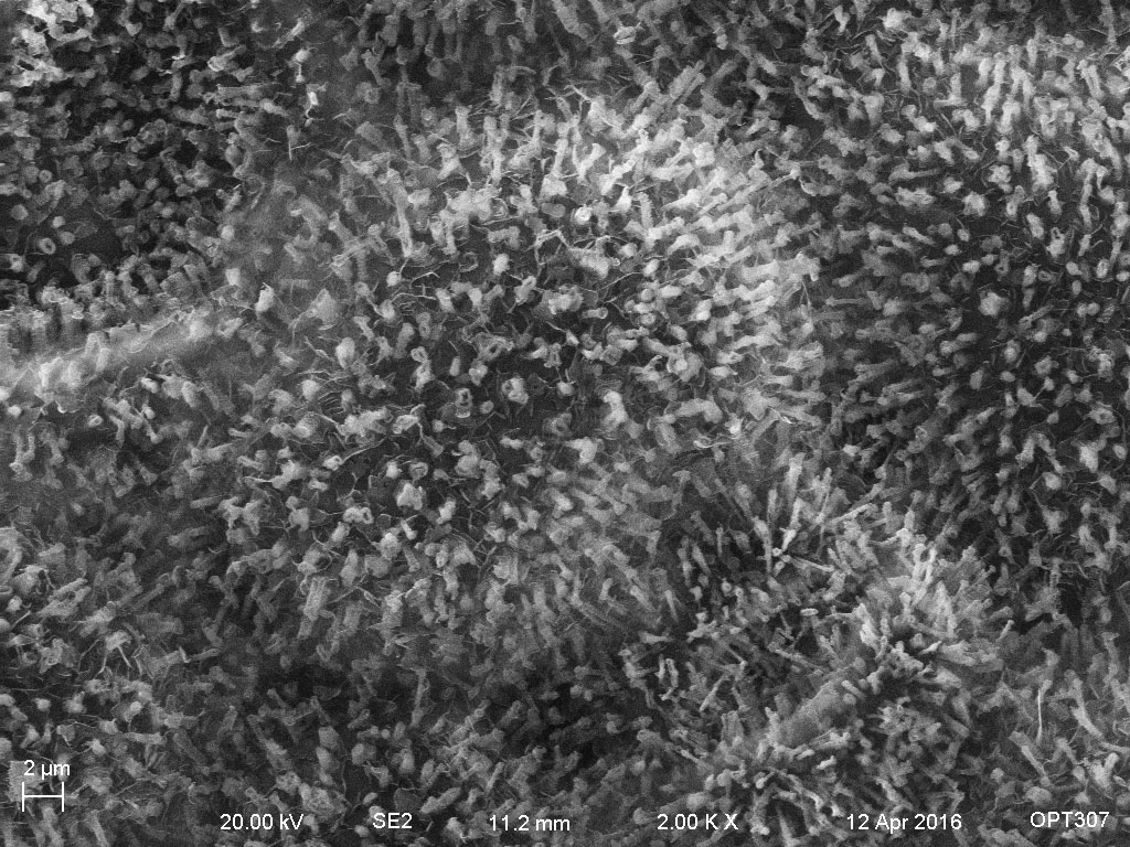
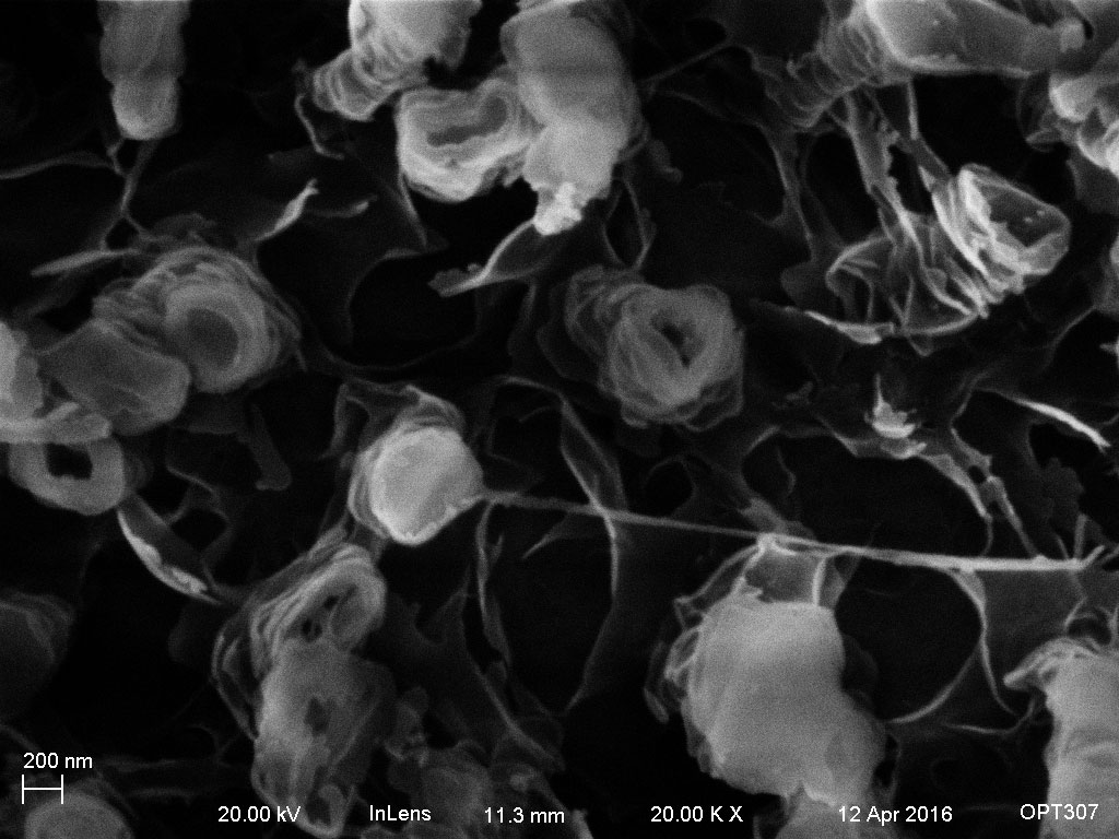
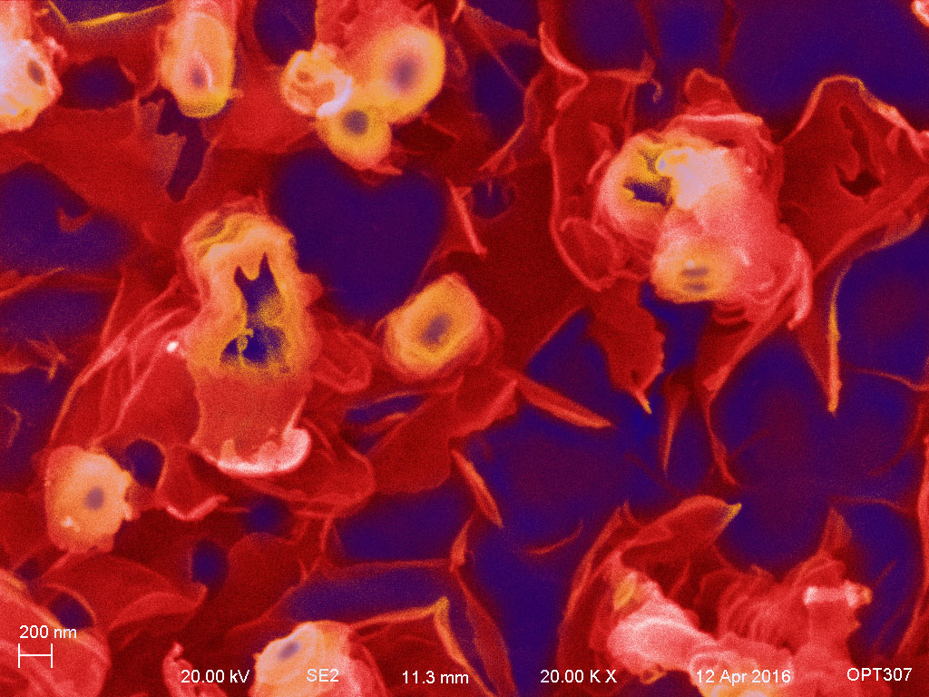
I found tons of tubes under high magnification SEM. (I named them “tubes” since I could not find relevant information online. If you know the professional name of them, please let me know. Thanks.)
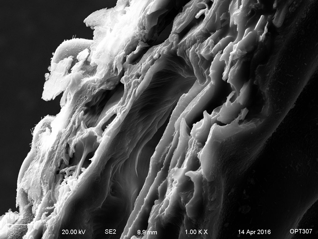
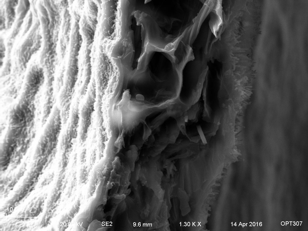
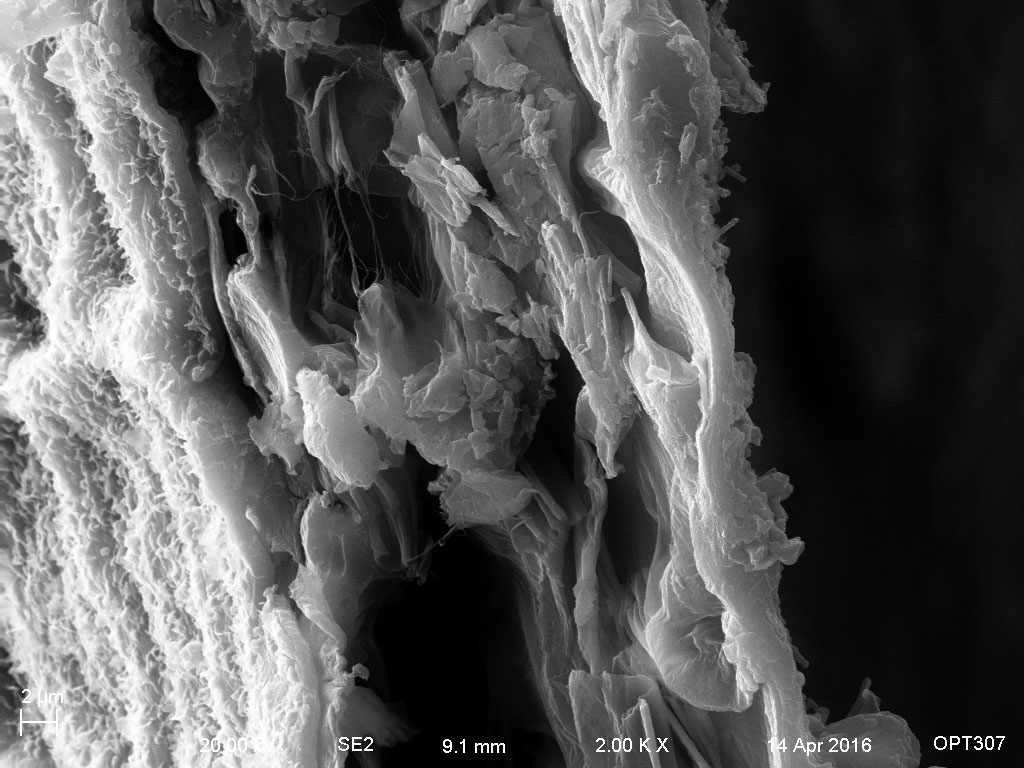
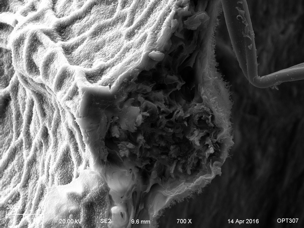
The cross-section of the leaves could show:
Cuticle
Upper Epidermis
Mesophyll: Palisade, Spongy
Lower Epidermis
Cuticle
Here is a figure that we could use to compare the structures.
http://www.enchantedlearning.com/subjects/plants/leaf/