

University of Rochester
Backscattered electrons are produced by elastic collisions between electron beams and nuclei of atoms in the specimens. Backscattered electrons are sensitive to the atomic mass of the nuclei they scatter from. BSE mode imaging provides compositional contrast information of samples.
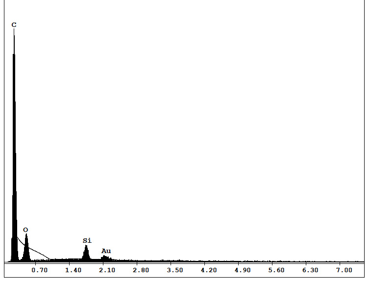
Backscattered electron images indicate the compositional information of samples. It usually works with energy dispersive X-ray spectroscopy(EDS) for elemental analysis. EDS analysis identifies elements and their distribution in samples. EDS works relying on the interaction of some source of X-ray excitation and the sample. Since each element shows a unique set of peaks on its electromagnetic emission spectrum resulting from their unique atomic structure, EDS analysis is able to collect this information and obtain the elemental composition of samples.
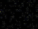
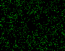
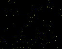
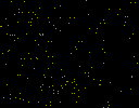
Figure 4 is a BSE image of Arborvitae and the elemental distribution map is shown in Figure 5. It is noted that there is a stone-like stuff in the stoma. To figure out the stone composition, EDS spectrum of the stone and its surrounding are demonstrated in Figure 6.
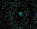

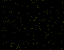
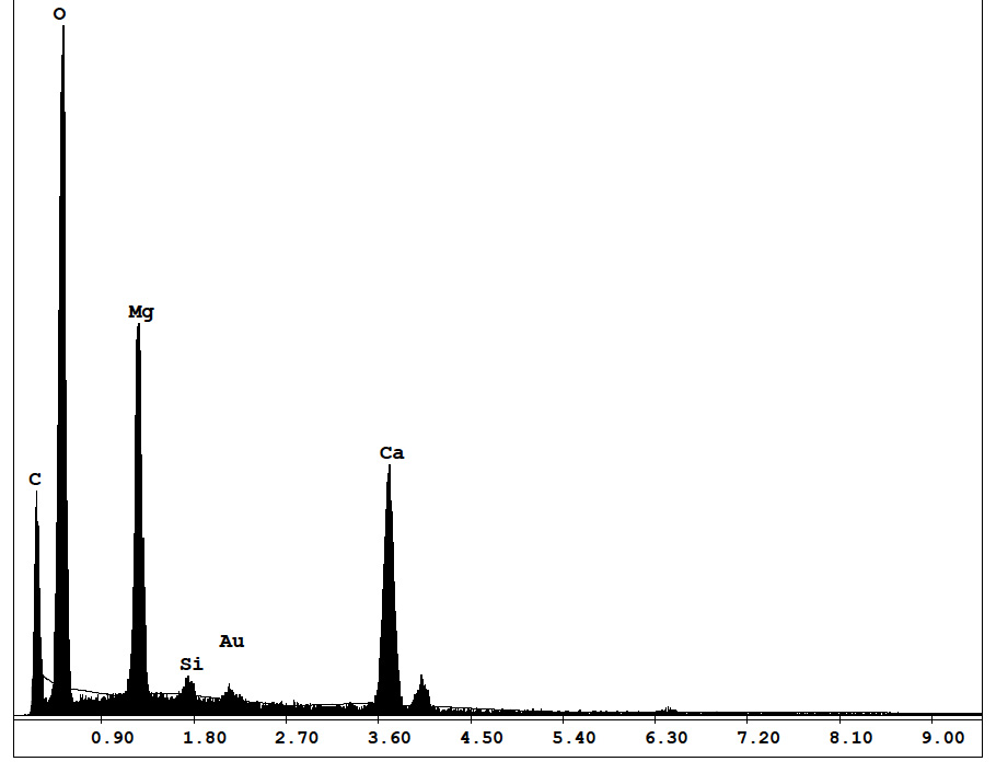
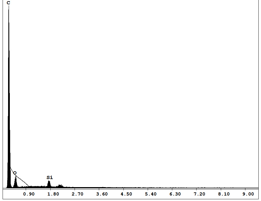
Figure 7 is a BSE image of Juniper, and Figure 8 shows the elemental distribution of it. The EDS spectra is shown as Figure 9.