
Gotcha! Rochester membranes help researchers capture tiny, telltale vesicles
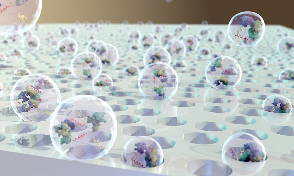
Researchers from the University of Rochester and University of Chicago teamed up for one of the first known projects to successfully isolate and study extracellular vesicles (EVs). Extracellular vesicles are tiny particles—as small as 40 nanometers in diameter—released by cells into the bloodstream and other fluid-filled cavities. EVs carry proteins, lipids, metabolites, and genetic material unique to the cells that release them. As a result, they could serve as valuable biomarkers for the early detection of diseases, including cancer—especially if single EVs could be assessed individually.
To this end, the researchers adapted nanomembranes from the lab of James McGrath, a professor of biomedical engineering at the University of Rochester, in a microfluidic cross-flow filtration system to capture and study individual EVs. Their findings appear in Communications Biology.