Biomedical Optics
What is Biomedical Optics?
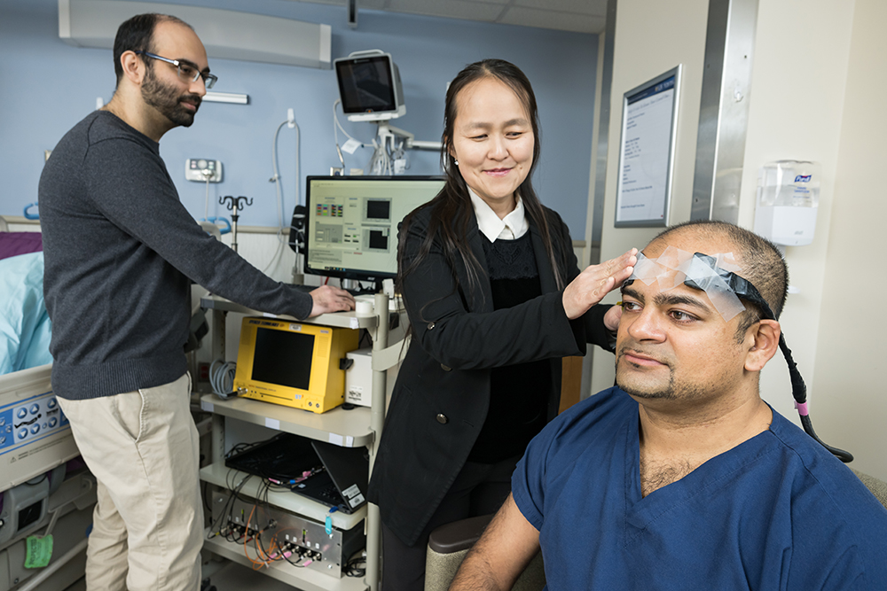
Biomedical optics uses the physical properties of light to design and apply advanced techniques to solve pressing problems in medicine and biology. Advanced optics can be used in many biomedical pursuits, including probing tumor pathology, diffusion of cell surface receptors, single molecule spectroscopy, and expanding the limits of human vision.
Areas of Focus
Some of the advanced optical techniques our biomedical engineers use include:
- Multiphoton laser-scanning microscopy: used to study living biological tissue.
- Diffuse optical tomography: a non-invasive way to examine tissues in vivo, particularly for tumor detection and chemotherapy response monitoring.
- Fluorescence recovery after photobleaching: used to study the movement of biomolecules through tissues or cells.
- Raman spectroscopy: a chemical analysis technique that identifies materials based on how light interacts with their chemical bonds.
- Near-field optics: a microscopy method that defies the laws of diffraction and allows for measurements at the nanometer scale.
- Adaptive optics: carefully deforming mirrors can compensate for distorted light, which is particularly helpful in retinal imaging systems.