
Research
Functional Ultrasound
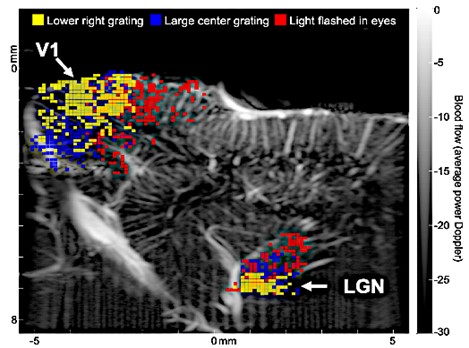
Functional magnetic resonance imaging (fMRI) is the current “gold standard” for human and primate brain imaging. However, employing fMRI in small animal research is challenging and very expensive. Additionally, fMRI’s low spatiotemporal resolution limits its effectiveness for small animal research. To deal with these issues, more researchers are now relying on optical imaging for basic neuroscience research in animals. Optical imaging has many attractive attributes, such as exceptional subcellular level spatial resolution and fast temporal resolution. However, the main challenge is that it cannot noninvasively visualize deep brain structures. Neuroimaging requires a different approach to tackle its main challenges, one that takes a fresh look at the critical issue – neither optical microscopy nor fMRI is ideal for live imaging of small animals’ brains. High frequency functional ultrasound (fUS) imaging is filling an essential gap in currently available functional brain imaging technology. fUS provides sufficient spatial and temporal resolution (~100 microns and tens of milliseconds) to image the activity of neurons while also enabling penetration into deep brain structures. Plane-wave imaging, the technology that underpins fUS, allows excellent separation of stationery and blood flow signal in small vessels from the surrounding tissues, which enables fUS to detect neural activity changes through neurovascular coupling. We are using fUS to study and understand the visual system. The figure shows fUS imaging of brain response to sensory stimuli. Gray map shows baseline image in sagittal plane of a ferret brain, including visual cortex (V1) and thalamus (LGN). Note the detailed vasculature. Following 30 seconds of patterned visual stimuli, increased signals were detected in V1 and LGN. Differently colored pixels show the highest activation correlated with different stimuli.
Journal Articles
- Functional ultrasound imaging reveals 3D structure of orientation domains in ferret primary visual cortex
W. Hu, S. Zhu, F. Briggs, and M. M. Doyley
NeuroImage 268 , pp. 119889-1 -119889-12 (2023). View Online