Why are they degrading and why must electron microscopy be used?
Since the image is formed by scattered nano-particles on the surface, it therefore makes sense that this surface would be very susceptible to physical, chemical as well as contamination.
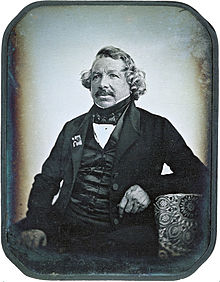
During a display showcasing hundreds of daguerreotypes from around globe at the International Center of Photography in New York, a number of these irreplaceable images began to show unexplainable signs of degradation.
Initial analysis has shown the culprit to be primarily biological in nature - fungi, taking over the surface. This came as a shock to many, as it was believed that the anti-microbial nature of Silver would protect the surface from such organisms.
Given the scale of the particles and organisms at work (often only 10s or 100s of nanometers), not only are daguerreotypes some of the first known forms of nano-technology (from the early 1800's, no less!) but also require current electron microscopy techniques to image the surface at a level adequat to provide useful data.
How are samples prepared for the Transmission Electron Microscope?
To accurately image and collect compositional x-ray data in low-nanometer resolutions a thin sample must be prepared for the TEM. Through a thin enough sample the electron beam effectively has a very small diamter ballistic trajectory, and eliminates resolution degradation as a result of large interaction volumes.
This thin sample is prepared using the Focused Ion Beam mode of a Scanning Electron Microscope. Similar to the way the electron beam is used, Gallium ions are instead used to ablate material leaving only a thin 'coupon' shape which can then be moved to a TEM grid and easily imaged in the TEM. |