Sputter Coating
Sputter coating is required for this sample due to electrical conductivity reasons. Sample are coated with 40 angstroms of gold particles.
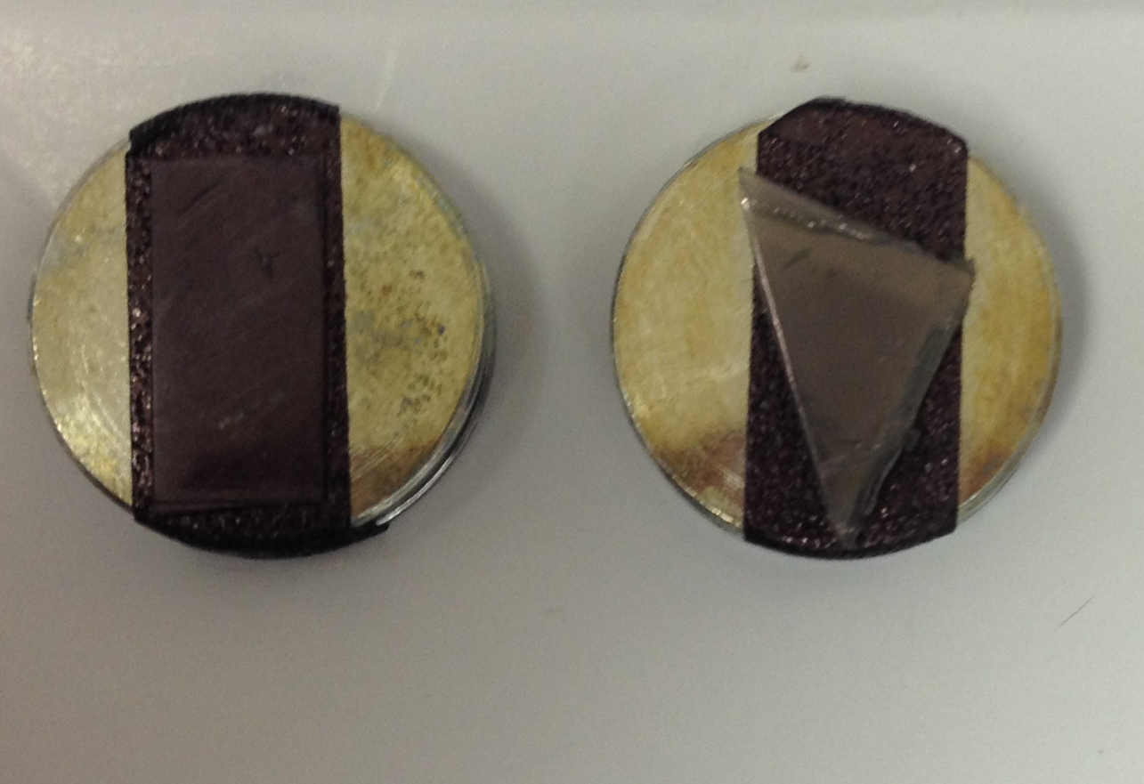 Figure 3.0: Gold coated
samples are ready for imagining in the
Scanning Electron Microscope.
Figure 3.0: Gold coated
samples are ready for imagining in the
Scanning Electron Microscope.
Secondary Electron Detector
The sample is imaged using the secondary electron detector within the scanning electron microscope. Figure 3.1 below illustrates the indentation at 397X magnification with 20kV accelerating voltage. Although it is an excellent image, the scanning electron microscope is not the best with determining the depth (Z height) and surface roughness of the sample; therefore, the Atomic Force Microscope is used for depth and surface roughness analysis.
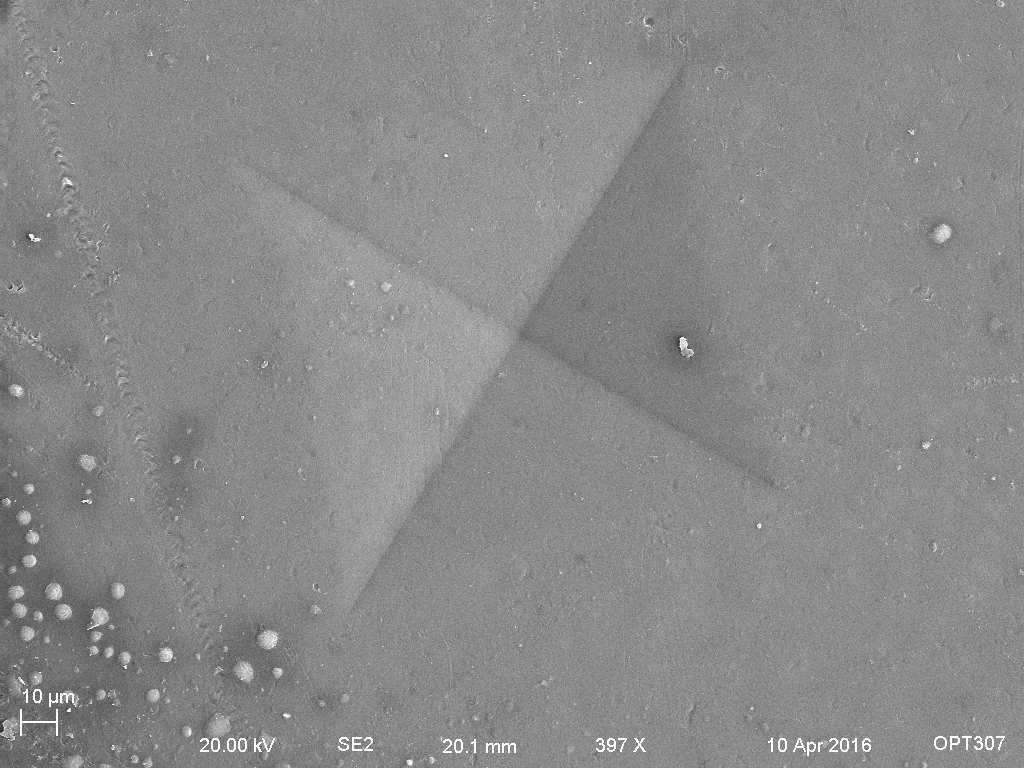 Figure 3.1: An image
of the indentation on the surface of the
sample generated via the secondary
electron detector.
Figure 3.1: An image
of the indentation on the surface of the
sample generated via the secondary
electron detector.
Energy Dispersive Spectrometry
Energy Dispersive Spectrometry or EDS is a detector that sits within the Scanning Electron Microscope. It is used to identify element composition and chemical characterization of a sample. Figure 3.2 below shows an EDS of the polymer sample. Carbon is the highest content within the sample and it is followed by Oxygen, Gold and Molybdenum. Moreover, EDS is capable of mapping out each element with colour, which is shown in Figure 3.2.
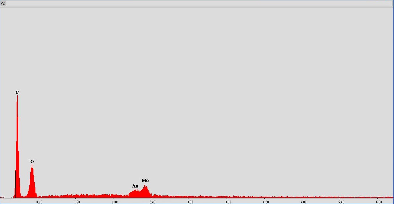 Figure
3.2: Energy-dispersive X-ray
spectroscopy of the sample.
Figure
3.2: Energy-dispersive X-ray
spectroscopy of the sample.
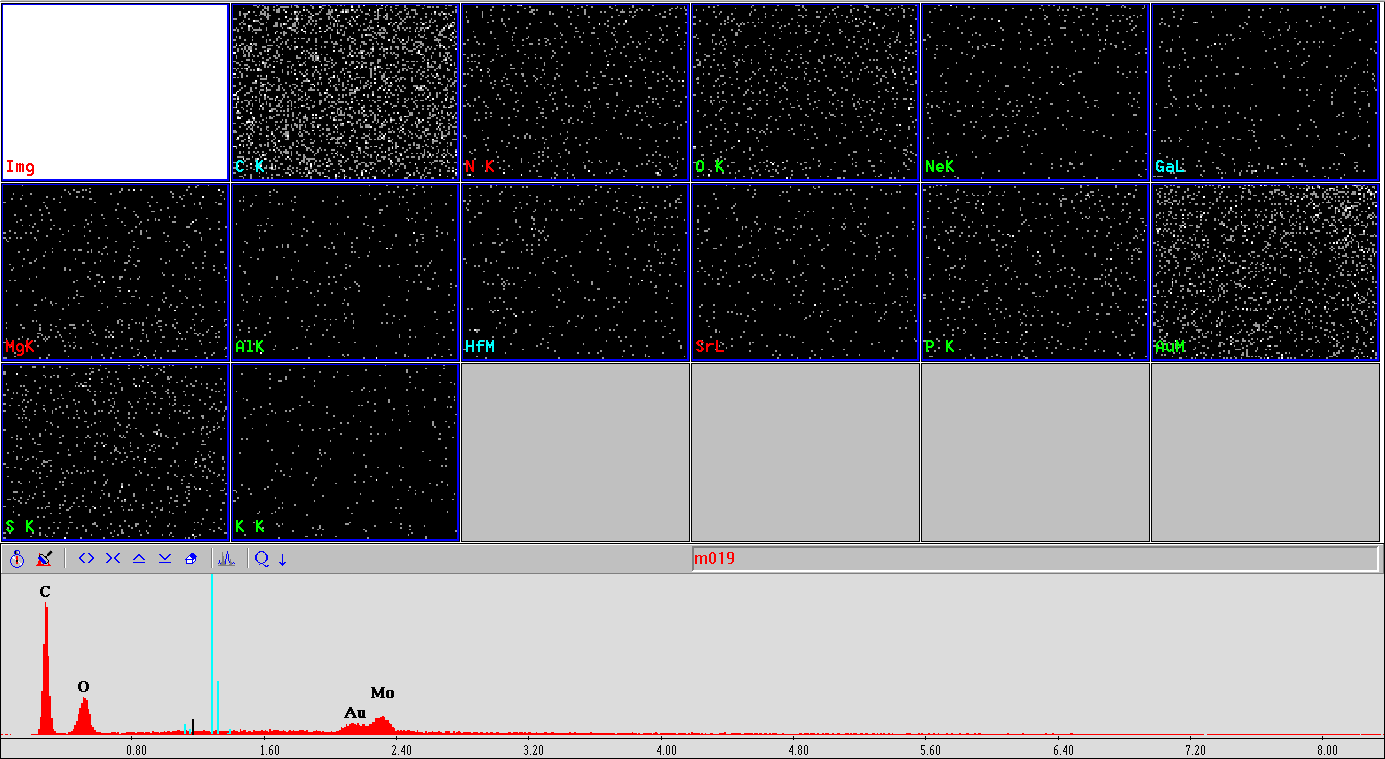 Figure
3.3: Elemental and chemical
composition maps of the sample.
Figure
3.3: Elemental and chemical
composition maps of the sample.