Light Microscopy
Light microscope image is taken via the Buehler micro-hardness indenter with 10X objective lens. Figure 1.0 below is generated immediately after the micro-indentation. A Vickers hardness test was performed on the sample with a load of 10g for 30 seconds. The diamond shape indentation provides a hardness value of the material.
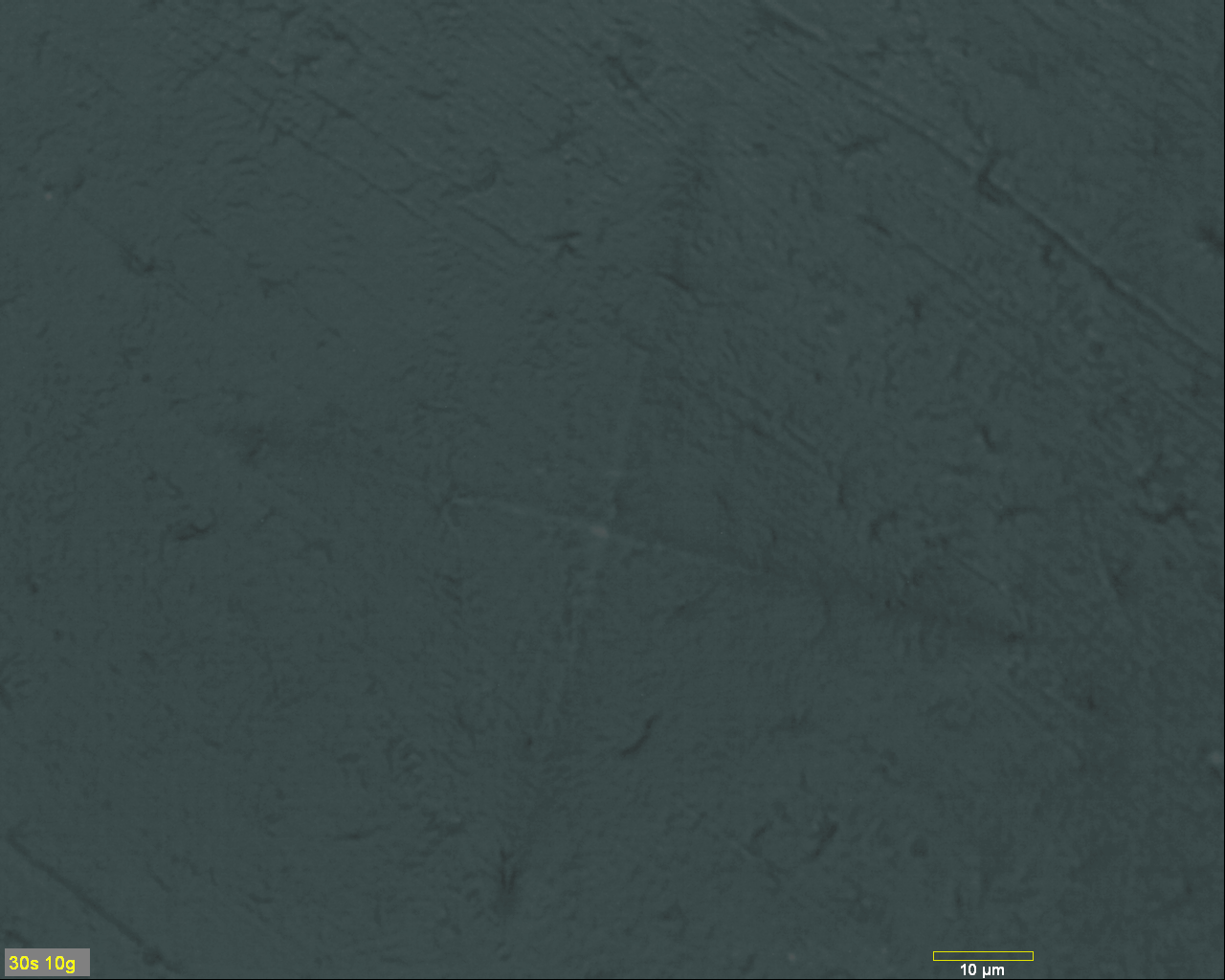 Figure 1.0: Light
microscope image of a micro indentation on the
surface of the sample.
Figure 1.0: Light
microscope image of a micro indentation on the
surface of the sample.
Atomic Force Microscopy
A NT-MDT NEXT atomic force microscope is used to generate a surface scan of an 85 by 85 micron area with the indentation in the middle. Figures 1.1, 1.2, and 1.3 below illustrates the indentation made by the Buehler micro-hardness machine. This image provides an excellent understanding of the depth (Z height) of the indentation. A section analysis of the indentation is shown in Figure 1.4, this particular feature calculates profiles from the image in certain orientations. Additionally, a 3D roughness view of the sample in a plane area is shown in Figure 1.5.
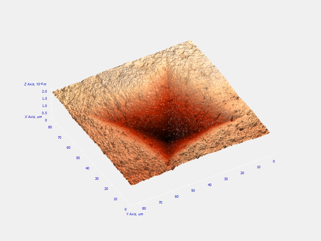 Figure 1.1: A top 3D
iso-view of the indentation from the
atomic force microscope.
Figure 1.1: A top 3D
iso-view of the indentation from the
atomic force microscope.
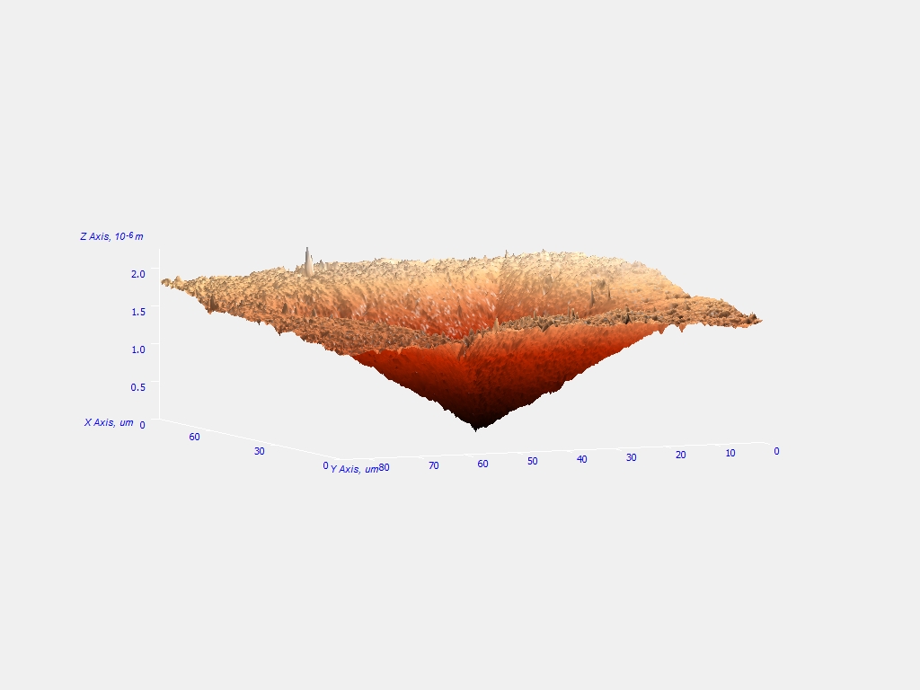 Figure
1.2: A side 3D iso-view of the indentation
from the atomic force microscope.
Figure
1.2: A side 3D iso-view of the indentation
from the atomic force microscope.
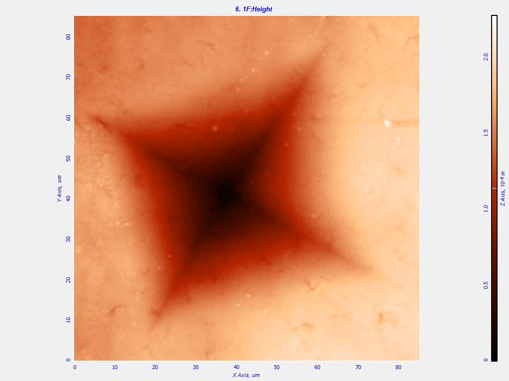 Figure 1.3: A 2D top
view of the indentation from the atomic
force microscope.
Figure 1.3: A 2D top
view of the indentation from the atomic
force microscope.
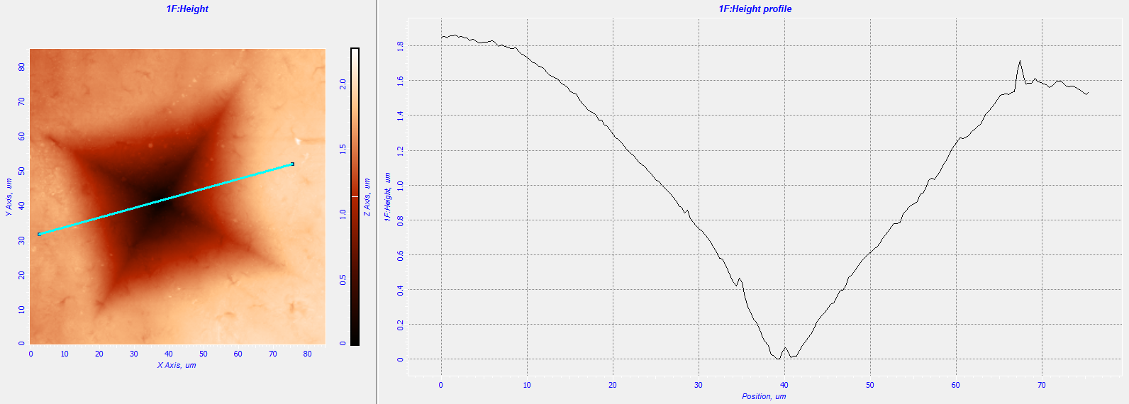 Figure
1.4: A section analysis of the
indentation. This particular analysis
illustrates the height profile of the
indentation.
Figure
1.4: A section analysis of the
indentation. This particular analysis
illustrates the height profile of the
indentation.
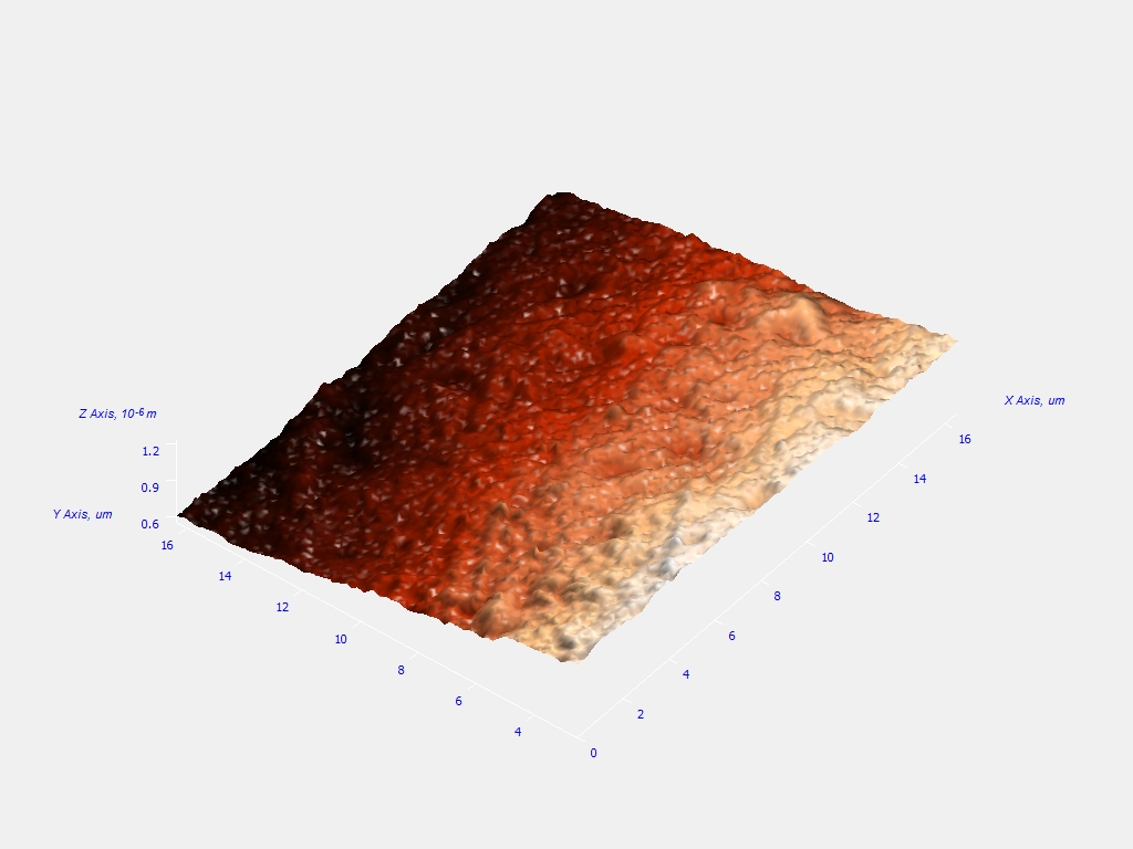 Figure 1.5: A 3D plane
area surface roughness view from the AFM.
Figure 1.5: A 3D plane
area surface roughness view from the AFM.