backscattered Imaging
Backscattered electrons consist of high-energy electrons originating in the electron beam that are reflected out of the specimen’s interaction volume. They are produced as a result of elastic collisions with the atoms of the sample and usually retain about 80% or more of their original energy. Because of their greater energy, backscattered electrons can escape from much deeper regions of the sample than can secondary electrons thus creating a larger region of excitation. The backscattered detector differs from secondary electron detectors in that backscatter detectors don’t attract the electrons. Only those electrons that travel in a straight path from the specimen to the detector go towards forming the backscattered image. Since heavy atoms with a high atomic number are stronger scattered than light ones, images with back-scattered electrons BSE contain compositional information. The number of backscattered electrons produced increases with atomic number of the specimen which is why some BSE images display differential contrast of the elements present despite a uniform surface topography. BSE analysis was performed on both, surfaces with and without cells.
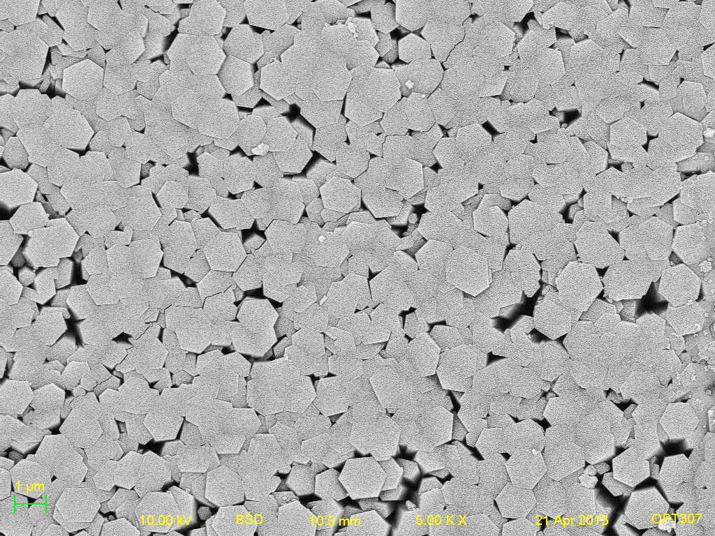
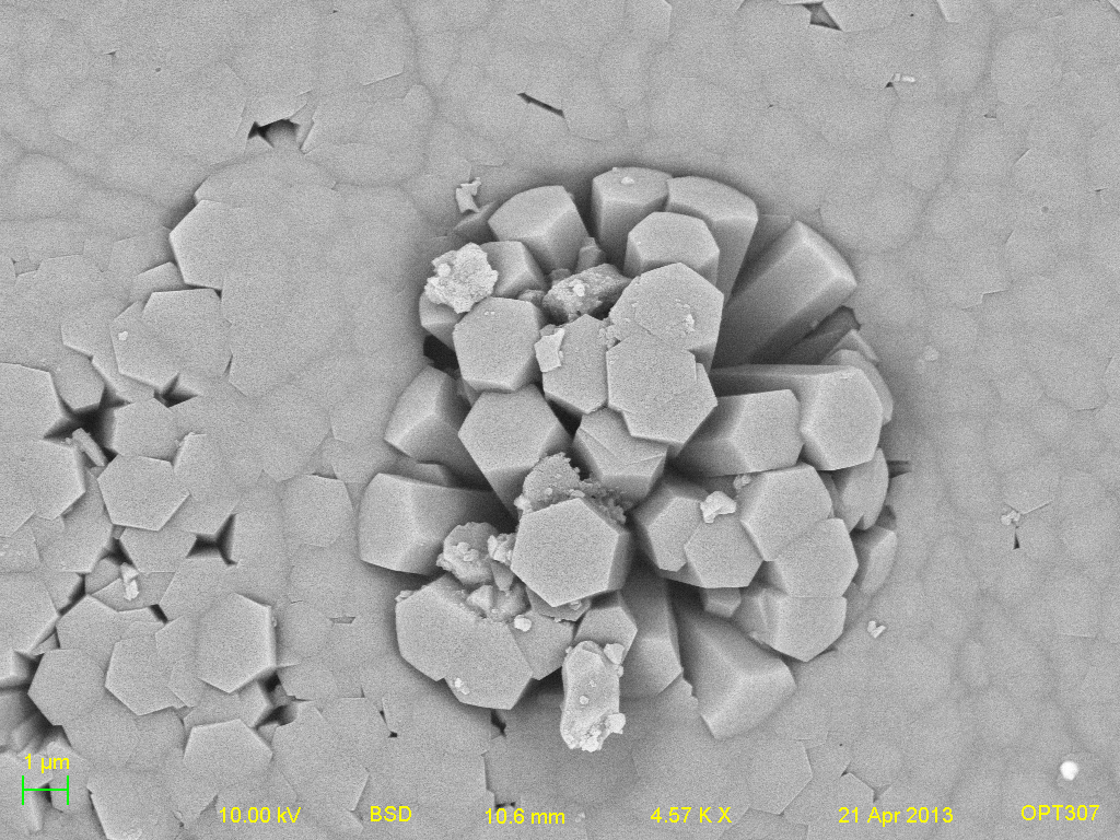
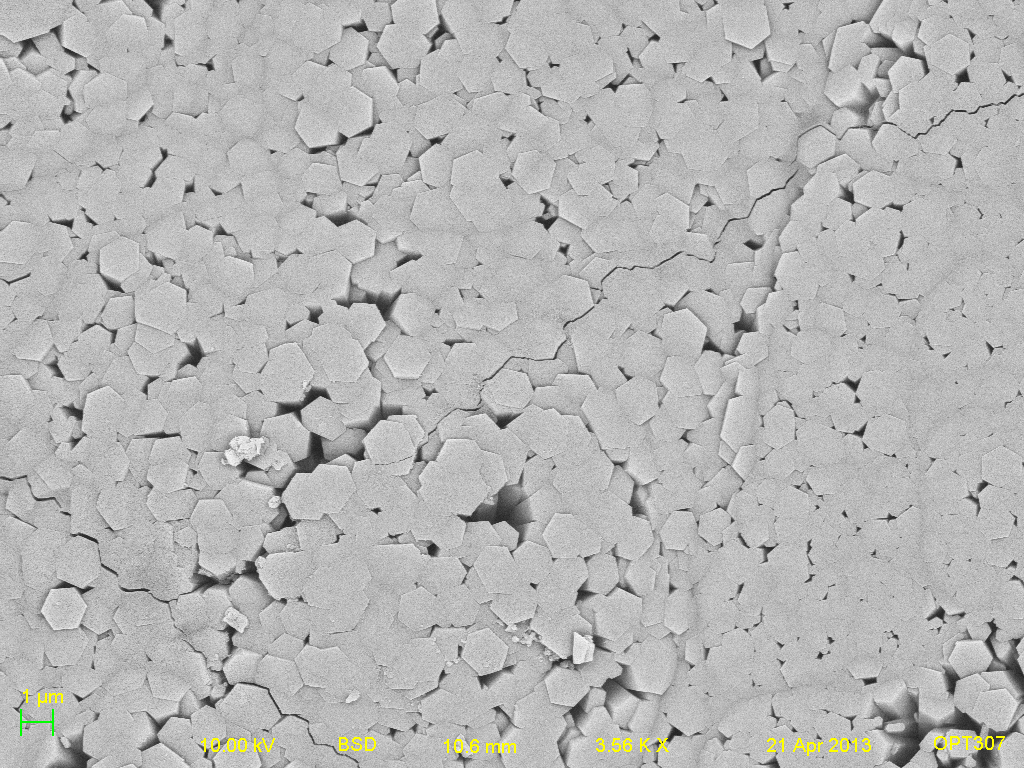
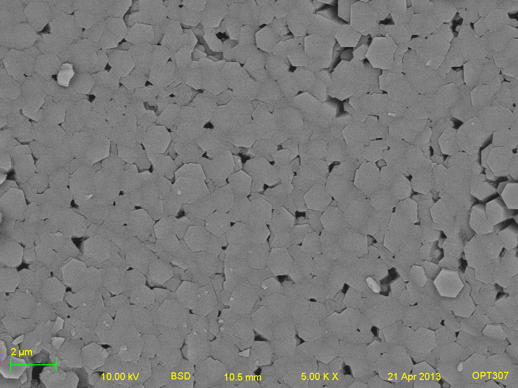
The following BSE images were taken for visual purposes. These images were taken at an accelerating voltage of 10kV, a 10mm working distance and a 5000x magnification. The bightness and contrast were also held constant for all surfaces.
Titanium samples- (left to right: Titanium, Positive HAP, Negative HAP, Heated HAP and HAP respectively).
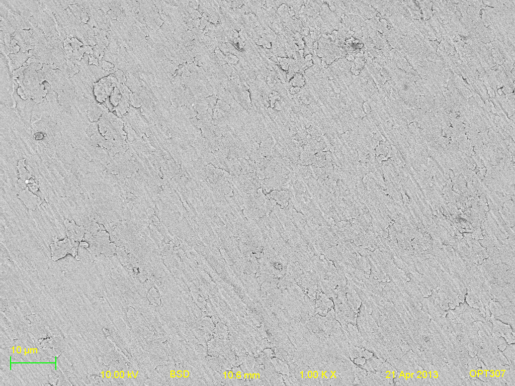
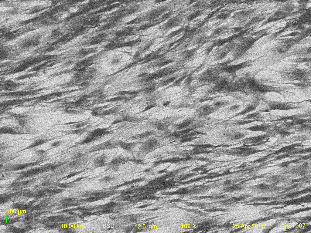
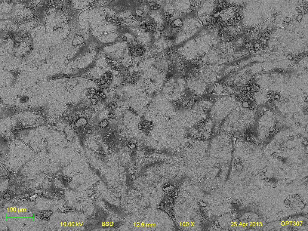
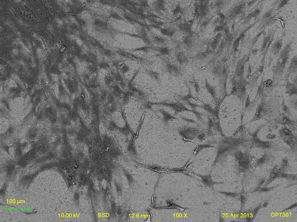
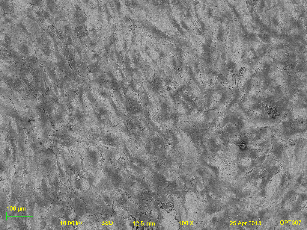
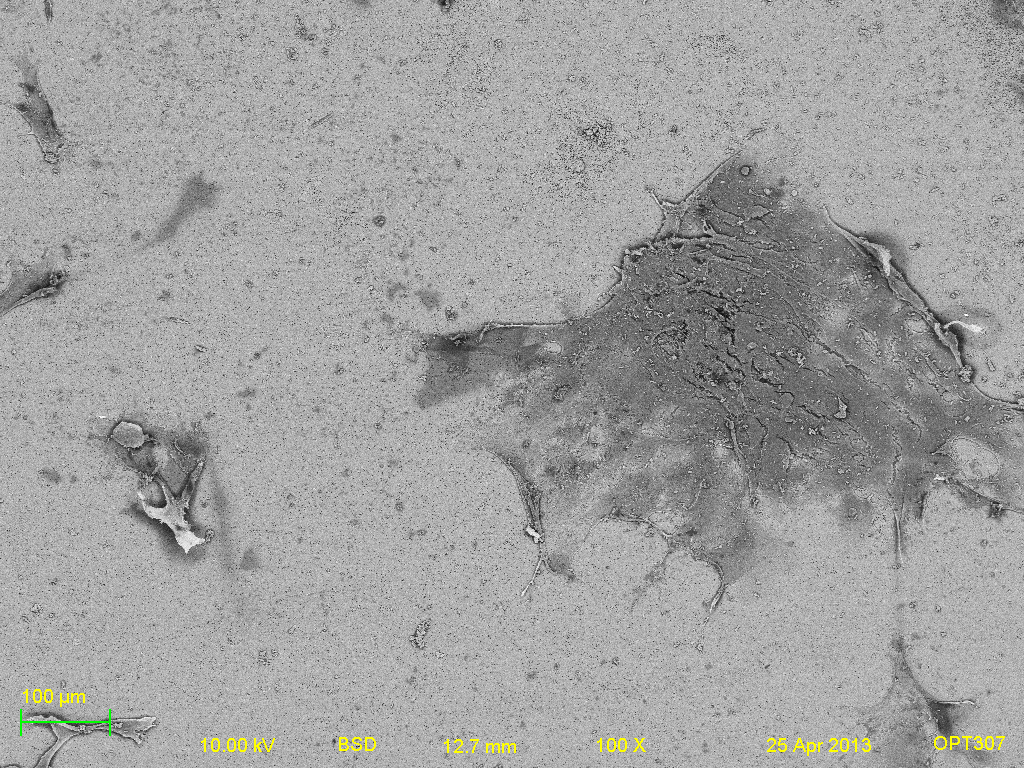
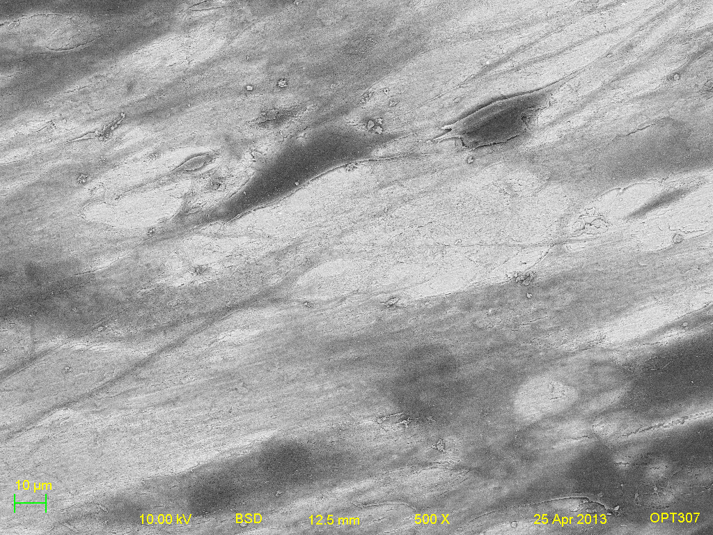
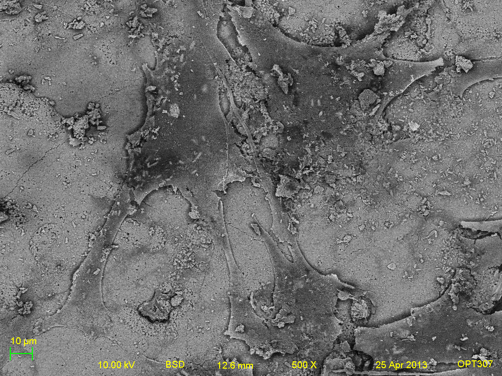
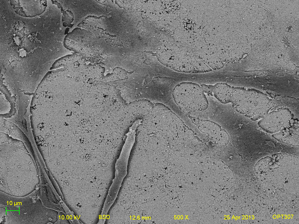
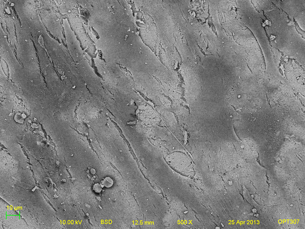
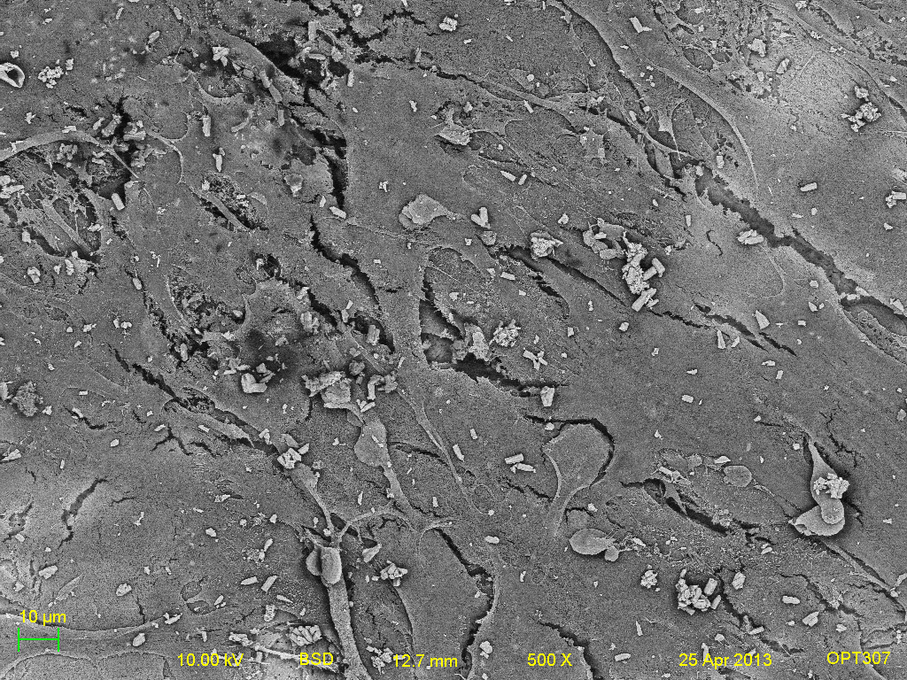
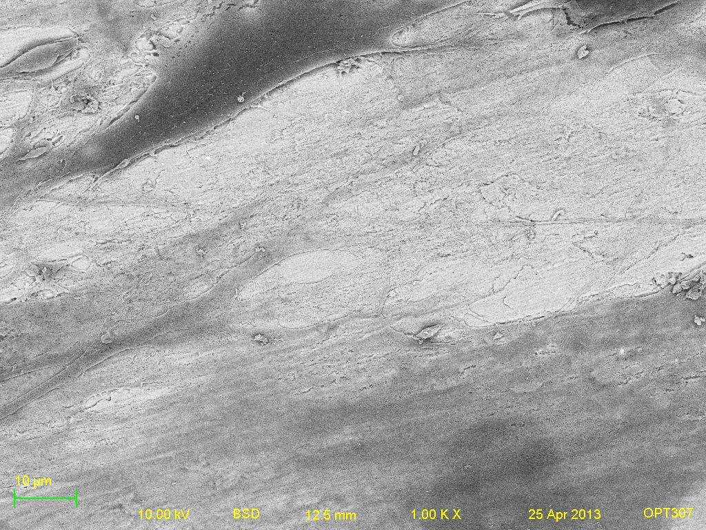
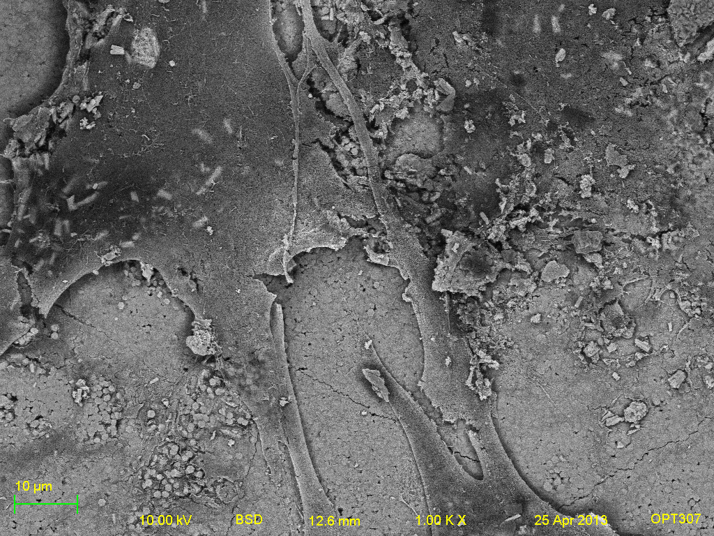
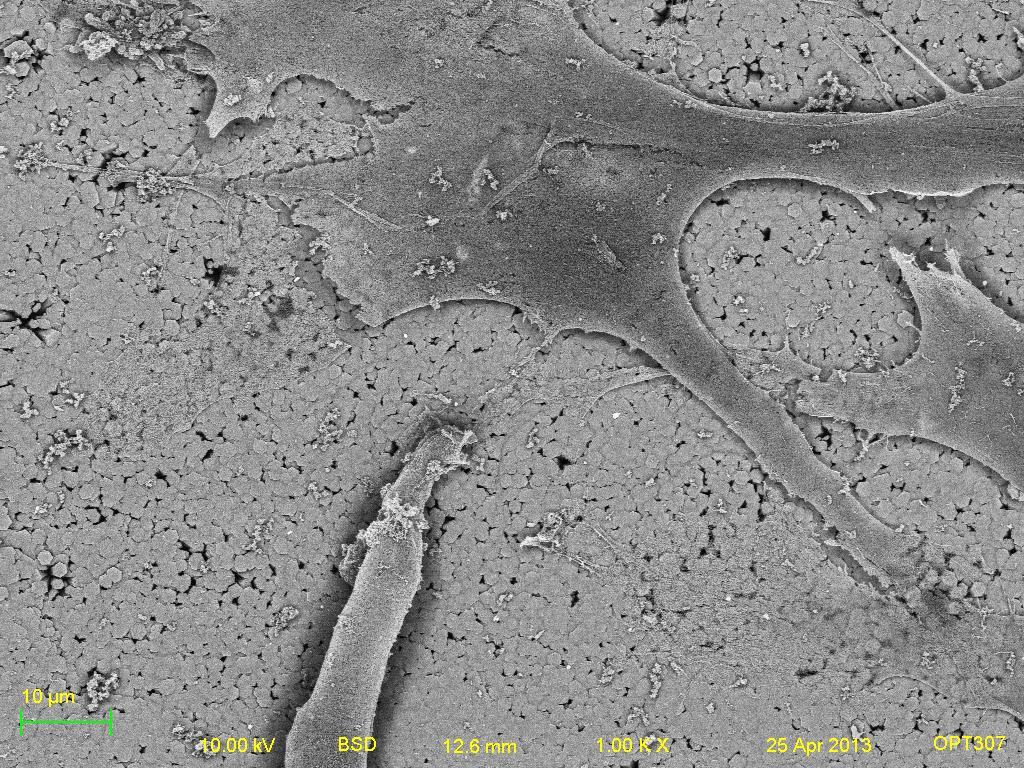
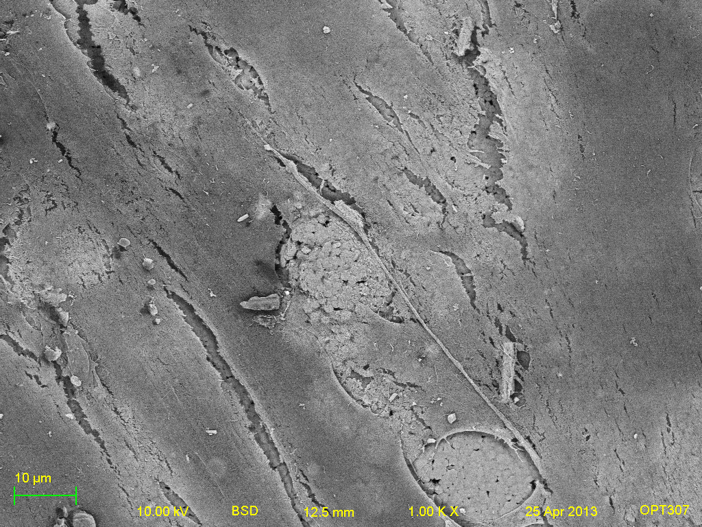
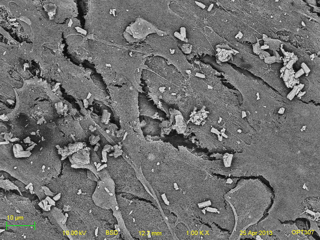
Mesenchymal stem cells seeded on the different titanium surfaces were also imaged under BSE parameters. In terms of comparison, the images were all taken at the same parameters of brightness, contrast, working distance, accelerating voltage and lens apperture and magnification.
Titanium samples- (right to left: Titanium, Positive HAP, Negative HAP, Heated HAP and HAP respectively. Top-bottom: 100x, 500x and 1000x).