sem imaging
Image formation in the SEM is dependent on the acquisition of signals produced from the interaction of the specimen and the electron beam. Interaction of the accelerated electron beam with a sample target produces a variety of elastic and inelastic collisions between electrons and atoms within the sample. The most common type of signal used in modern SEMs is the secondary electron emission signal. A secondary electron is produced when an electron from the primary beam collides in an inelastic manner with an electron from a specimen’s atom and loses energy. From this collision an electron may be emitted if charge on the atom needs to be re-established. In order to detect the secondary electrons that are emitted from the specimen a specialized detector is required. A scintillator- photomultiplier first converts the energy of secondary electrons into photons which travel down a light pipe to be converted into an electrical signal by the way of a photocathode and photomultiplier. SEM analysis was performed on both, surfaces with and without cells.
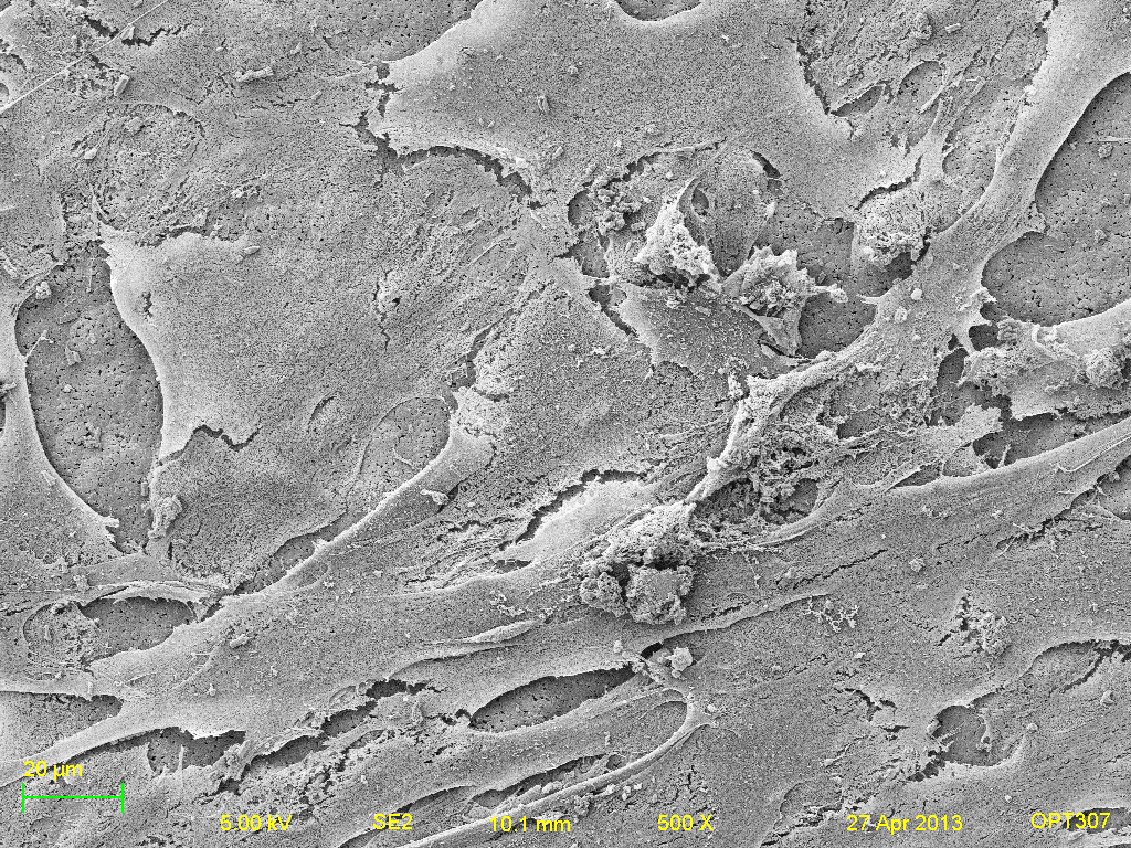
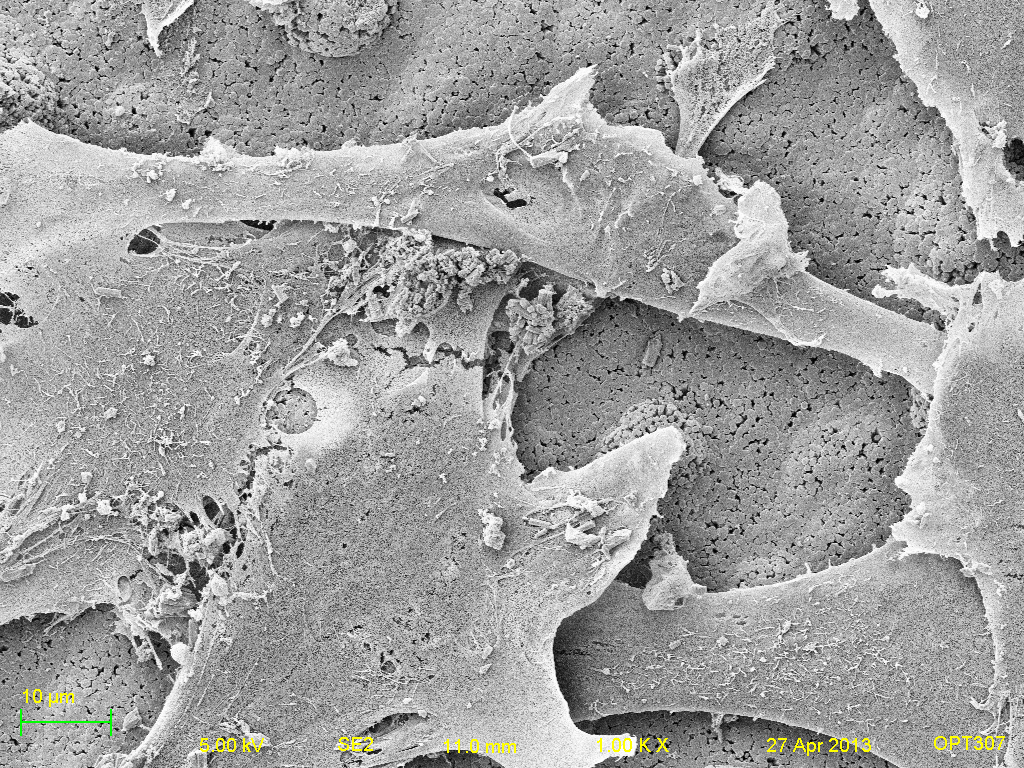
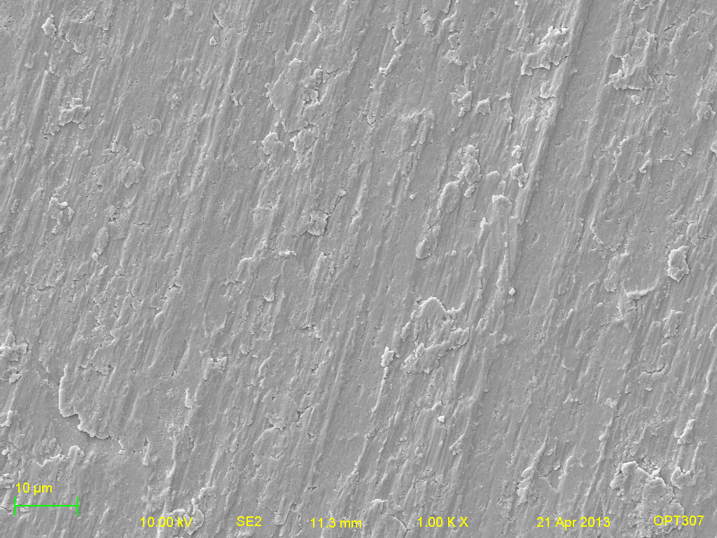
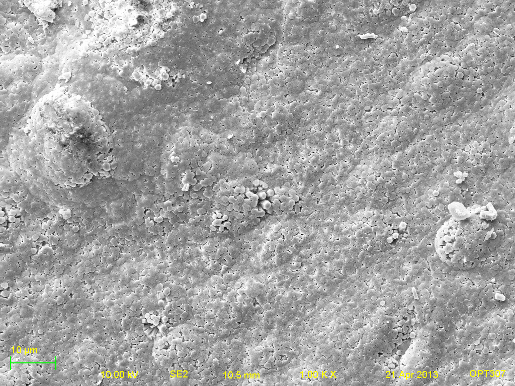
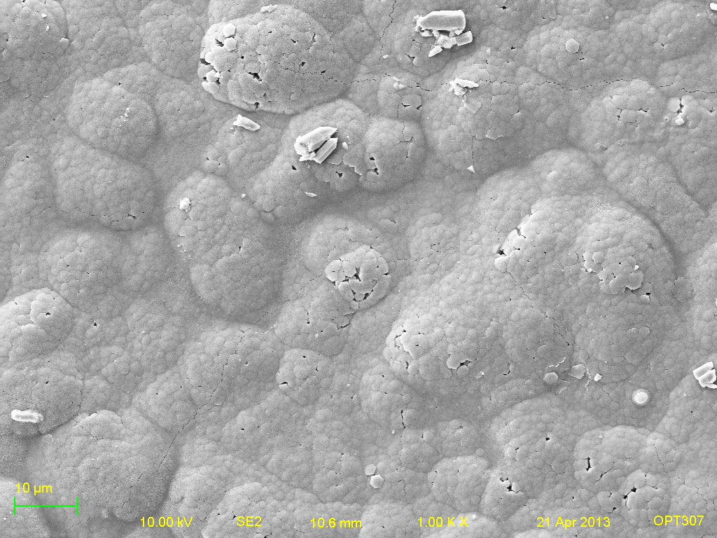
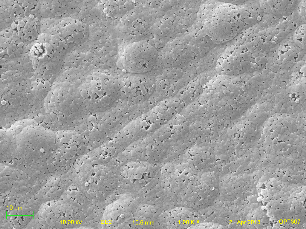
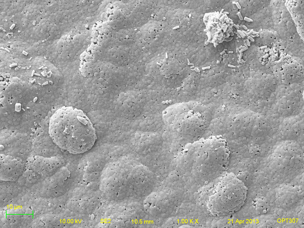
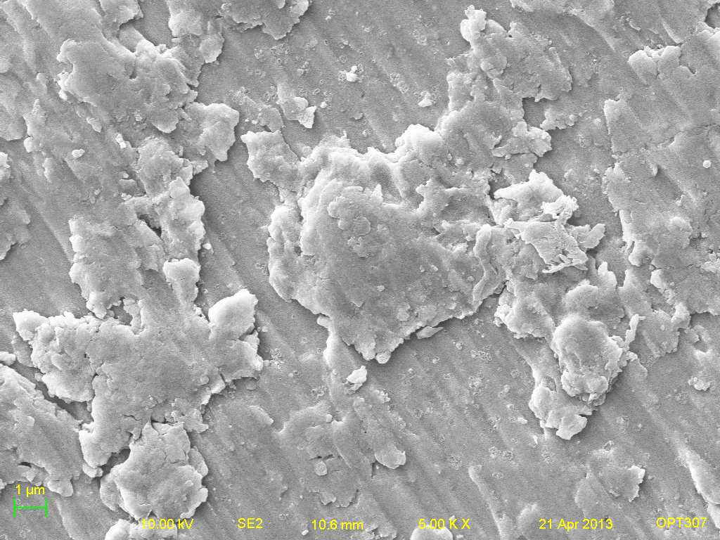
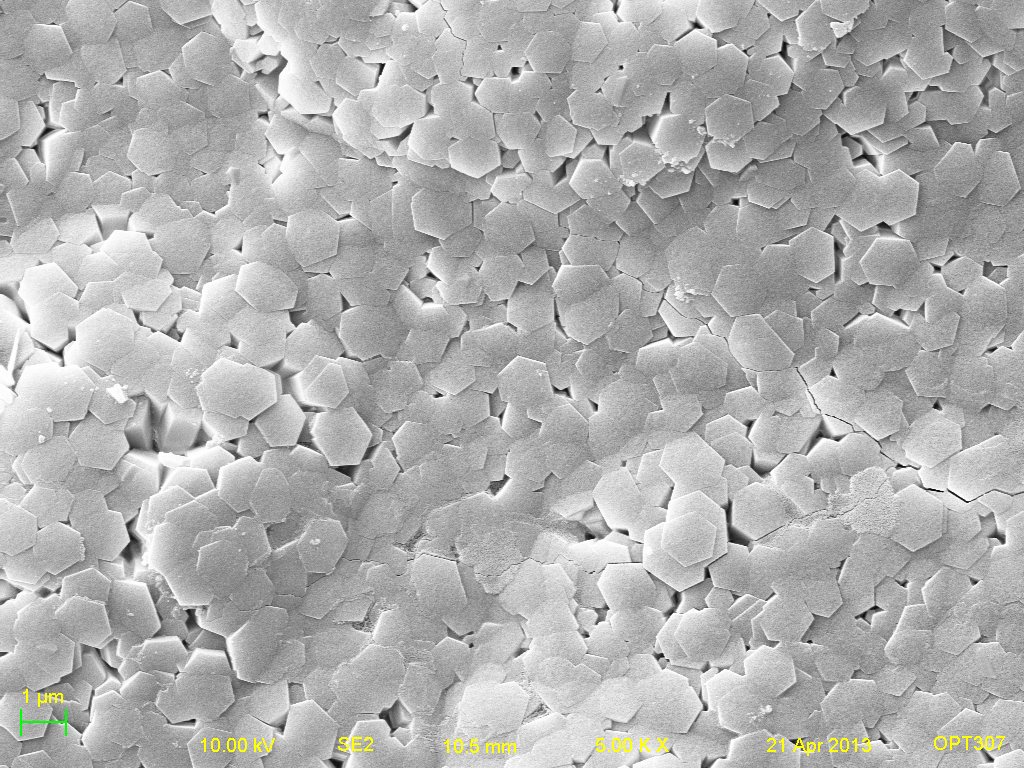
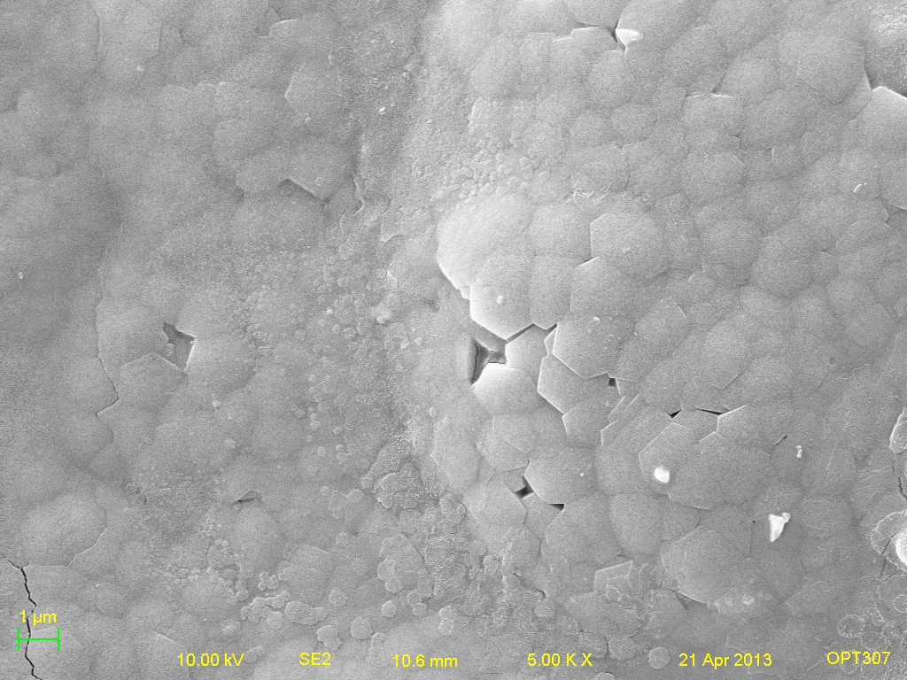
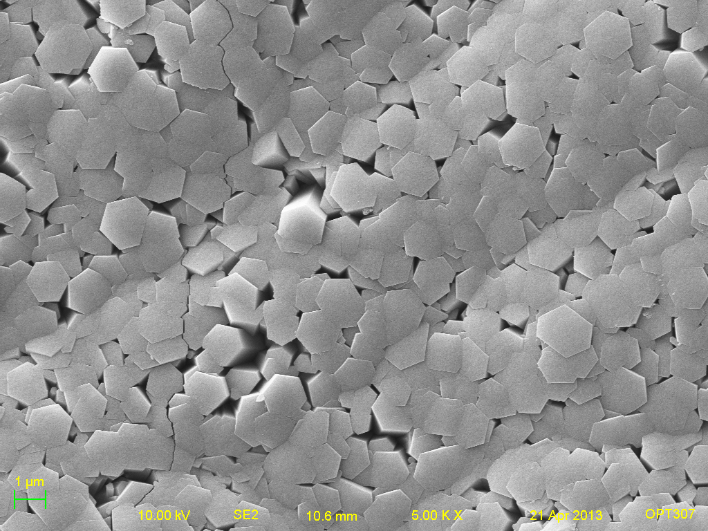
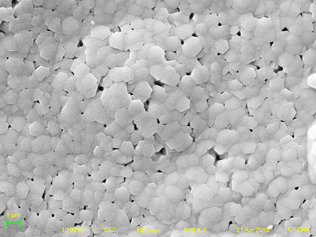
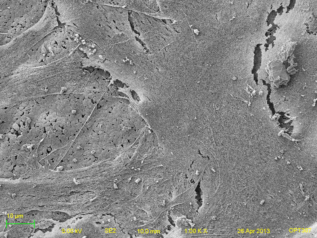
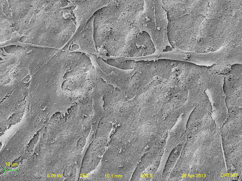
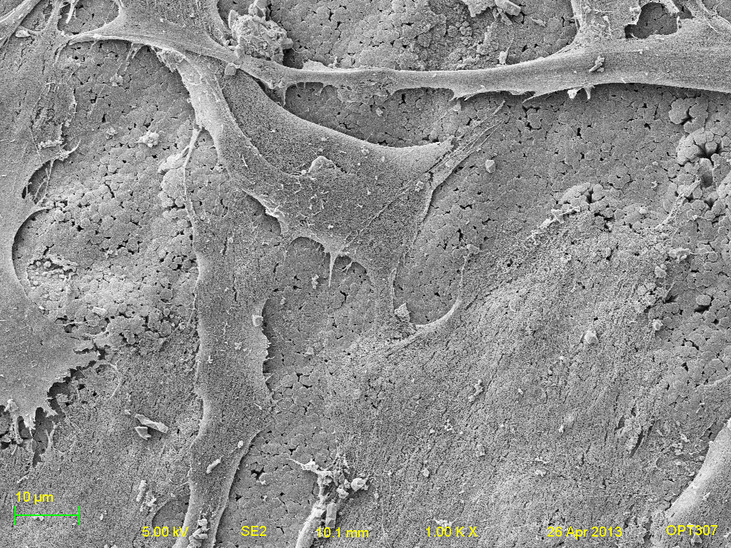
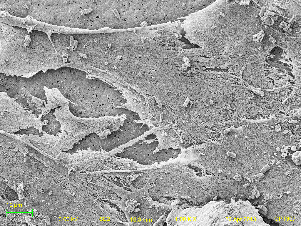
The following SEM images were taken for visual purposes. These images were taken at an accelerating voltage of 10kV, a 10mm working distance and magnification of 1000x and 5000x. The bightness and contrast were also held constant for all surfaces.
Titanium samples- (left to right: Titanium, Positive HAP, Negative HAP, Heated HAP and HAP respectively. Top-bottom: 1000x and 5000x respectively).
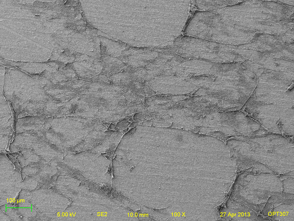
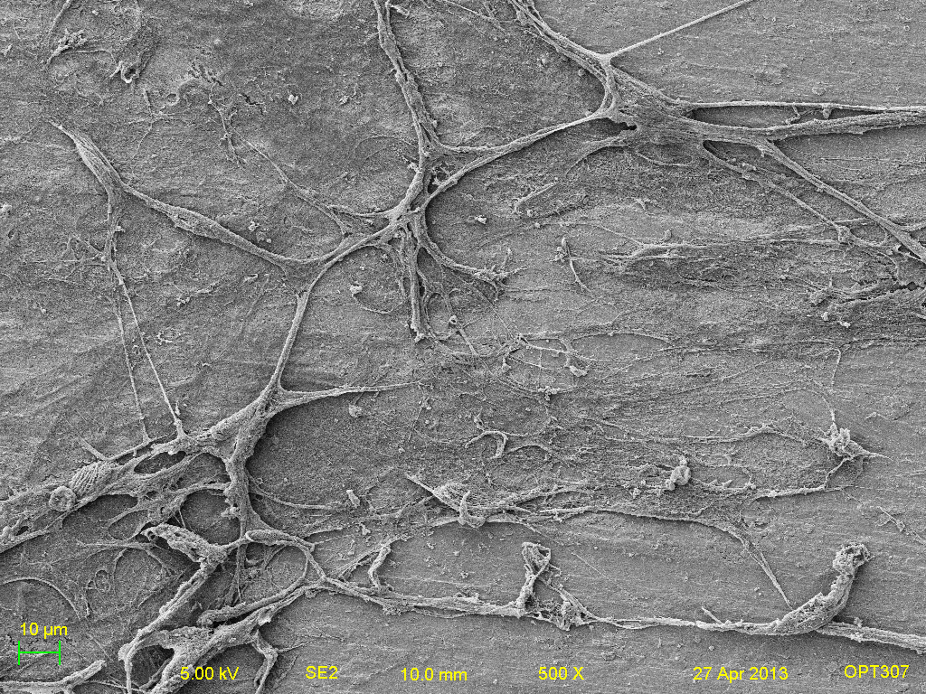
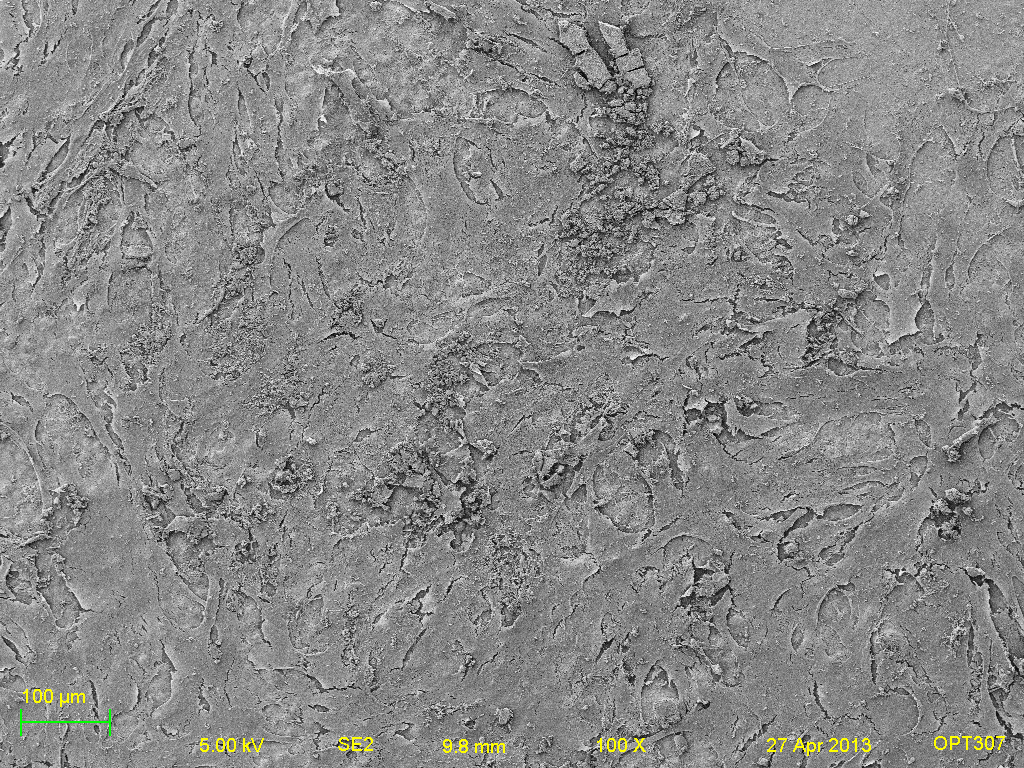
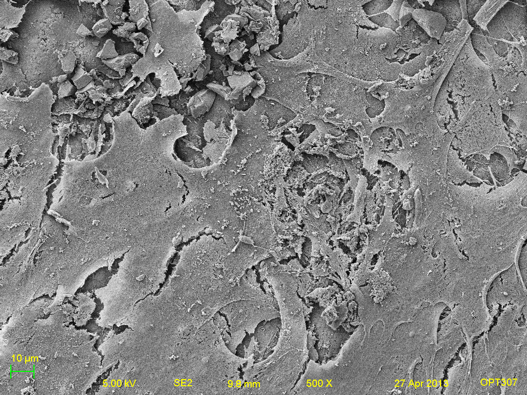
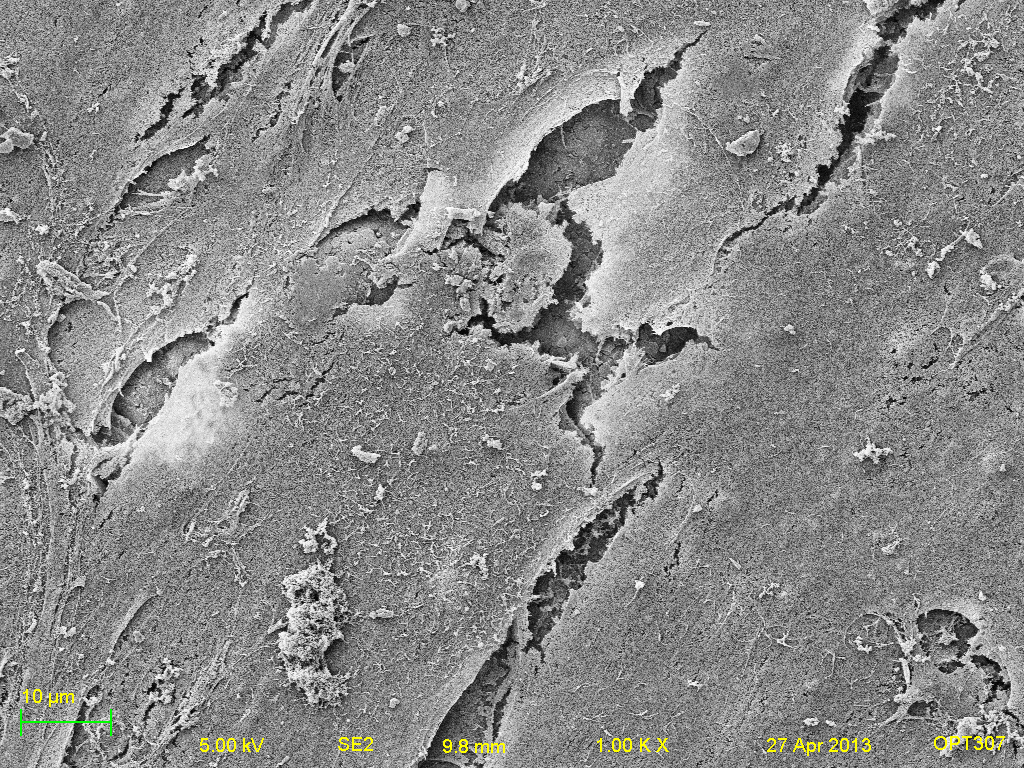
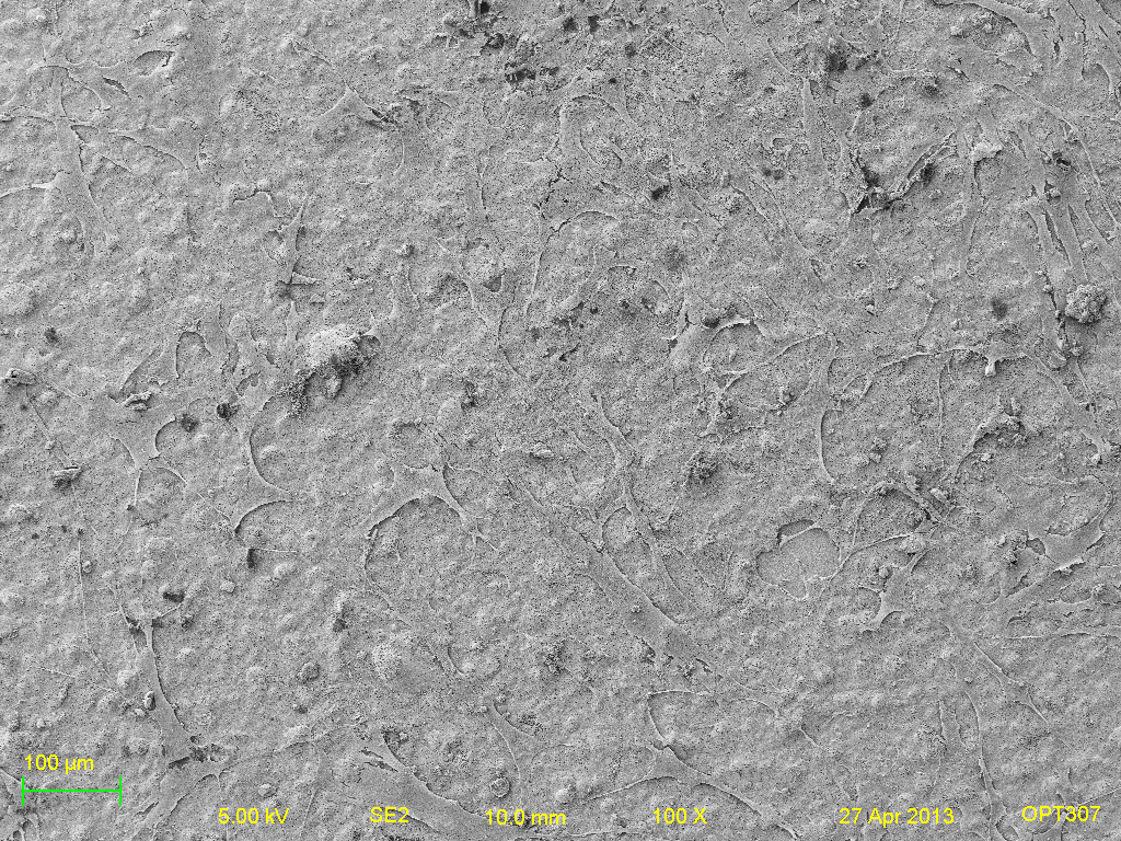
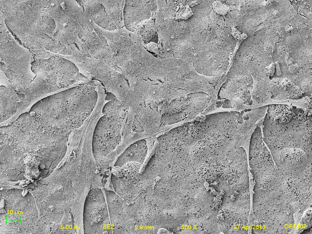
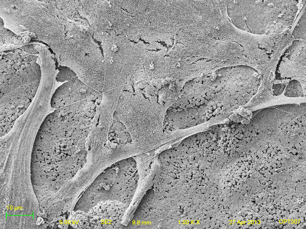
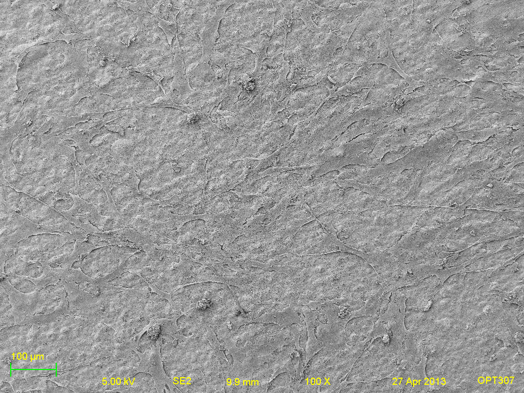
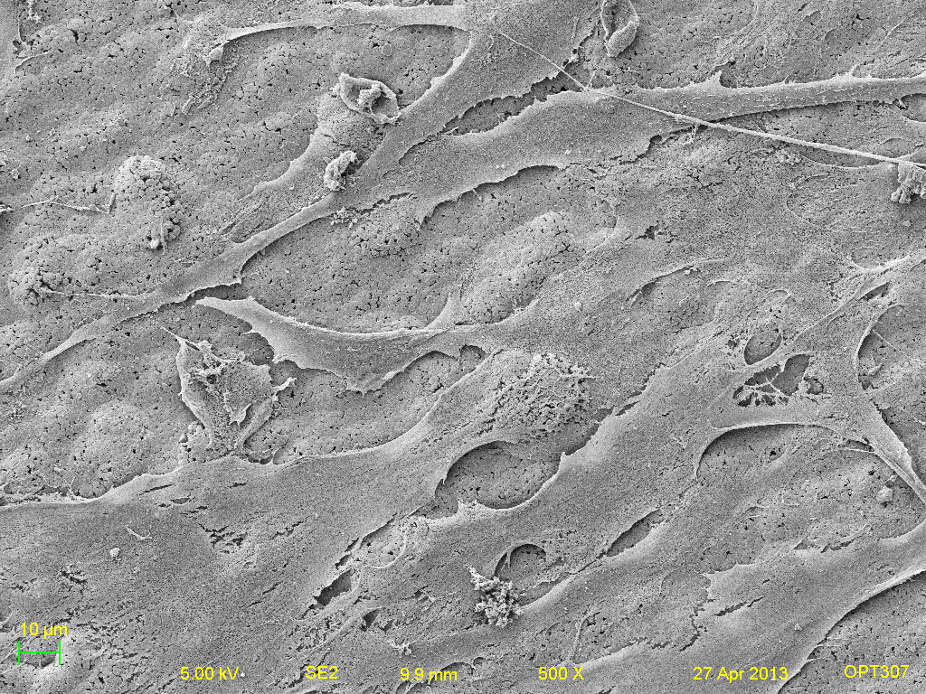
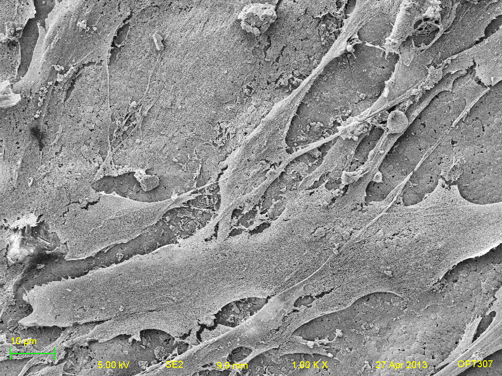
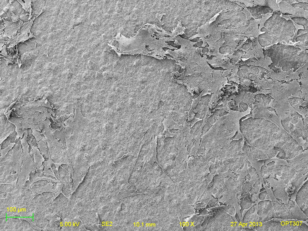
The next set of SEM images are divided into three set of groups. Within each group, images of the five different surfaces were recorded at 100x, 500x and 1000x. In terms of comparison, the images were all taken at the same parameters of brightness, contrast, working distance, accelerating voltage and lens apperture and magnification.
Set 1 of Titanium samples- (left to right: 100x, 500x, and 1000x respectively. Top-bottom: Titanium, Positive HAP, Negative HAP, Heated HAP and HAP respectively).
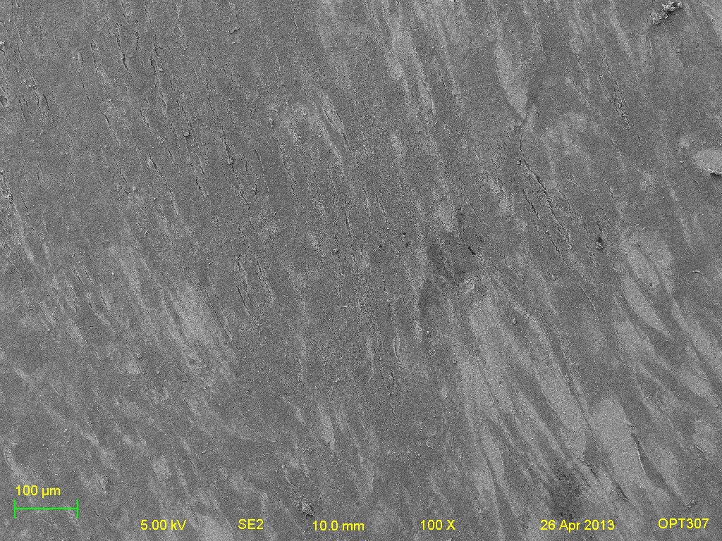
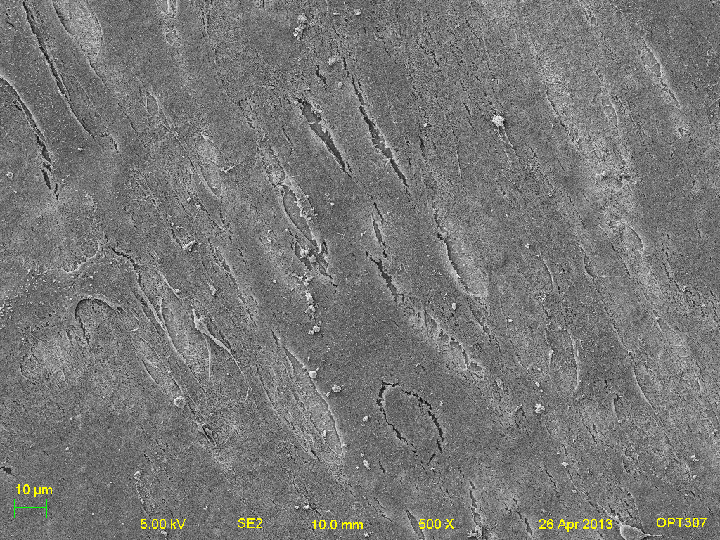
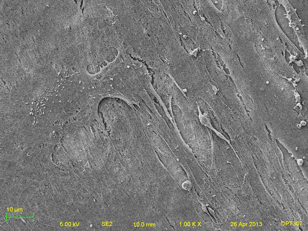
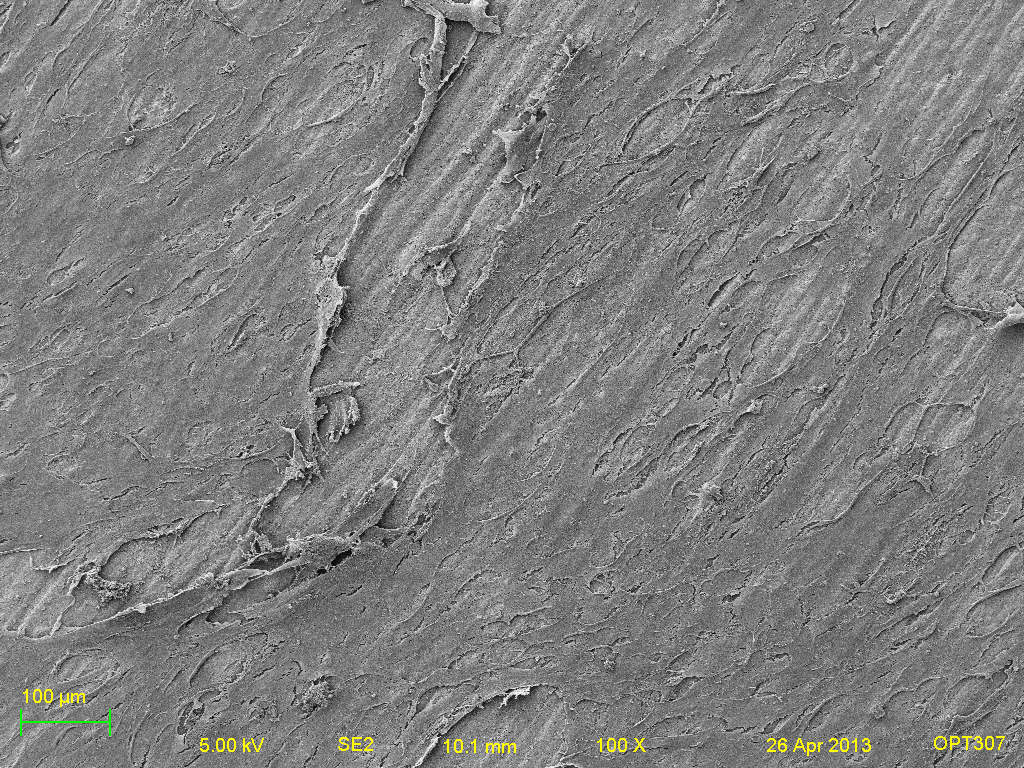
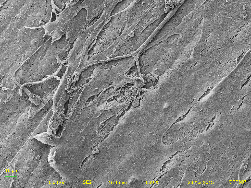
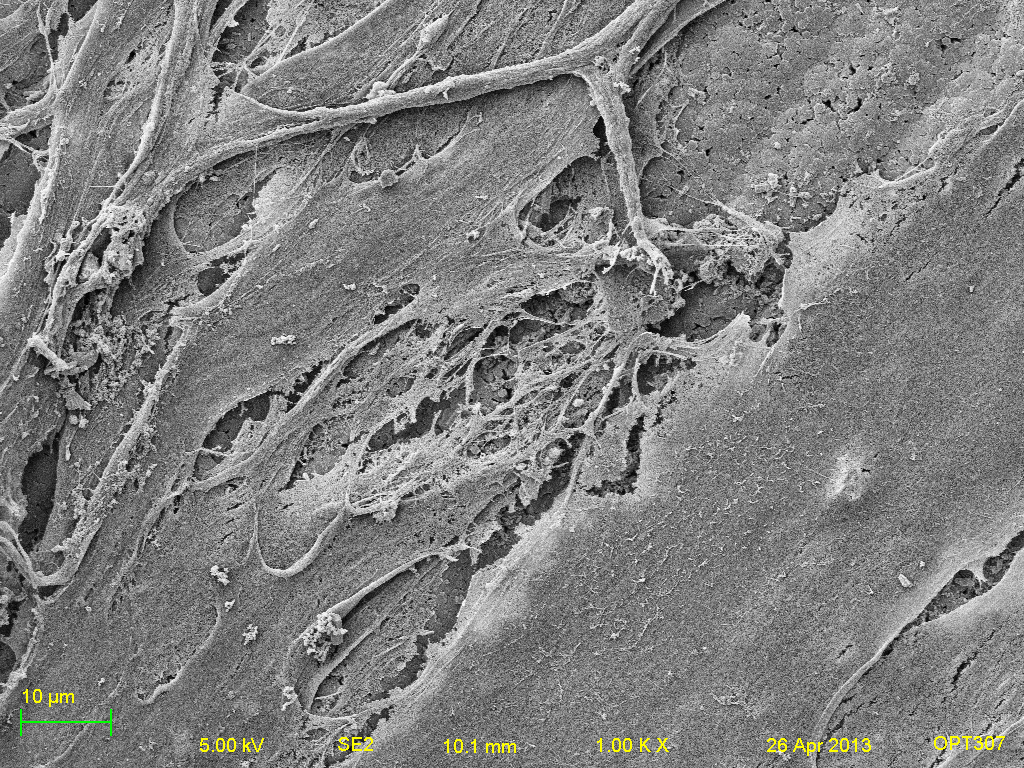
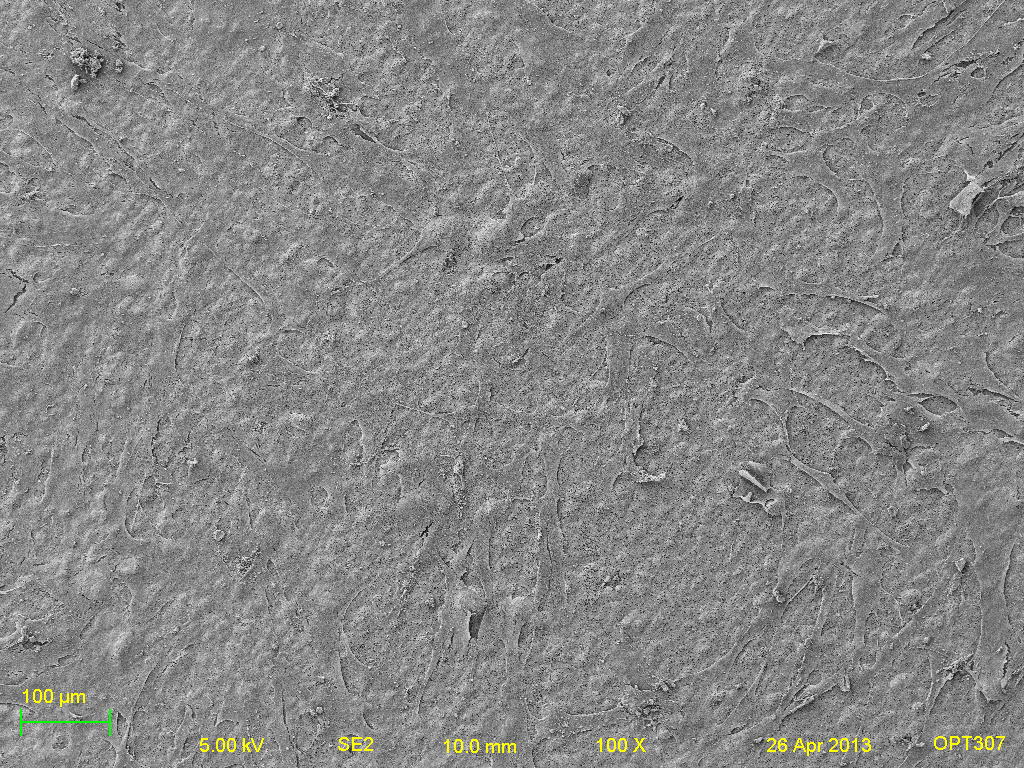
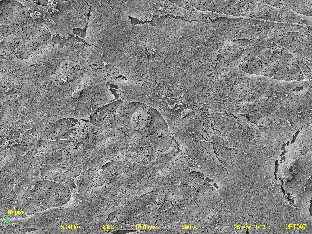
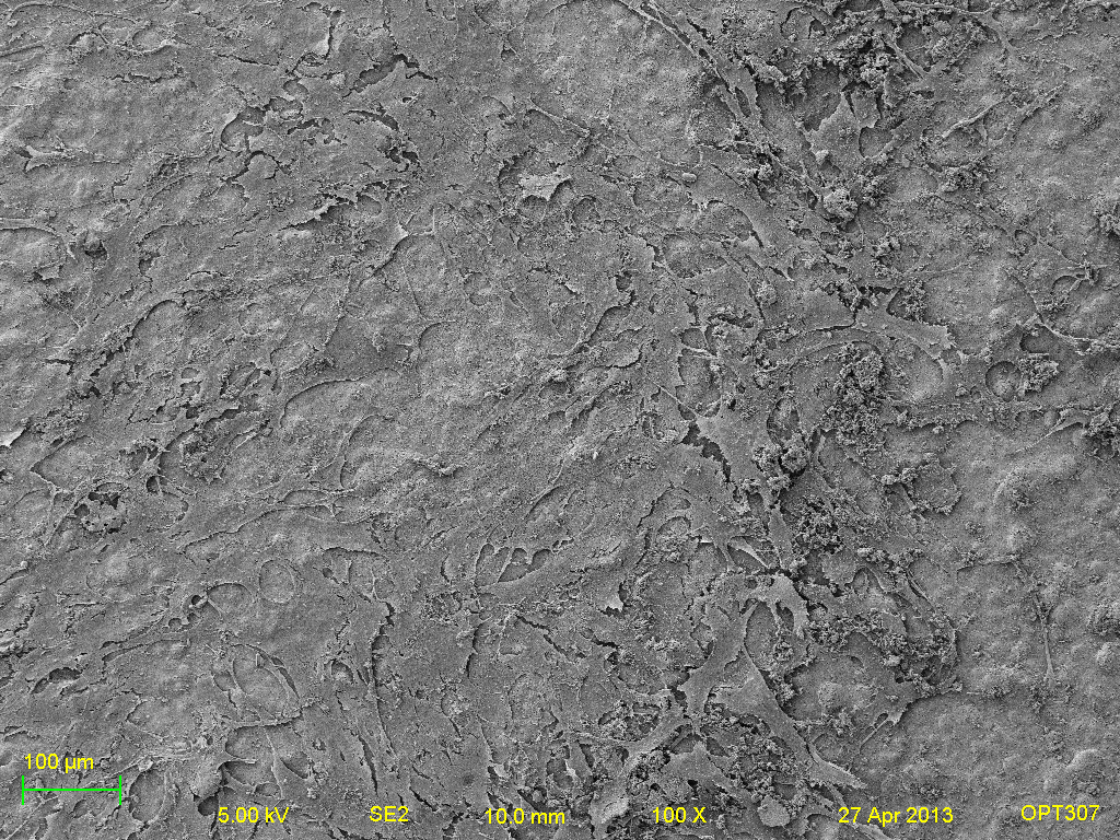
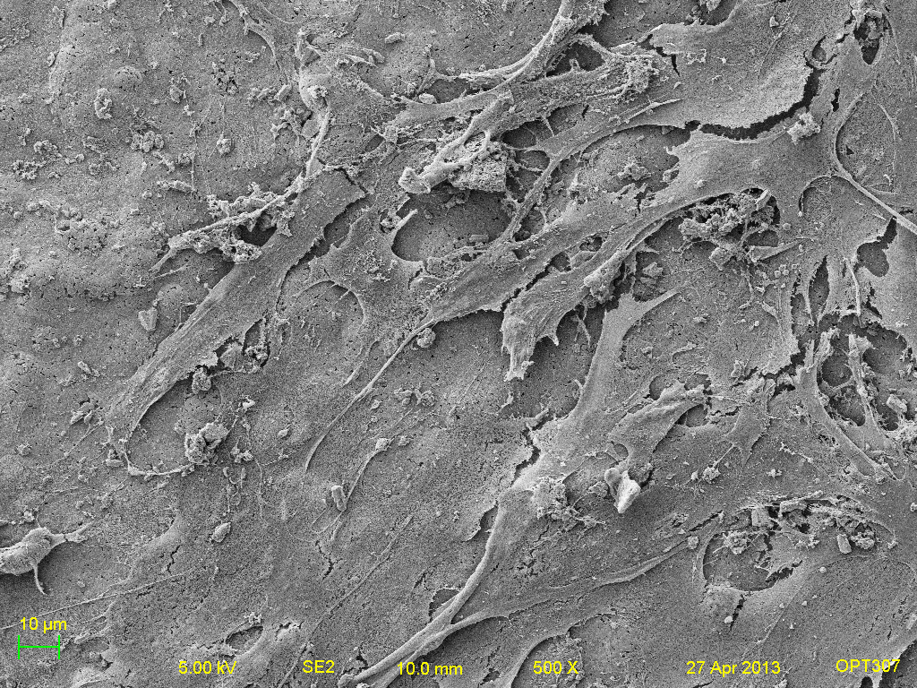
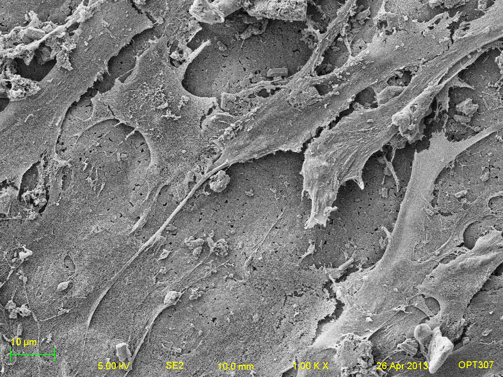
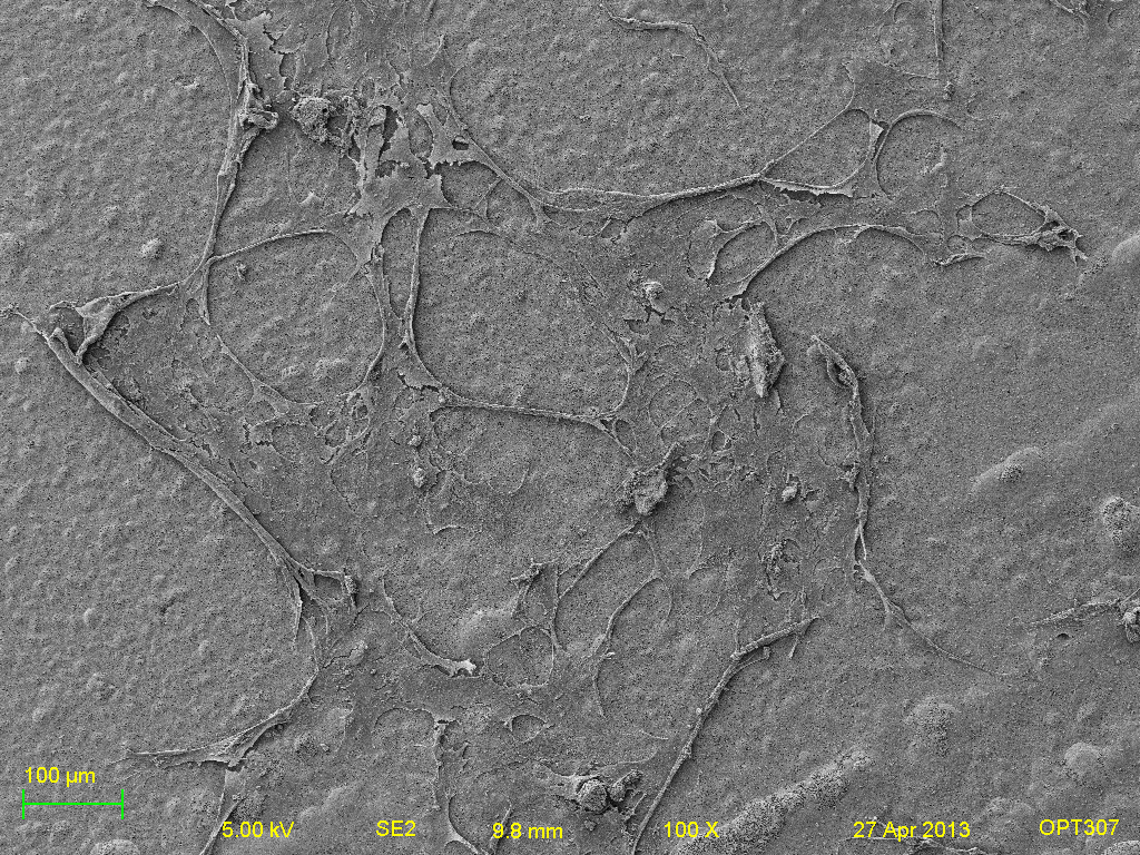
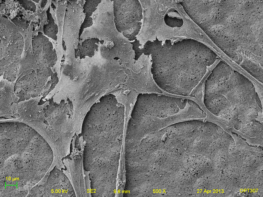
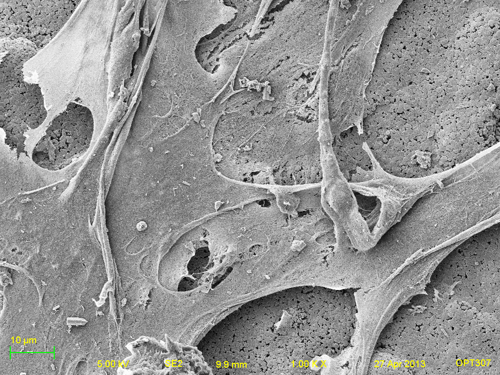
Set 2 of Titanium samples- (left to right: 100x, 500x, and 1000x respectively. Top-bottom: Titanium, Positive HAP, Negative HAP, Heated HAP and HAP).
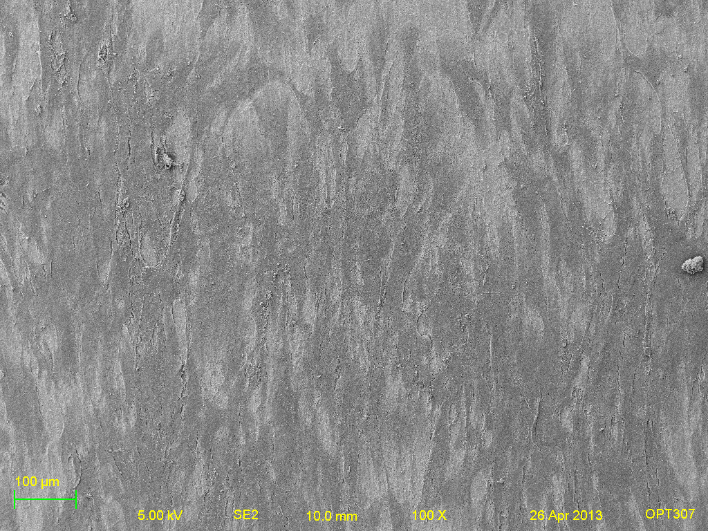
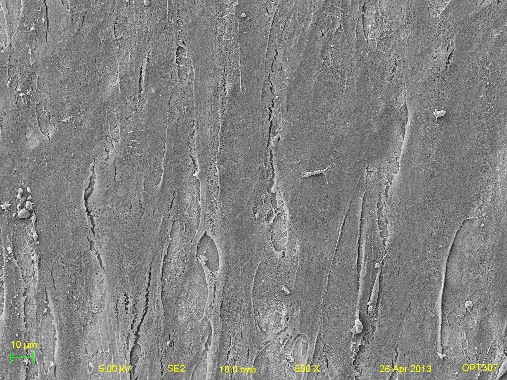

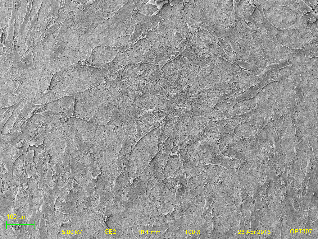
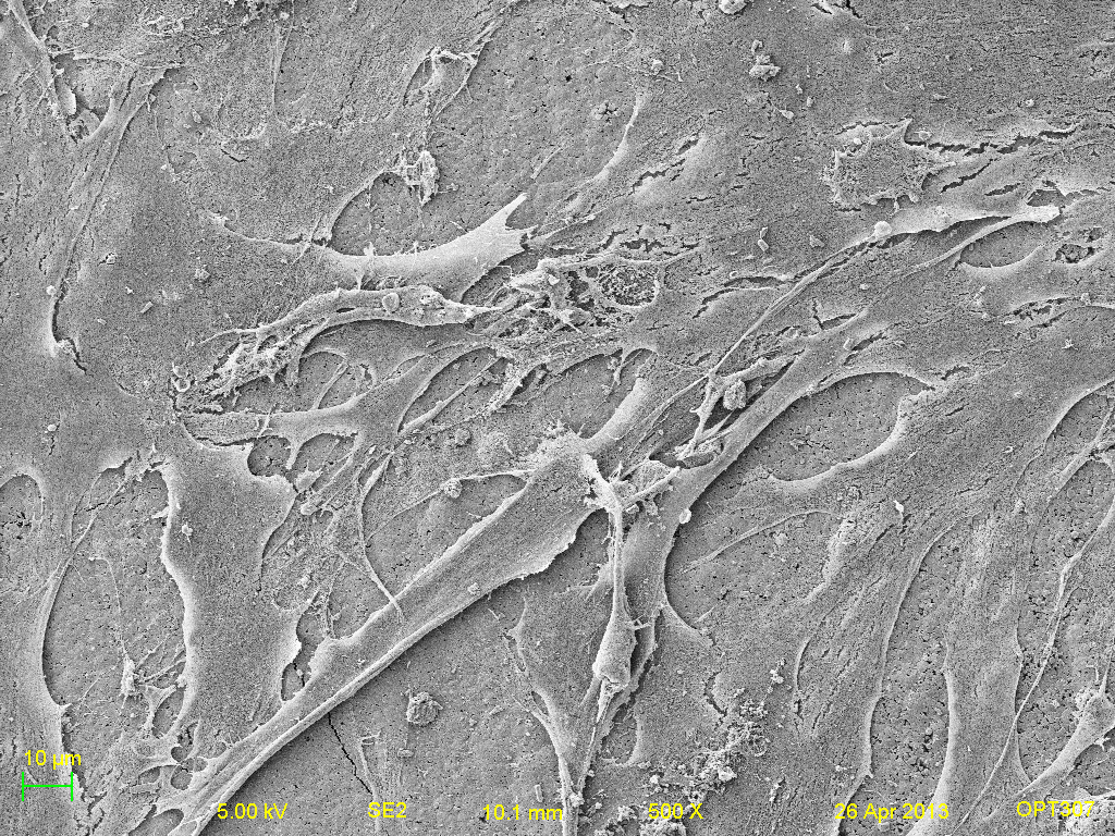
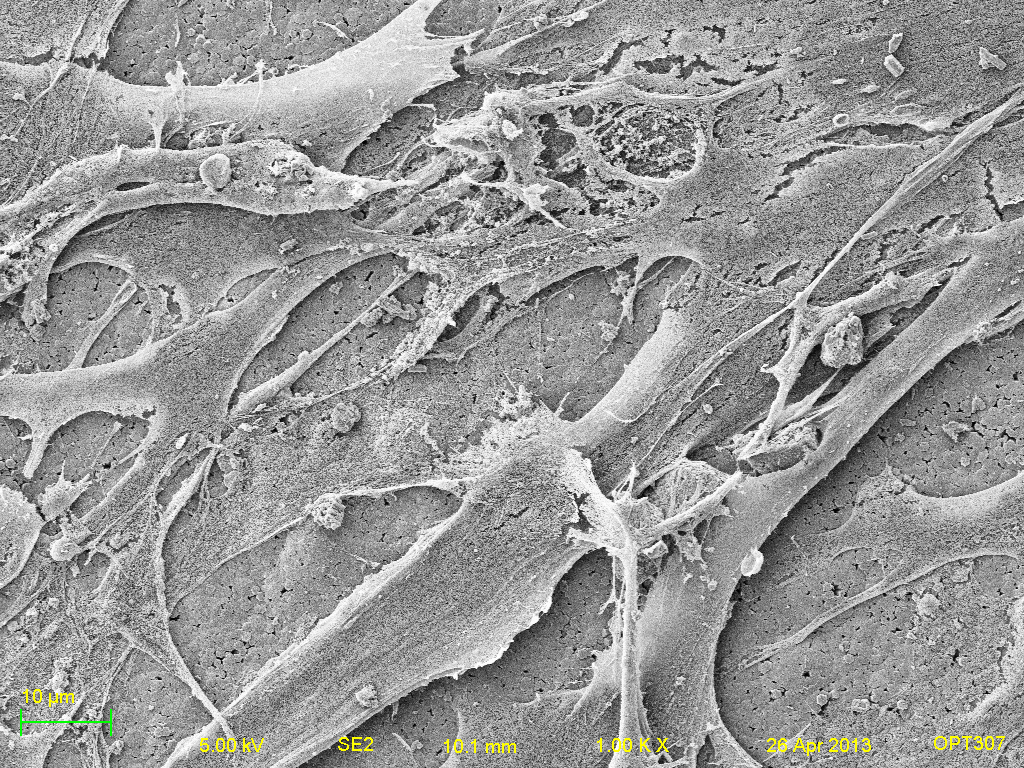
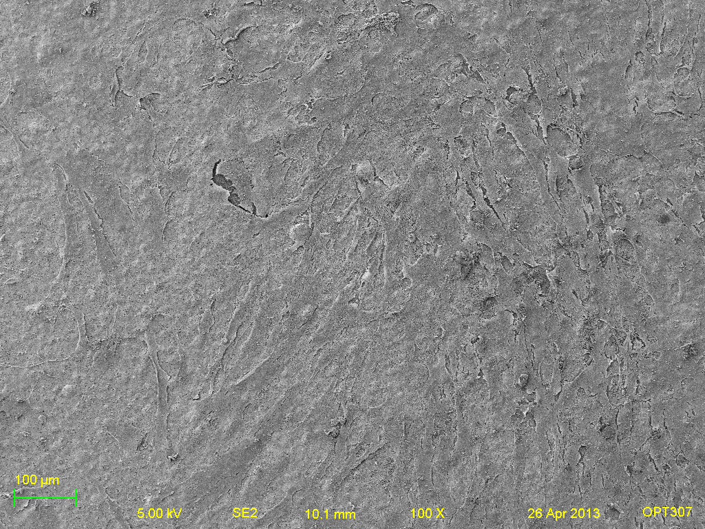
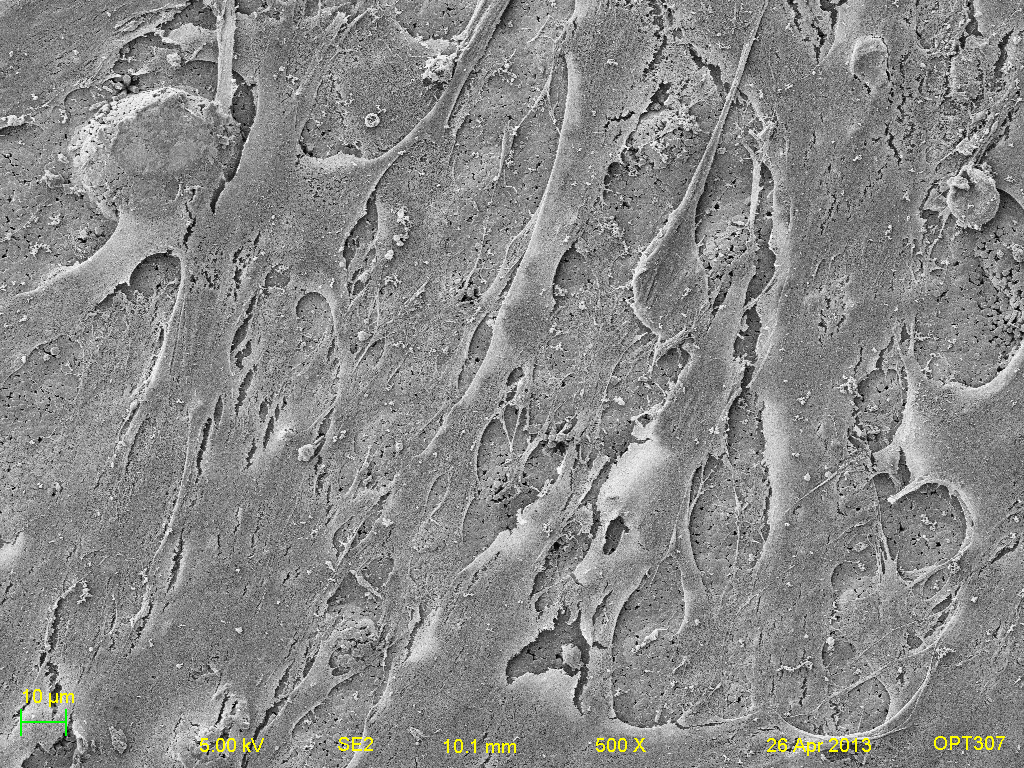
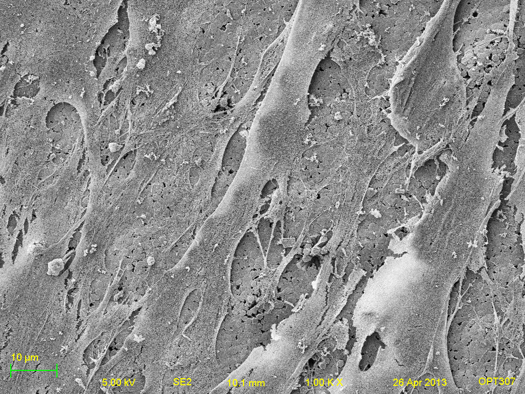
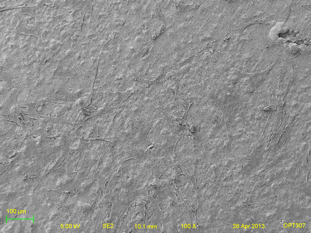
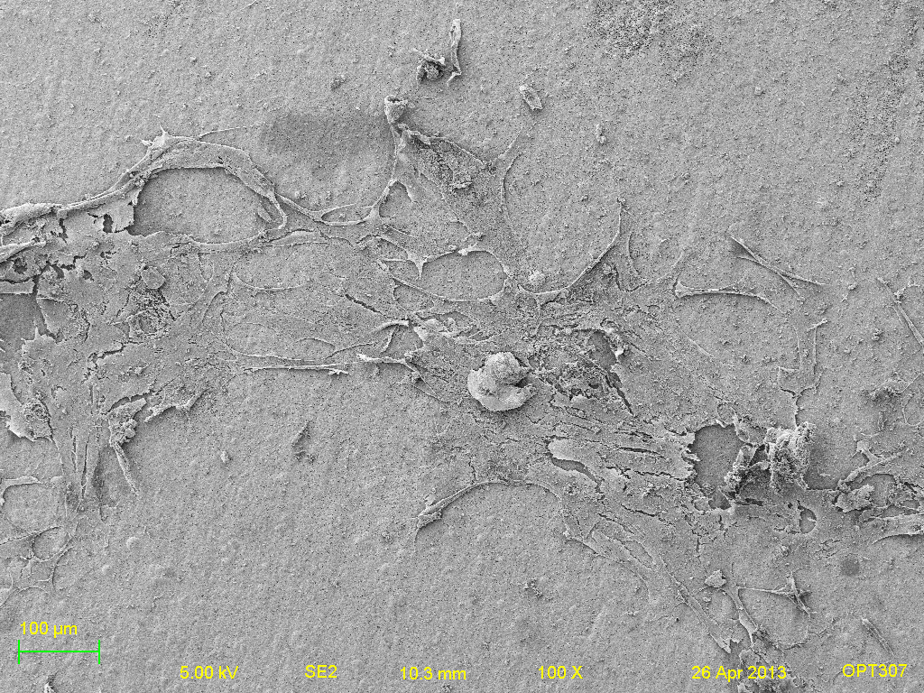
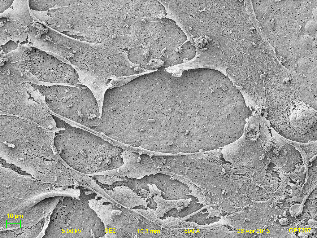
Set 3 of Titanium samples- (left to right: 100x, 500x, and 1000x respectively. Top-bottom: Titanium, Positive HAP, Negative HAP, Heated HAP and HAP).