 Cell Culturing and Immunofluorescence
Mesenchymal stem cells, initially identified in bone marrow, are non- hematopoietic stem cells that can differentiate into tissues of mesodermal origin, such as bone- forming osteoblasts [2]. These cells were first seeded for 2 hours onto fifteen titanium discs provided by Dr. Yates' group and cultured for 48 hours in 48- inch wells with growth medium. The titanium discs were divided into three sets, with each set containing five different surfaces. The surfaces included: titanium, titanium plus HAP with positive charge, titanium plus HAP with negative charge, titanium plus heat treated HAP and titanium plus HAP.
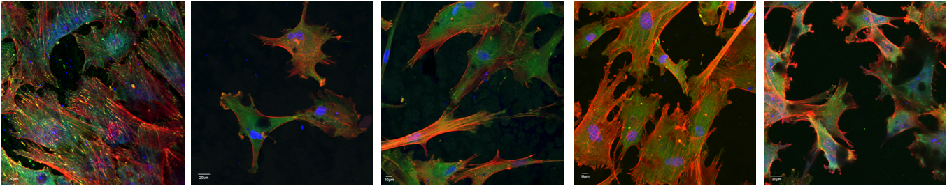
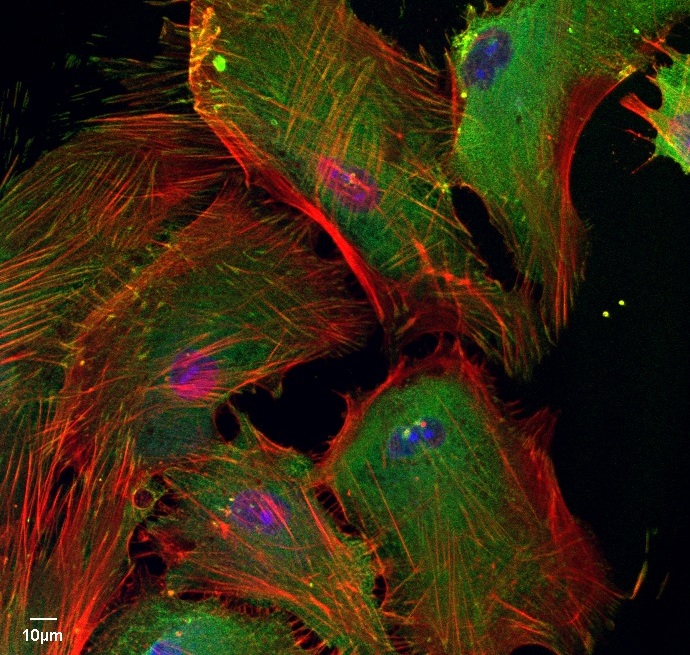
Confocal laser scanning microscopy was used to examine the cells morphology and cytoskeletal arrangement on the different surfaces. After the 48- hour incubation, the cell growth medium was removed and the wells were rinsed three times with phosphate buffer saline (PBS). The cells were fixed with 4% paraformaldehyde and then washed three more times to remove the excess paraformaldehyde. In order to reduce non-specific background, the samples were blocked and permeabilized with PBS containing 5% normal goat serum and 0.5% Triton X. For vinculin staining,, the cells were incubated with a mouse monoclonal anti-human vinculin antibody followed with Alexa Fluor 488 goat anti-mouse IgG (H+L). To detect polymerized actin, the cells grown on the titanium discs were incubated with tetramethyl rhodamine isothiocyanate (TRITC)-conjugated phalloidin. Nucleus staining was achieved by incubating the cells with To-Pro 3.
The image at the top- left shows the staining results. The nuclues is blue, the actin is expressed in red and the vinculin in green as seen in the images below. After analyzing the samples with the confocal laser scanning microscope, the samples were prepered for SEM analysis.