|
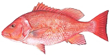
Red Snapper
Light Microscopy
SEM
AFM
|
Light Microscopy |
 |
|
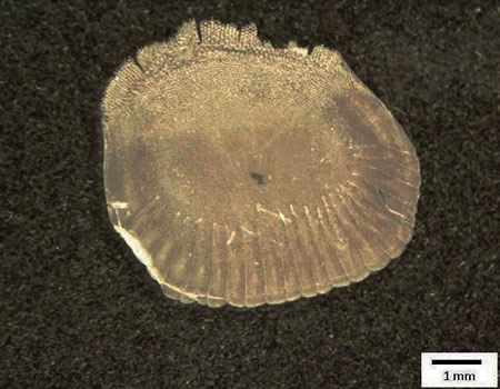
Fig.1 Light microscopy image of the red snapper scale
|
|
SEM
|
|
|
|
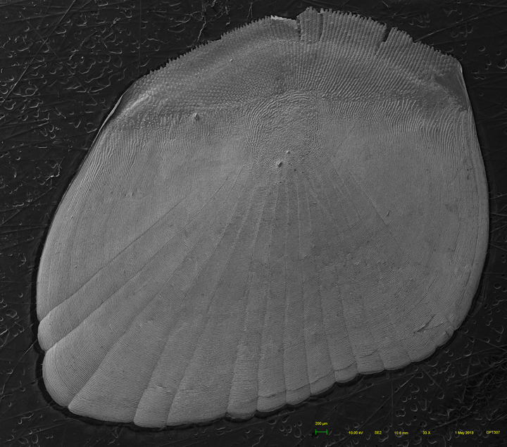
Fig.2 Panorama SEM image of the red snapper scale
(press ctrl + for mare details)
|
  |
|
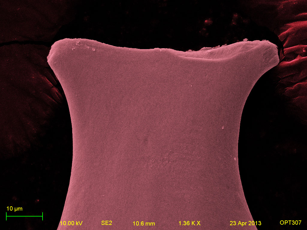
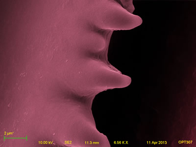
Fig.3 First domain of the red snapper scale (left: ctenii;
right: tip of the ctenii)
|
  |
|
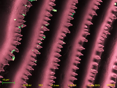
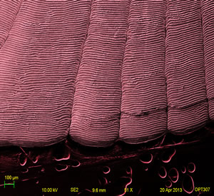
Fig.4 Second domain of the red snapper scale (striped pattern
with teeth)
|
  |
|
Fig.5 Third domain of the red snapper scale (interradial
denticles)
|
|
Fig.1-5 show the morphology of the red snapper
scale. The red snapper scale also has a "toothed" outer that is
similar with the striped bass scale. Furthermore, the red snapper
scale has three domains in the micrograph as well: one is featured
the bony texture with the tooth-shaped ctenii (Fig.3). It should
be pointed out that the tip the ctenii of the red snapper differs
from that of the striped bass scale. The second one has striped
pattern with teeth but no radii (Fig.4); and the third one has the
interradial denticles. Additionally, in fig.4 and 5, impurities
are found on the sample surface (green spots). In fig.5, protein
is also found on the surface (blue spots).
|
| AFM |
  |
|
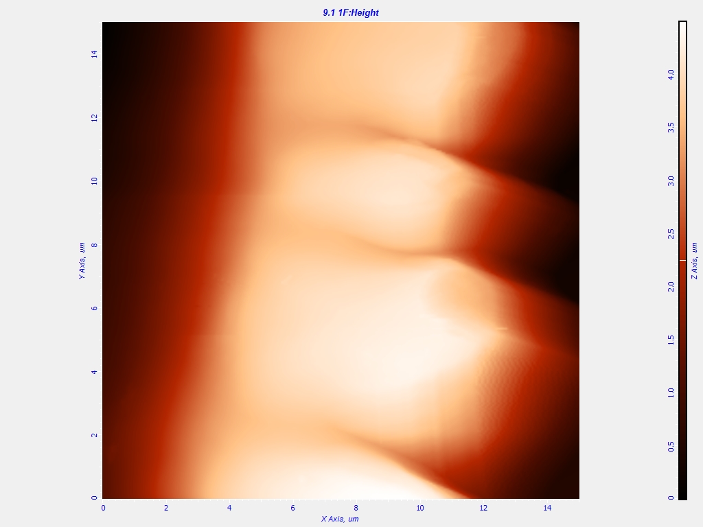
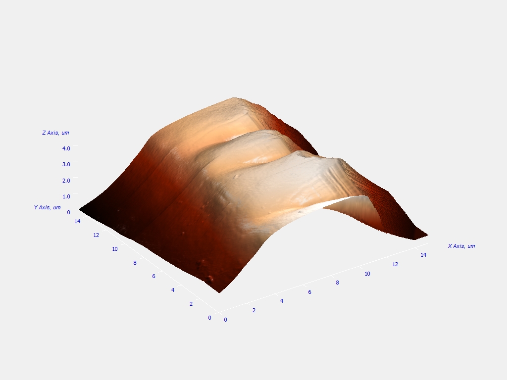
Fig.6 AFM results of the red snapper scale sample,
(left, 2D view; right, 3D view).
|
|
Fig.6 shows the height profile of a tooth the red
snapper scale, obtained with AFM. From the figures, some "teeth"
with a height about 4.0 μm are observed, which is also seen in the
SEM image (see Fig.4). According to the right 3D view, it is more
obvious to see the "tooth-shaped" pattern on the red snapper
scale.
|
| |
| |