|
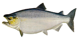
Salmon
Light Microscopy
SEM
AFM
|
Light Microscopy |
 |
|
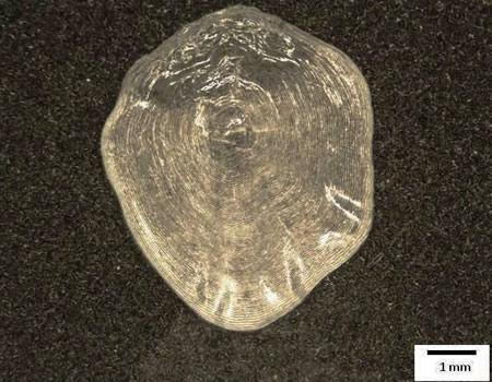
Fig. 1 Light microscopy image of the salmon scale
|
|
SEM
|
|
|
|
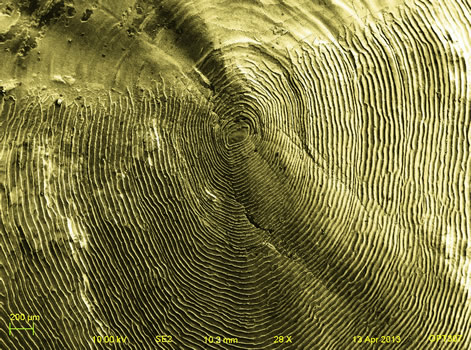
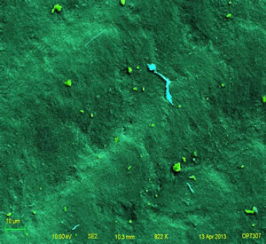
Fig.2 SEM picture of the salmon scale with natural
colorization
|
  |
|
Fig.3 First domain of the salmon scale (striped pattern)
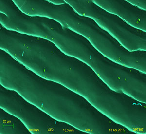
Fig.4 Second domain of the salmon scale (smooth surface)
|
|
Fig.1-4 show the morphology of the salmon scale. The salmon scale
has a smooth edge without any "tooth-shaped" ends. This is
distinct from the other three samples in this project. The salmon
scale has two domains in the micrograph: one is relatively smooth
without any texture which takes about one third area of the
sample; the other one is characterized with the oval types focus
and texture. In fig.4, the tiny blue strip represents the protein
found on the sample surface. And the green spots are impurities.
|
| AFM |
  |
|
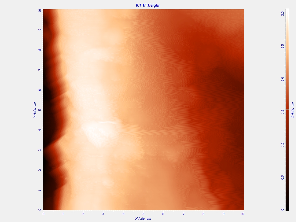
Fig.5 AFM results of the salmon scale sample,
(left, 2D view; right, 3D view).
|
|
Fig.5 is observed the height profile of a
certain area on the salmon scale, obtained with AFM. From the
figures, a ridge with a height about 3.0 μm is observed, which
is also seen in the SEM image (see Fig.3). According to the
right 3D view, more pattern details are observed on the salmon
scale.
|