|
Sea Bass
Light microscopy
SEM
AFM
|
Light Microscopy |
 |
|
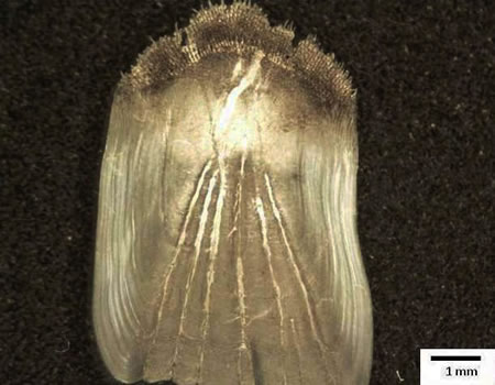
Fig.1 Light image of the sea bass scale
|
|
SEM
|
|

|
|
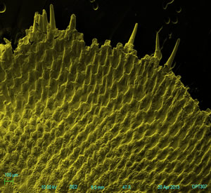
Fig.2 SEM picture of the sea bass scale with natural
colorization
|
  |
|
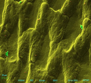
Fig.3 First domain of the sea bass scale (ctenii)
|
 |
|
Fig.4 The focus of the sea bass scale
|
  |
|
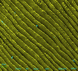
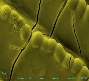
Fig.5 The second domain of the sea bass scale (striped pattern
with teeth)
|
|
Fig.1-5 show that how the sea bass scale looks like.
It is noticed that the sea bass scale also has a "toothed" outer .
Furthermore, the striped bass scale has two domains in the
micrograph: one is featured the toothed edge (Fig.3);
the second one has striped pattern but no radii (Fig.5). In fig.5,
several cracks are intruduced by surface tension during air
drying. In addition, in fig.3, impurities and protein are found on
the sample surface (red and green respectively). In fig.5, protein
is found and colorized with blue.
|
| AFM |
  |
|
Fig.6 AFM results of the sea bass scale
sample, (left, 2D view; right, 3D view).
|
| Fig.6 is observed the height profile of a
certain area of the first pattern on the sea bass scale, obtained
with AFM. From the figures, a ridge with a height about 3.0 μm is
observed. |
| |
| |
| |