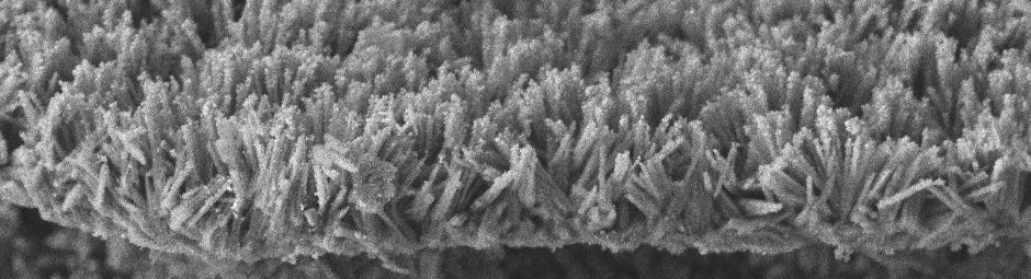
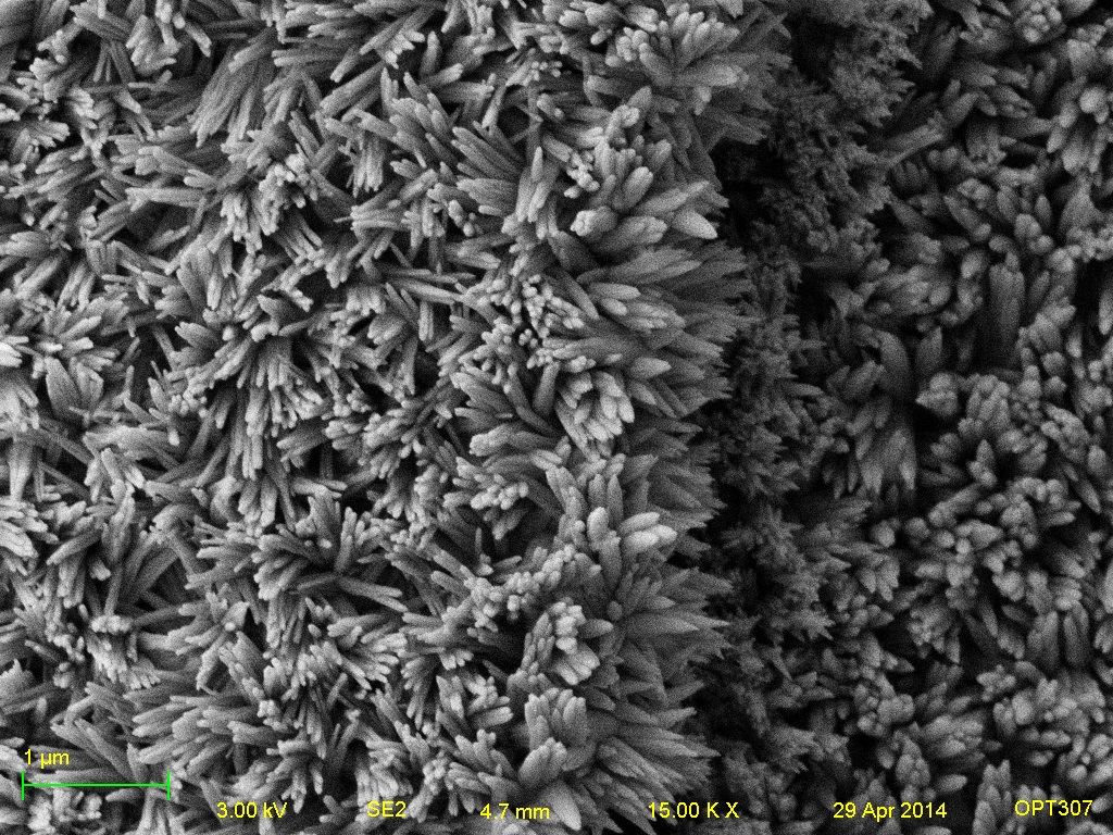
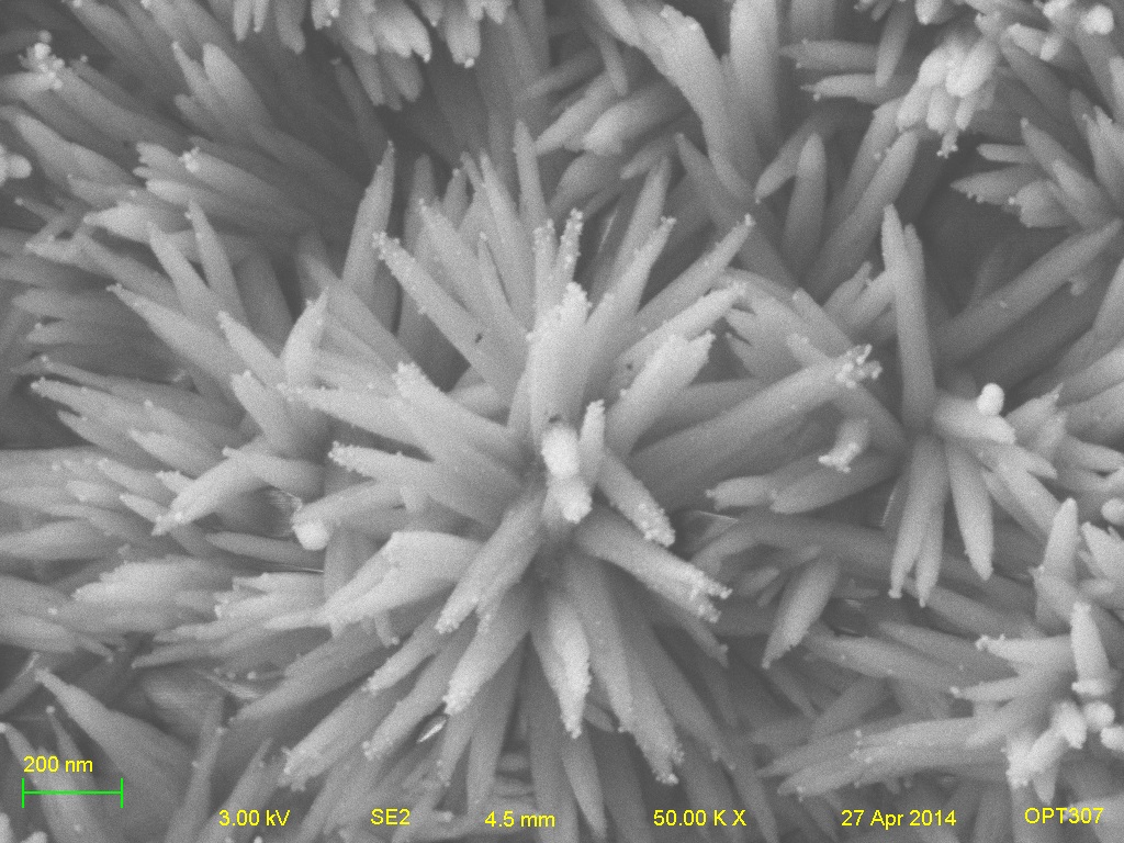
Abnormalities and Defects

During the electrochemical deposition of the hydroxyapatite coating, occasionally, due to local variations in the process, the crystal structures and morphology are altered in some areas. Additionally, due to the release of hydrogen gas at the titanium substrate during deposition, occasionally, due to poor coating adhesion, chunks of the coating will be dislodged, allowing for new crystal growth.

Various defects in the crystal morphology or coating layer


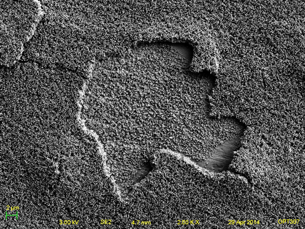
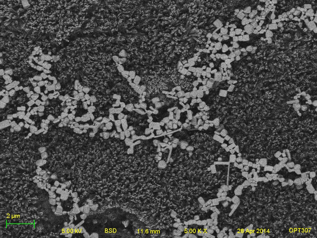
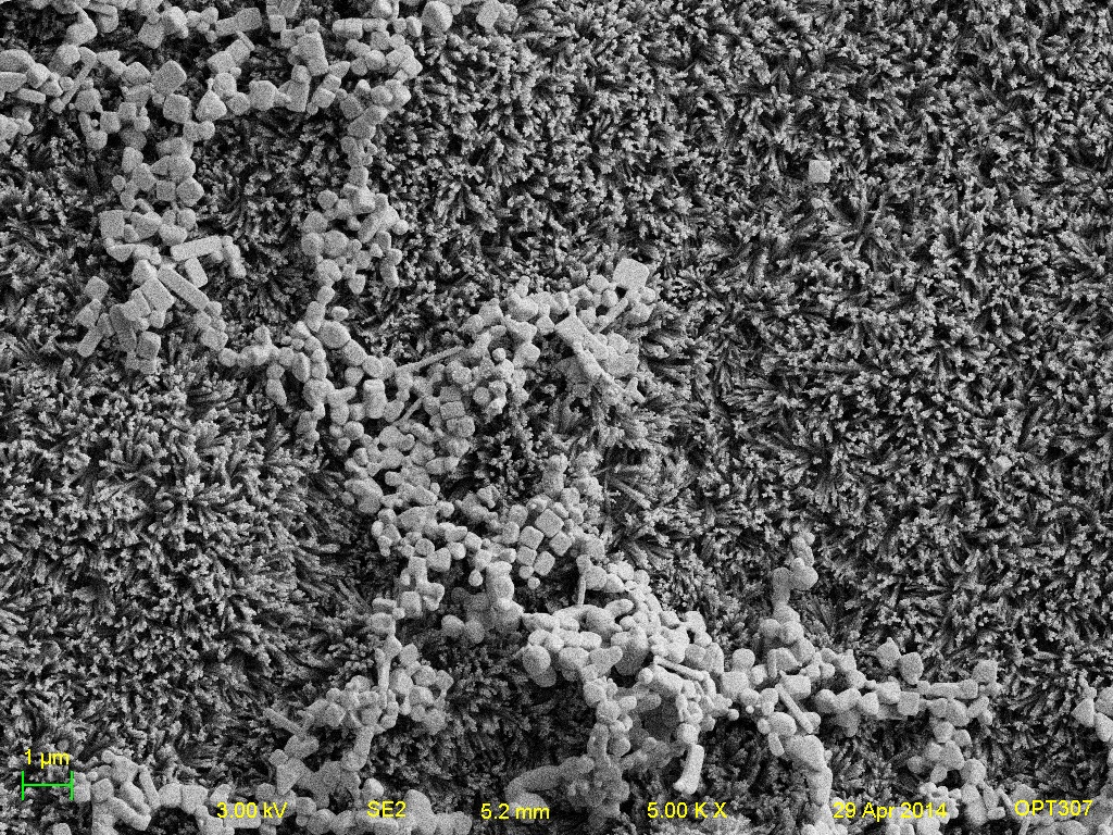
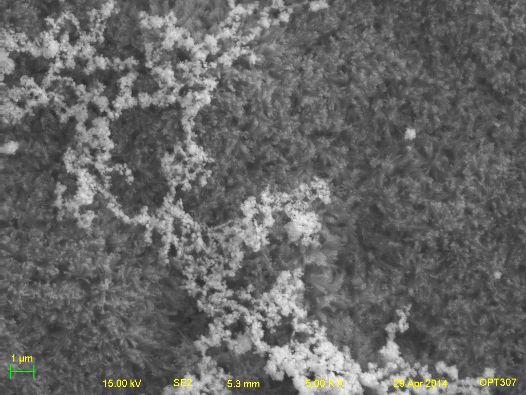
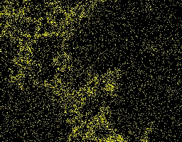
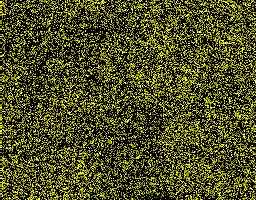
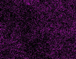
It was observed that a conglomeration of silver metal would begin to form occasionally in fractal patterns across the HAP coating surface during the electrochemical deposition of silver process.

Conglomeration of silver particles on HAP (1) BSD 5kx (2) SE2 5kx (3) SE2 20kx


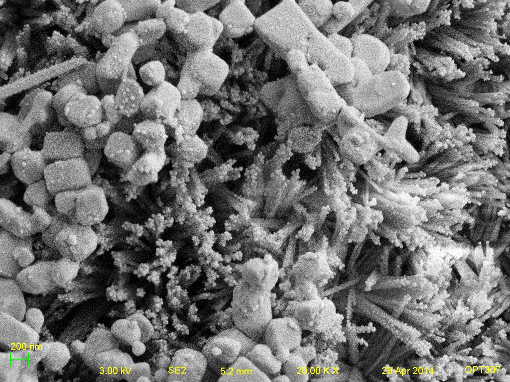
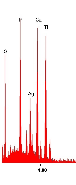
The conglomerates were verified to be silver using EDS X-Ray Spectrum Analysis and Mapping.

EDS Microanalysis (1) Area of silver conglomeration imaged (SE2 15kx) (2) EDS Spectra (Energy in kV vs Intensity)


EDS Mapping of Signal by Element (1) Ag (2) Ca (3) O (4) P (5) Ti


The relative percentage of silver in reference to the HAP present in these areas was calculated by EDS X-Ray Microanalysis and found to be much larger than the relative percentages of silver in the uniformly deposited samples.

Relative Abundance of Elements by Weight %


In fact, in many of the comparisons, this conglomeration of Ag is 4-6 times more relatively abundant to its surroundings than the depositions should have been.

Relative Silver Abundance
  Ag:O (Wt%) Ag:P (Wt %) Ag:Ca (Wt %)
Deposited Ag 0.095 0.32 0.22
Sputtered Ag 0.099 0.20 0.077
Ag Conglomeration 0.43 1.3 0.80


In synopsis, the electrochemical deposition process of both the hydroxyapatite and the silver nanoparticles must be explored and investigated further to achieve better uniformity.

Conclusion

This project successfully produced characteristic images of the electrochemically deposited hydroxyapatite coatings. The coating was found to be consistently 1 um thick, but some focus should be placed upon studying the process to ensure consistency of result. In conclusion, it is a viable structure onto which silver nanoparticles can be deposited. Both techniques to produce nanoparticles, electrochemical reduction and sputter coating, were effective. Through the use of secondary electron imaging and backscattered electron imaging, it was found that the former produced much larger and more pervasive microstructures of silver than sputter coating. In addition, the sputter coating’s nanoparticles were of particular difficulty to image and characterize.

X-ray microanalysis verified the HAP coating’s composition and the presence of silver. It was noted that, with regards to relative elemental compositions, the electrochemically reducing technique had higher relative amounts of silver. Atomic force microscopy found that the HAP coating has a roughness between 221 and 275 nm, and the deposited silver particles have a roughness around 30 nm. Colorization and anaglyph creation of the images from this project proved to be a creative and effective way of communicating the characteristics of these coatings and their nanoparticles. .

Acknowledgements

The author of this project offers his sincere gratitude to the Institute of Optics at the University of Rochester for use of its microscope. In particular, the author thanks Brian McIntyre for his invaluable instruction and never-ending guidance throughout the research process. The helping hand and watchful eye of grad student Diana Olvera was also greatly appreciated. Additional thanks goes to Professor Matthew Yates for use of his laboratory, equipment, and procedure in preparing samples.

References

Ghani, Y., Coathup, M. J., Hing, K. A. and Blunn, G. W. (2012), Development of a hydroxyapatite coating containing silver for the prevention of peri-prosthetic infection. J. Orthop. Res., 30: 356-363.

Dongxia Liu, Keith Savino, Matthew Z. Yates, Coating of hydroxyapatite films on metal substrates by seeded hydrothermal deposition, Surface and Coatings Technology, Volume 205, Issue 16, 15 May 2011, Pages 3975-3986.

Savino, Keith. (2013). Microstructured Hydroxyapatite Membranes for Ion Conducting and Orthopedic Applications. (Doctoral dissertation). Retrieved from UR Resarch. (LC Call No. [AS38.695])