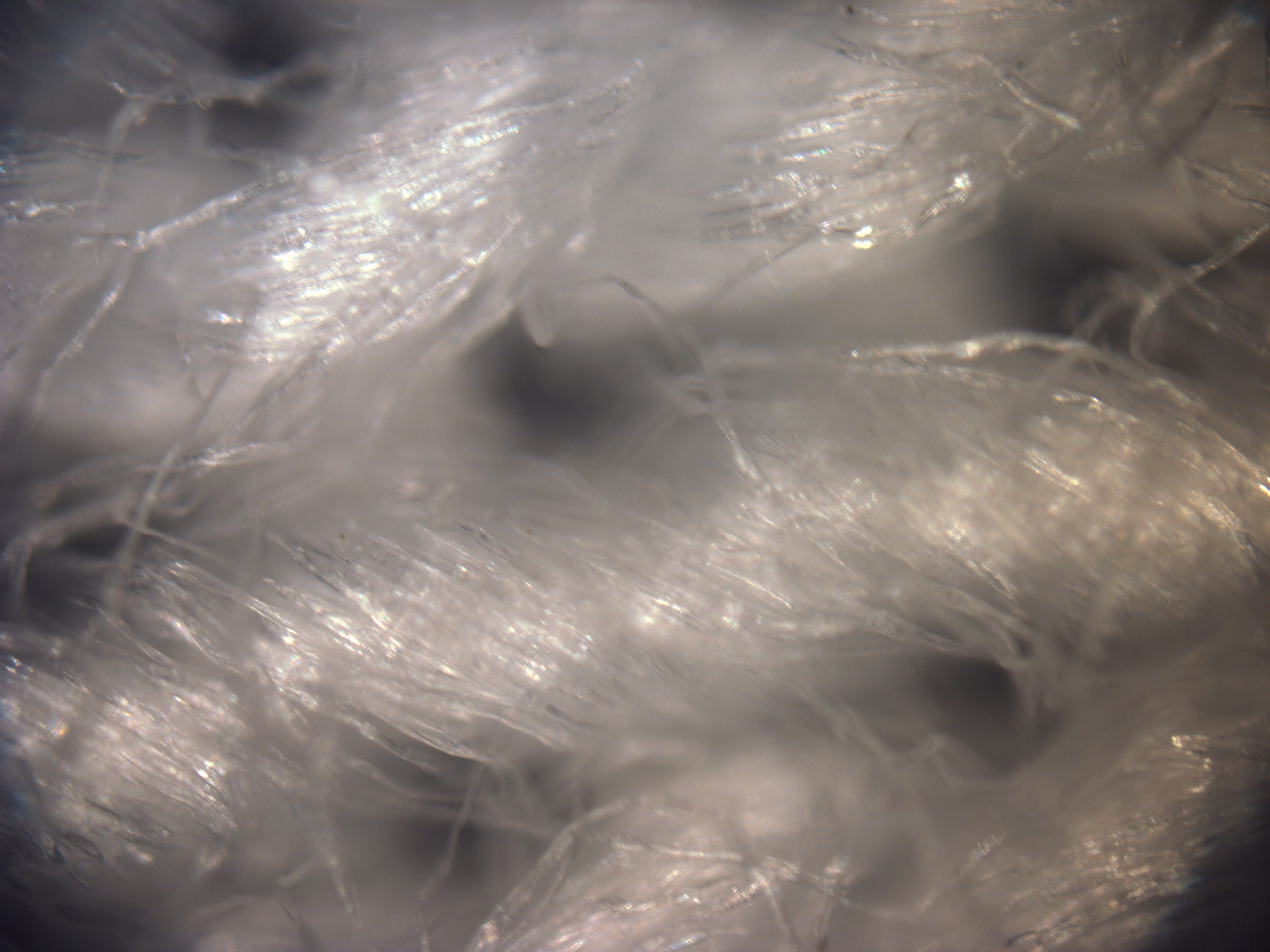
Figure 2.1 Optical Microscope image of the sample with a 10X objective. The image shows the interwoven sturture of the fibers.
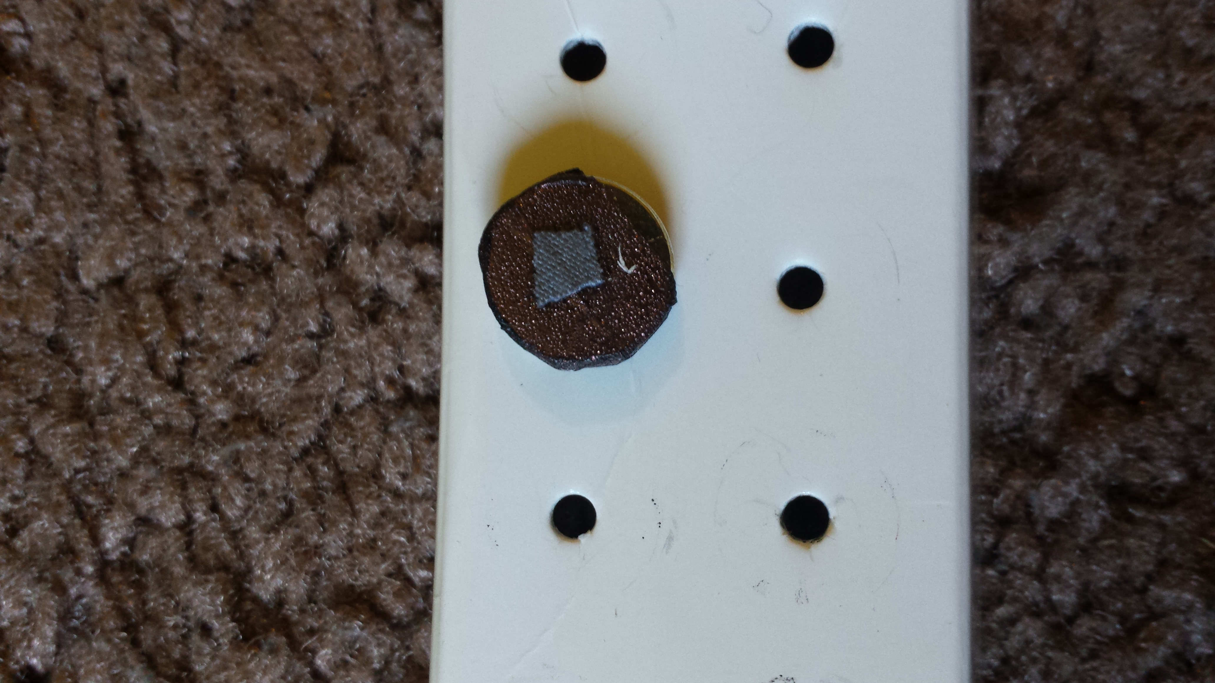
Figure 2.2 Sample after sputter coating with a layer of gold (~18nm)
SEM
imaging was carried out by loading the sample into
Zeiss- Auriga SEM. The sample was imaged using the SE2 detector at an
accelerating voltage of 20kV and a working distance of around 13mm.
Even after
deposition of gold of considerable thickness on the surface of the
sample,
imaging was difficult since it was not possible to evenly coat the
surface
owing to the interwoven structure of the fibers in the sample.

Figure 2.3 SE2 image of the sample at 20kV and 13mm working distance.