An analysis of hydrophobic fabric using electron microscopy

Sanjay Lakshmanan, Mechanical Engineering, University of Rochester
A single thread of the hydrophobic t-shirt was mounted on a stub and placed inside the NTMDT AFM for atomic force microscopy. The biggest challenge faced while imaging with AFM was in landing the tip on a fiber so as to not capture the transition of the tip from one fiber to the other. The set point was kept at a high value of 8 and imaging was carried out at a low gain of 0.1. A scan rate of 0.1 over a small area of 5µm x 5µm provided good results.

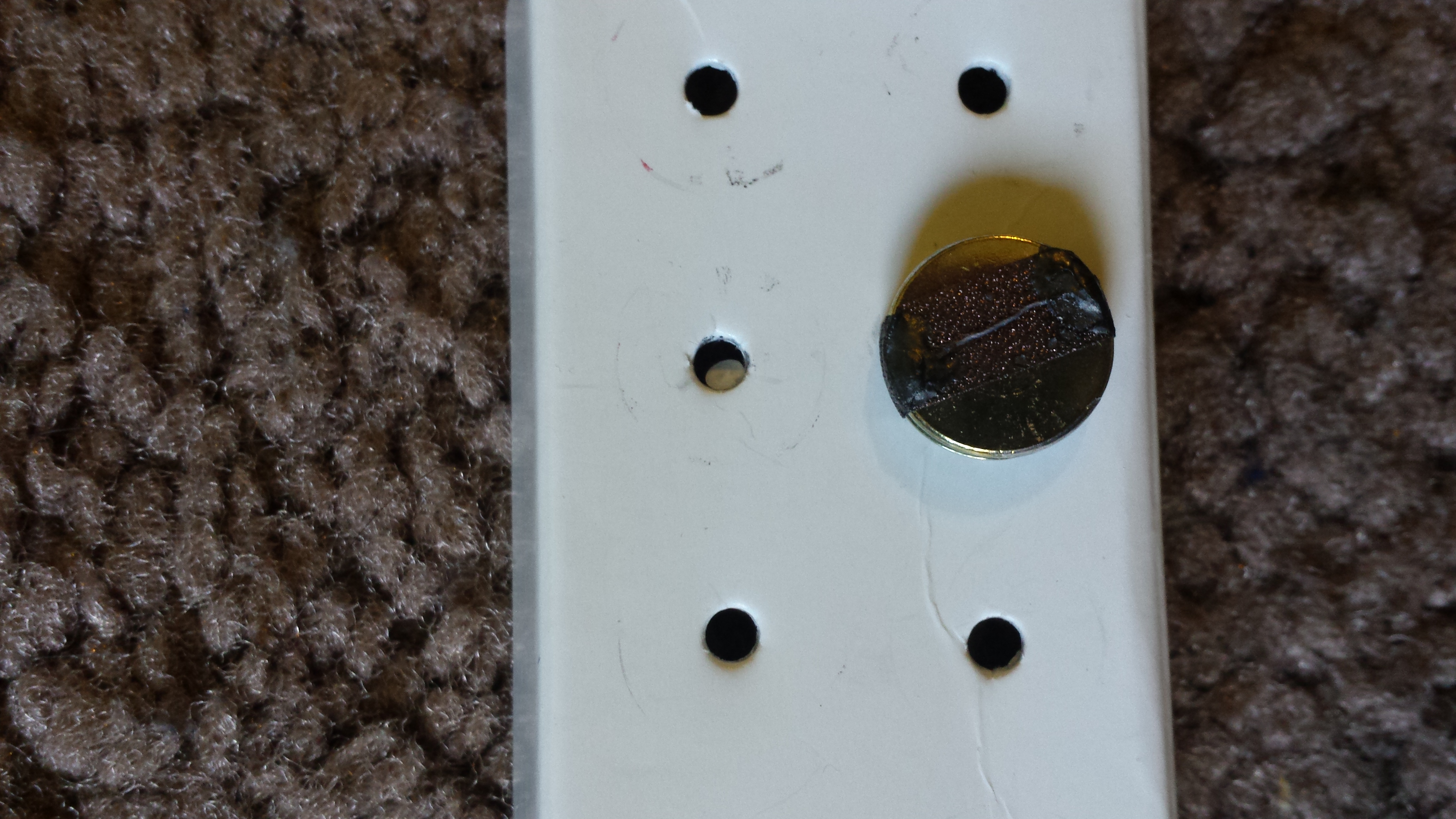
Figure 5.1 The AFM sample consisting of a single thread of the hydrophobic t-shirt

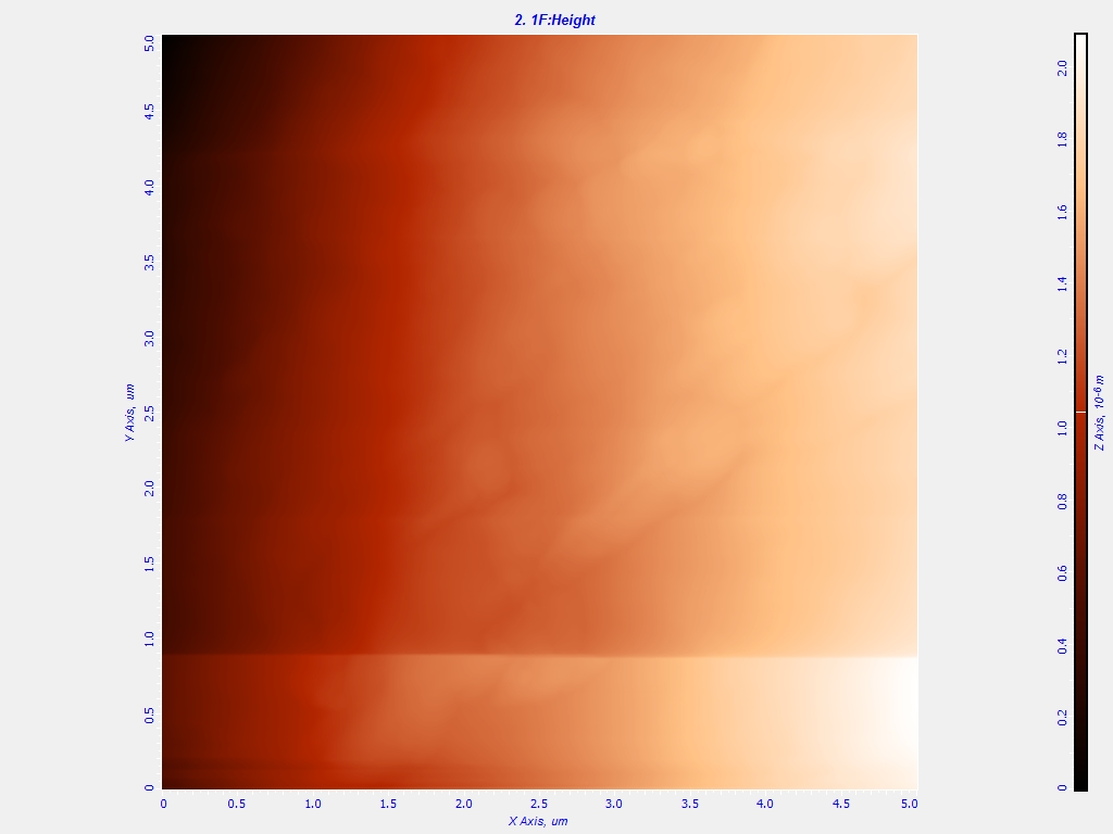
Figure 5.2 A 2D AFM scan of a 5x5 micron area of the sample

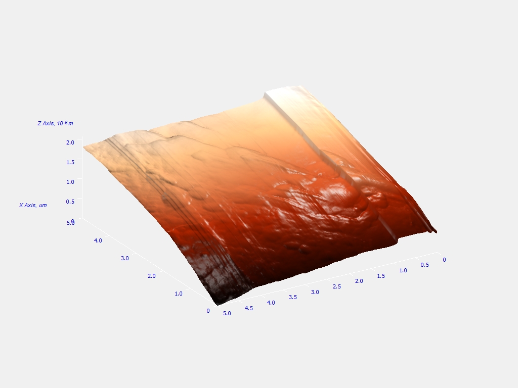
Figure 5.3 A 3D AFM scan of a 5x5 micron area of the sample


Figure 5.4 Sectional Analysis of the topography of the sample reveals no nanostructures on the surface


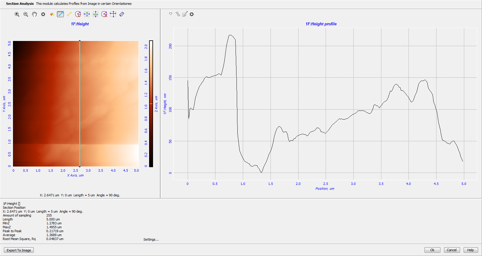
Figure 5.5 Sectional Analysis of the topography of the sample along the y-axis.
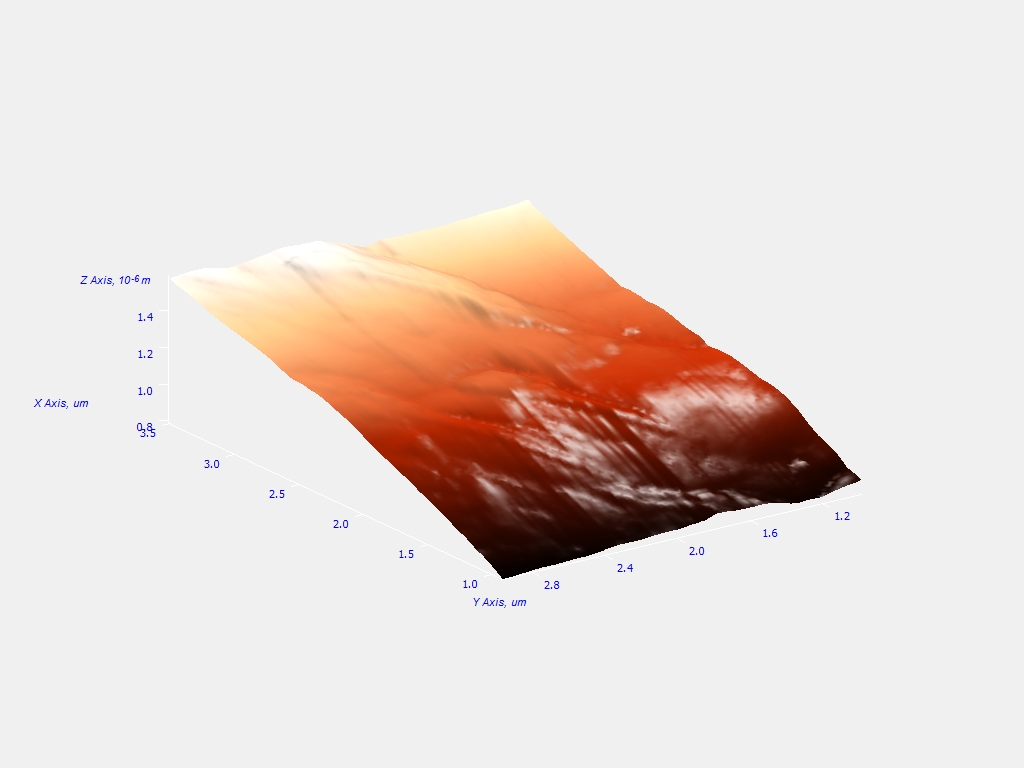
Figure 5.6 3D scan of a 3x3 micron area of the sample

Please enter any comments, criticisms, questions, etc. below.

Your name:

Email address: