|

Menu:
Home
Introduction
Procedure
SEM
TEM
X-ray
Electron
Flight Simulation
AFM
Colorization
Conclusion
Acknowledgments
References
|
AFM
AFM is a
mode
of scanning probe microscopy. In the
AFM, the tip is mounted on a cantilever and the tip scanned the sample
surface
line by line, which is a raster scan. Then
a laser beam senses the deflection of the
cantilever and the signal
is detected. The advantage of using the
AFM is to provide measurements in three dimensions. The
results for the nanoparticles are not high
quality, however it is seen that there are some particles with a height
of 50 nm
on the surface of the copper TEM grid. The
3D image was fit to plane, but the TEM grid may not have been
completely flat
and smooth which means that the particle height is closer to 20-30 nm
instead
of 50 nm.

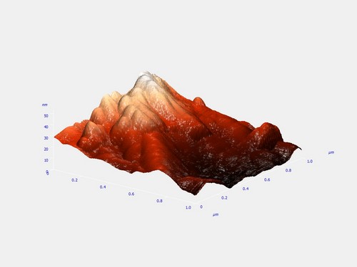
Figure
23:Low concentration Ag-Pd NPs Atomic Force Microscopy 2D
and 3D images
|