
Atomic Force Microscopy
Atomic force microscopy (AFM) was used to scan and characterize the topographical features across the boundary layer. Although, the scan was done on polished section, I was able to distinguish betwen higher iron oxide crystals and flatter clay particles. 3 point levelling was done after scanning the section to correct for any slope in the sample.
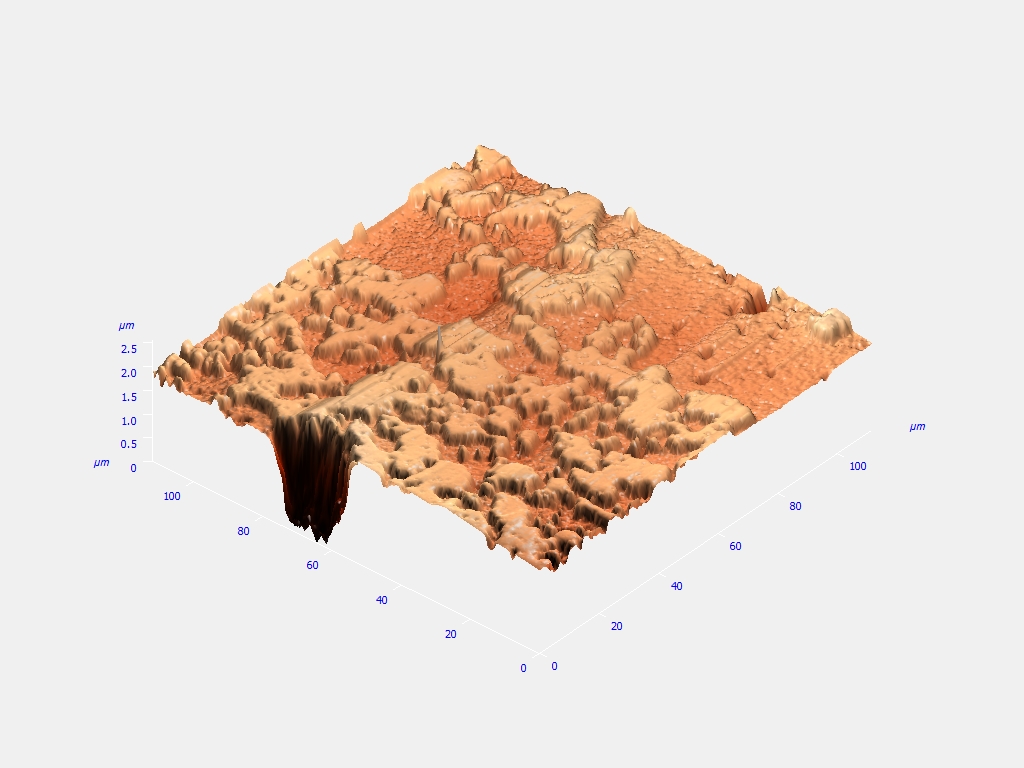
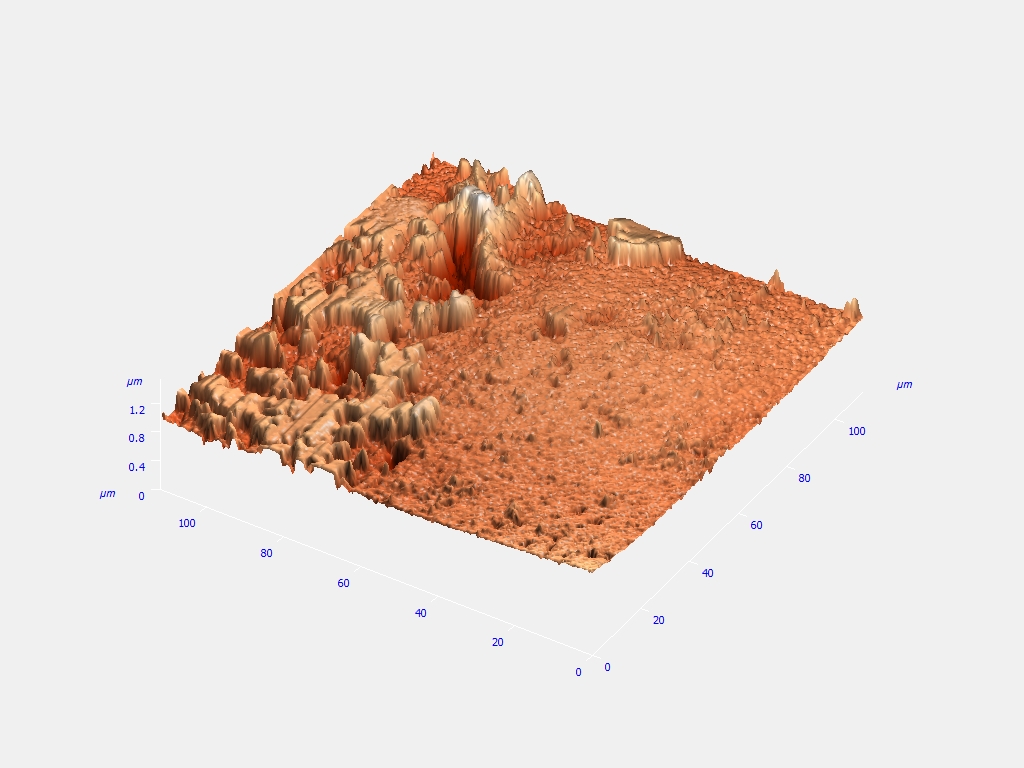
Fig.1: An AFM image of a polished section of the clay layer showing variable topography, arising due to difference in mineralogical compositions.
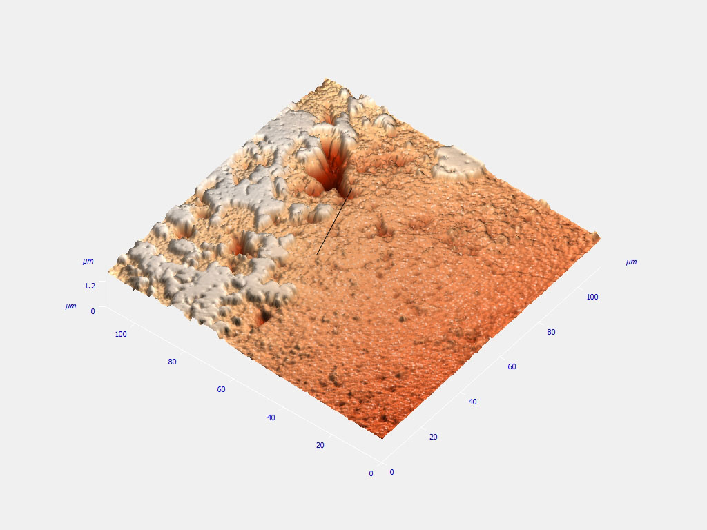
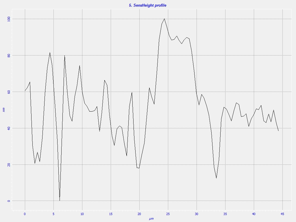
Fig.2: A sens height profile of a segment (above) from the polished section of the clay layer showing crests and troughs, indicative of the variable topography.