
Scanning Electron Microscopy
Scanning electron microscopy was used to identify and image structural features covering the entire section from the Permian Shales to the Triassic limestones. Backscattered detector was useful to distinguish betwen the shale (darker) and the pyrite (brighter). Based on the images obtained (below), we can conclude that we can use SEM techniques to identify textural changes or the appearance and disappearance of structues that may reflect changing environmental conditions.
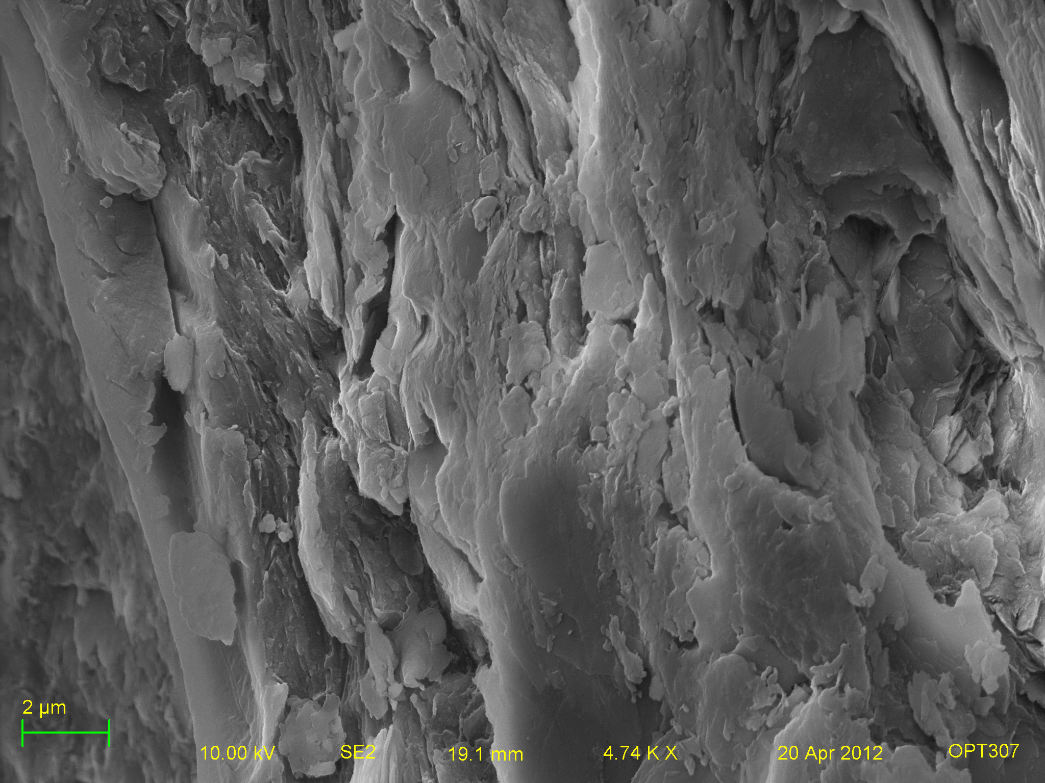
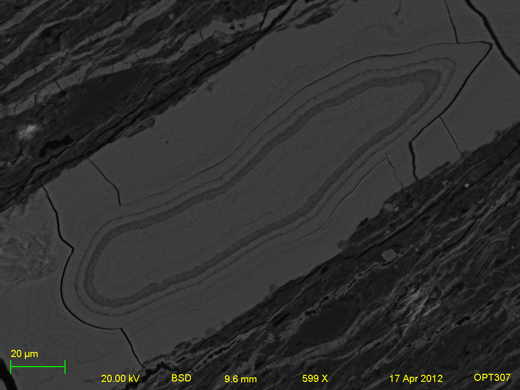
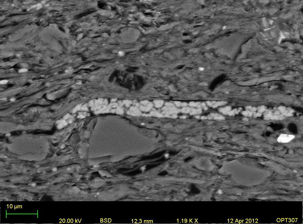
Fig.1: Scanning electron (SE2 and BSD) micrographs of the Permian Shale layer showing distinct clay layers (left), textural zonation (center) and pyrite framboid beads (right) embedded in them.
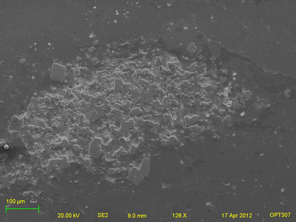
Fig.2: Scanning electron (SE2) micrograph of pyrite crystals found at the beginning of the Triassic limestone layer.
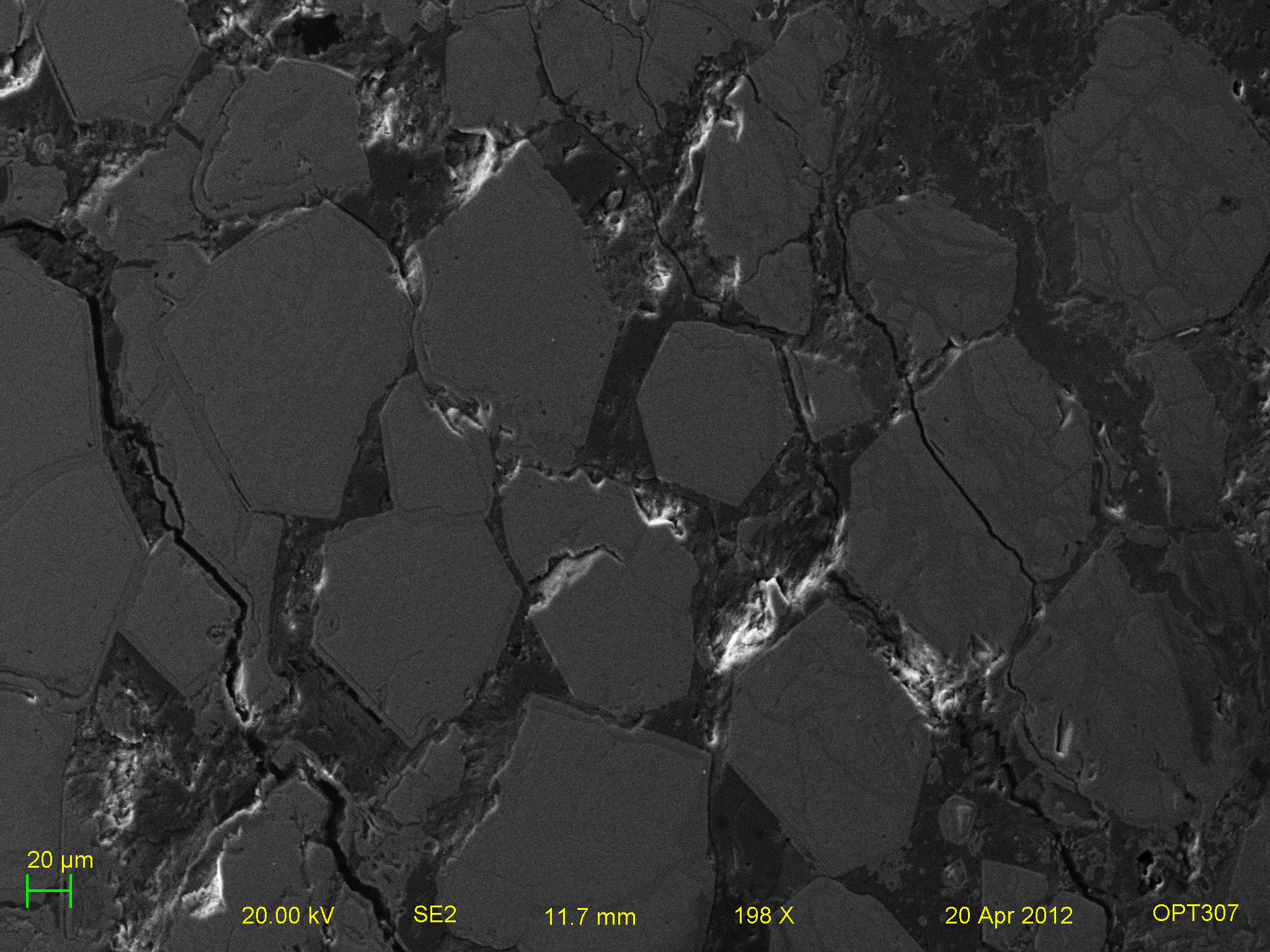
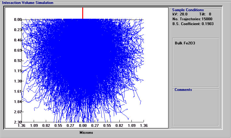
Fig.3: SE2 electron micrograph of hexagonal iron oxy-hydroxide crystals from the ferruginous layer of the Mud Section in Spiti (top). Electron flight simulation of electron interaction with the iron rich crystals.