
Light Microscopy
Light microscopy was used for preliminary study and selection of specific rock thin sections. A combination of transmitted light, polarized light and Differential Interference Contrast (DIC) Reflected light was used for distinguishing between mineral crystals, fossils and other textural features. A set of fossil imprints and mineral crystals were imaged. Comparing the imprints with fossil records elsewhere will give a biostratigraphic control on the Permian - Triassic boundary. For example, the appearance of an ammonoid Otoceras woodwardi reflects the beginning of the Triassic period.
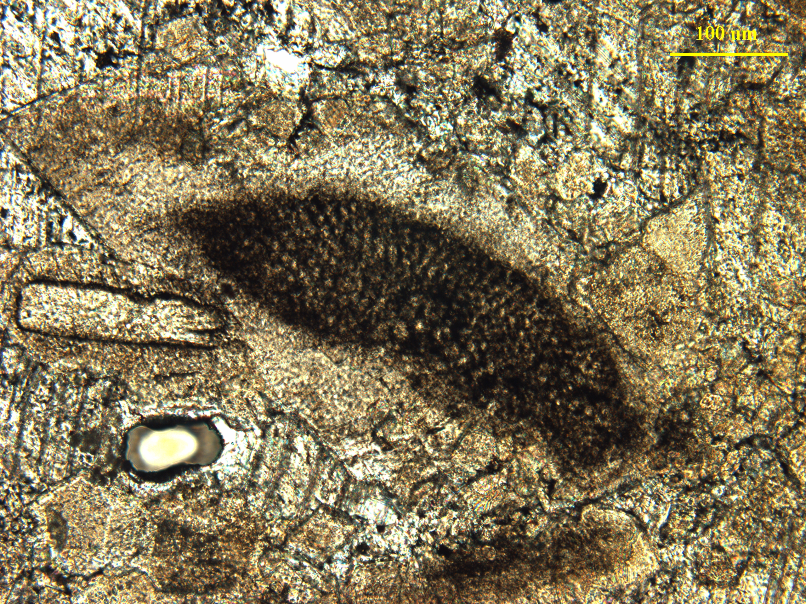
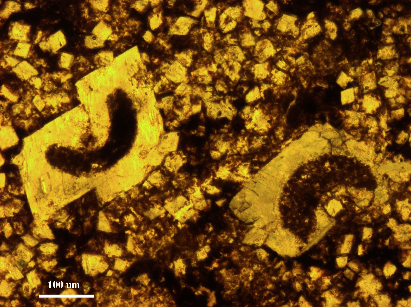
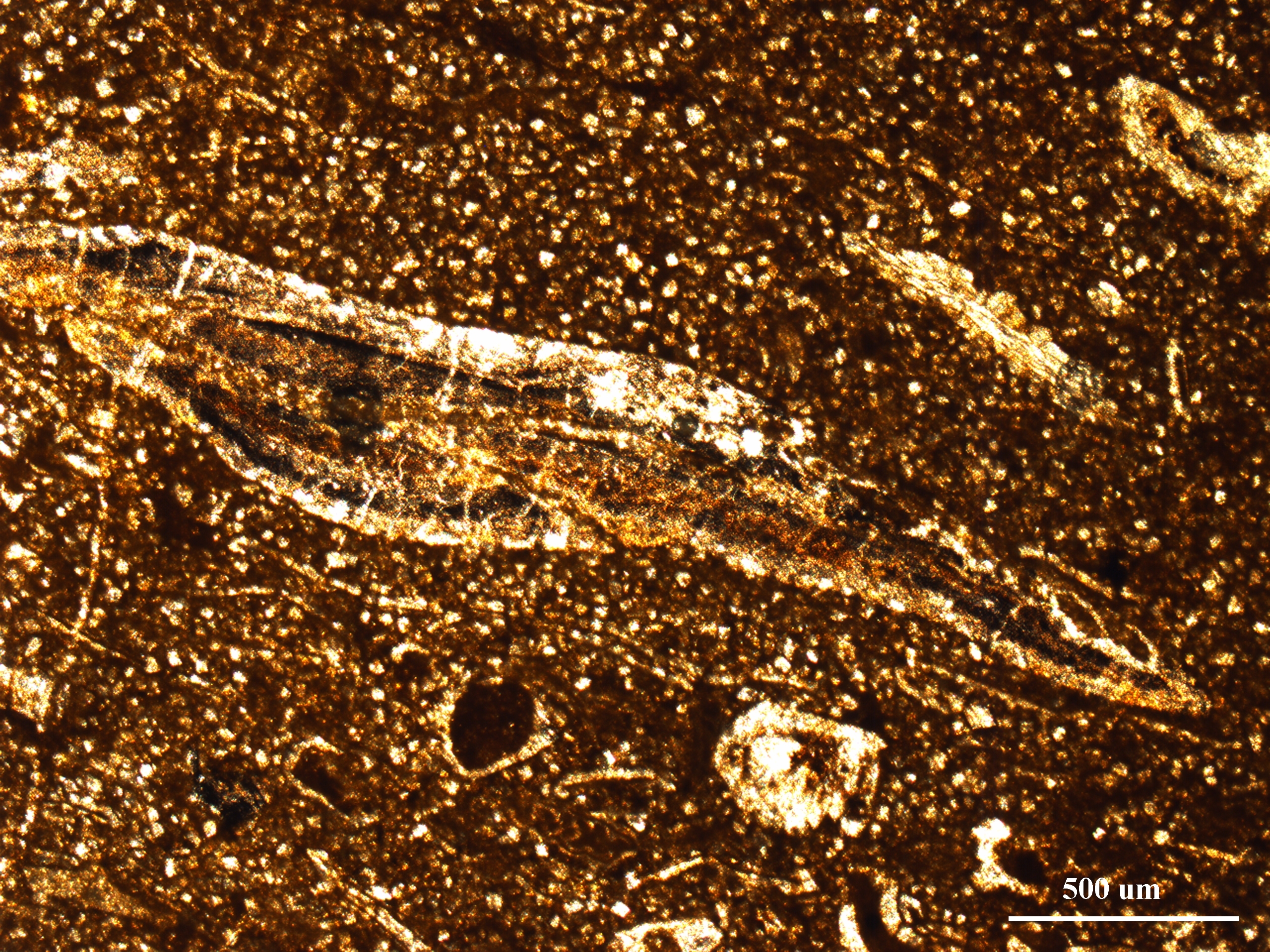
Fig.1: Polarized and transmitted light micrographs of calcified fossil imprints (Ammonoids, Conodonts etc.) found in the Triassic limestone rock sections.
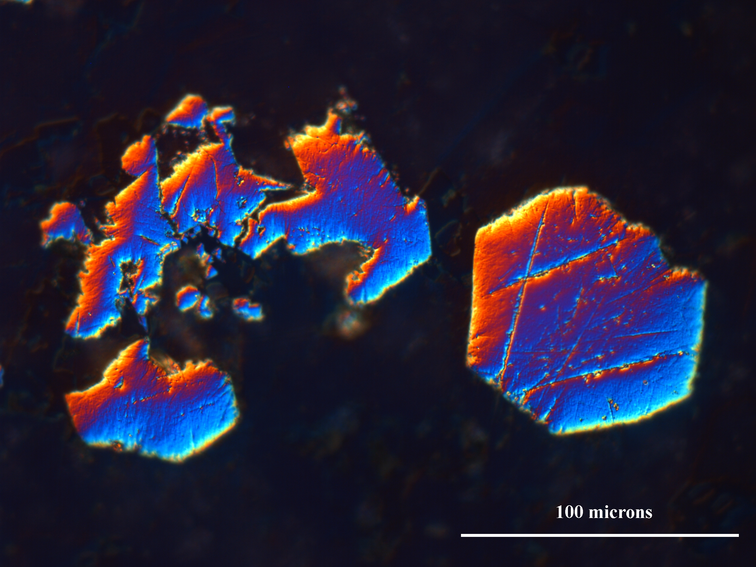
Fig.2: DIC reflected light micrograph of pyrite crystals in a Triassic limestone rock section.