
Energy Dispersive X-Ray Spectrometry (EDS)
Energy Dispersive Spectroscopic (EDS) methods were used on SEM images of structural features in order to characterize them compositionally. A combination of X-ray spectra and X-ray mapping was carried out to identify the distribution of major elements in unique structures, found in the rock sections. A gastropod fossil obatined from a Permo-Triassic section in Vietnam was also imaged.
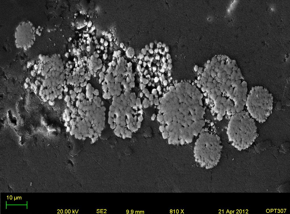
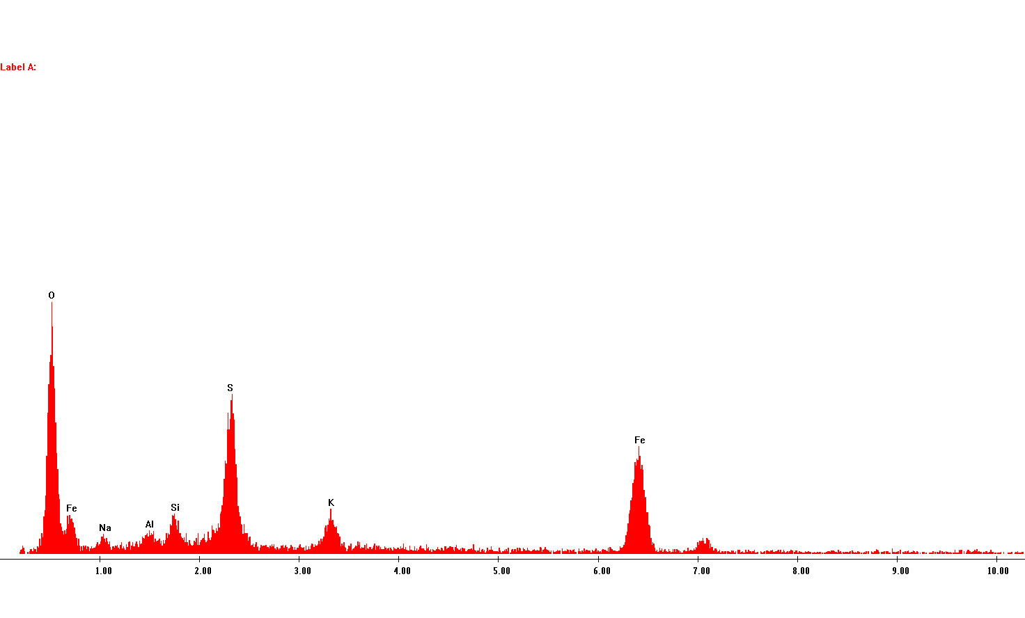
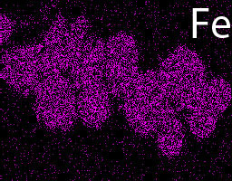
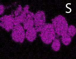
Fig.1: SE2 electron micrograph, X-ray maps and spectra of spherical framboids (>6 microns) of pyrite crystals in the ferruginous layer. Framboidal pyrites are typical of coastal and marshy sediments where microorganisms form these spehrical aggregates. The diameter of framboids may reflect changing oxidation state of the environment.
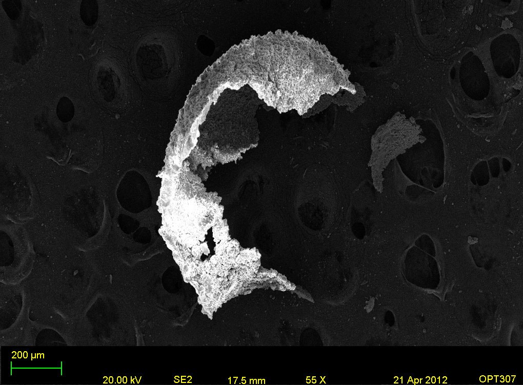
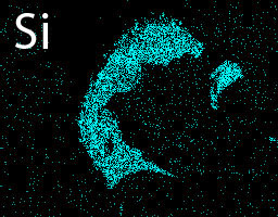
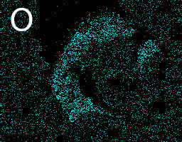
Fig.2: SE2 electron micrograph and X-ray maps of a Gastropod fossil fragment, obtained from a Permo-Triassic section in Vietnam. Unlike carbonate fossils found in marine environments, these gastropods are silicon rich.