
The H-scan for Identification of Scatterers
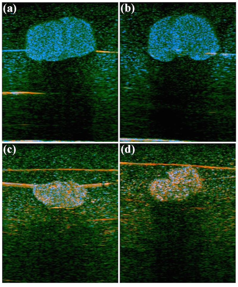
The H-scan is based on a simplified framework for characterizing scattering behavior, and visualizing the results as color-coding of the B-scan image. The methodology begins with a standard convolution model of pulse-echo formation from typical situations, and then matches those results to the mathematics of Gaussian Weighted Hermite Functions. In this framework, echoes can be classified as returning from specific categories of scatterers, and these can be conveniently displayed as colors. Thus, some information not evident in conventional grayscale pulse-echo images can be visualized in the H-scan format. Learn more about the H-scan at the links below and the journal articles following.
- The University of Rochester Newscenter
- Video at the University of Rochester YouTube channel
- Aunt Minnie, a national radiology forum
Journal Articles
- A first-in-human study of quantitative ultrasound to assess transplant kidney fibrosis
E. Hysi, J. Baek, A. Koven, X. He, L. U. Severino, Y. Wu, K. Kek, S. Huang, A. Krizova, M. Farcas, M. Ordon, K. H. Fok, R. Stewart, K. T. Pace, M. C. Kolios, K. J. Parker, and D. A. Yuen
Nature Medicine, vol. 3, no. 3 , pp. 970 -978 (2025). View PDF - A multi-parametric model for progression of metabolic dysfunction-associated steatohepatitis (MASH) in humans
J. Baek, S. Sanabria, I. Oyarzabal, J. Echevarria-Uraga, C Quesada, J. Dahl, and K. J. Parker
Proceedings, IEEE Ultrasonics, Ferroelectrics, and Frequency Control Joint Symposium, doi: 10.1109/UFFC-JS60046.2024.10793524 , pp. 1 -3 (2024). View PDF - Frequency estimator to improve H-scan tissue characterization
J. Baek, T. Brevett, D. Hyun, A. El Kaffas, K. J. Parker, and J. Dahl
Proceedings, IEEE Ultrasonics, Ferroelectrics, and Frequency Control Joint Symposium, doi: 10.1109/UFFC-JS60046.2024.10793751 , pp. 1 -3 (2024). View PDF - Development of a multiparametric ultrasound image using an integrated system and method to assess hepatic steatosis
L. Basavarajappa, M. Khairalseed, K. J. Parker, and K. Hoyt
Proceedings, IEEE South Asian Ultrasonics Symposium, doi: 10.1109/SAUS61785.2024.10563849 , pp. 1 -4 (2024). View PDF - Multiparametrics quantification and visualization of liver fat using ultrasound
J. Baek, A. El Kaffas, A. Kamaya, K. Hoyt, and K. J. Parker
WFUMB Ultrasound Open, vol. 1, no. 1 , pp. 100045-1 -100045-9 (2024). View PDF - H-scan discrimination for tumor microenvironmental heterogeneity in melanoma
J. Baek, S. S. Qin, P. A. Prieto, and K. J. Parker
Ultrasound Med Biol, vol. 50, no. 2 , pp. 268 -276 (2024). View PDF - Multiparametric ultrasound imaging for early-stage steatosis: comparison with magnetic resonance imaging-based proton density fat fraction
J. Baek, L. Basavarajappa, R. Margolis, L, Arthur, J. Li, K. Hoyt, and K. J. Parker
Med Phys, vol. 51, no. 2 , pp. 1313 -1325 (2023). View PDF - Detecting kidney fibrosis using H-scan
J. Baek, E. Hysi, X. He, D. A. Yuen, M. C. Kolios, and K. J. Parker
Proceedings, 2022 IEEE International Ultrasonics Symposium, doi: 10.1109/IUS54386.2022.9957217 , pp. 1 -4 (2022). View PDF - Breast lesion detection and visualization utilizing artificial intelligence and the H-scan
J. Baek, A. M. O'Connell, and K. J. Parker
Proceedings, 2022 IEEE International Ultrasonics Symposium, doi: 10.1109/IUS54386.2022.9957217 , pp. 1 -4 (2022). View PDF - Improving breast cancer diagnosis by incorporating raw ultrasound parameters into machine learning
J. Baek, A. M. O'Connell, and K. J. Parker
Mach Learn Sci Technol, vol. 3, no. 4 , pp. 045013-1 -045013-19 (2022). View PDF - Disease-specific imaging utilizing support vector machine classification of H-scan parameters- assessment of steatosis in a rat model
J. Baek, L. Basavarajappa, K. Hoyt, and K. J. Parker
IEEE Trans Ultrason Ferroelectr Freq Control, vol. 69, no. 2 , pp. 720 -731 (2022). View PDF - H-scan imaging and quantitative measurement to distinguish melanoma metastasis
J. Baek, S. S. Qin, P. A. Prieto, and K. J. Parker
Proceedings, 2021 IEEE International Ultrasonics Symposium, DOI: 10.1109/IUS52206.2021.9593760 , pp. 1 -4 (2021). View PDF - Early detection of liver steatosis using multiparametric ultrasound imaging
L. Basavarajappa, J. Li, H. Tai, J. Song, K. J. Parker, and K. Hoyt
Proceedings, 2021 IEEE International Ultrasonics Symposium, DOI: 10.1109/IUS52206.2021.9593500 , pp. 1 -4 (2021). View PDF - Disease-specific imaging with H-scan trajectories and support vector machine to visualize the progression of liver diseases
J. Baek and K. J. Parker
Proceedings, 2021 IEEE International Ultrasonics Symposium, DOI: 10.1109/IUS52206.2021.9593627 , pp. 1 -4 (2021). View PDF - H-scan trajectories indicate the progression of specific diseases
J. Baek and K. J. Parker
Med Phys, vol. 48, no. 9 , pp. 5047 -5058 (2021). View PDF - Clusters of ultrasound scattering parameters for the classification of steatotic and normal livers
J. Baek, S. S. Poul, L. Basavarajappa, S. Reddy, H. Tai, K. Hoyt, and K. J. Parker
Ultrasound Med Biol, vol. 47, no. 10 , pp. 3014 -3017 (2021). View PDF - Multiparametric ultrasound imaging for the assessment of normal versus steatotic livers
L. Basavarajappa, J. Baek, S. Reddy, J. Song, H. Tai, G. Rijal, K. J. Parker, and K. Hoyt
Nature Scientific Reports, vol. 11, no. 1 , pp. 2655-1 -2655-11 (2021). View PDF - Scattering signatures of normal versus abnormal livers with support vector machine classification
J. Baek, S. S. Poul, T. A. Swanson, T. A. Tuthill, and K. J. Parker
Ultrasound Med Bio, vol. 46, no. 12 , pp. 3379 -3392 (2020). View PDF - H-scan, shear wave and bioluminescent assessment of the progression of pancreatic cancer metastases in the liver
J. Baek, R. Ahmed, J. Ye, S. A. Gerber, K. J. Parker, and M. M. Doyley
Ultrasound Med Biol, vol. 46, no. 12 , pp. 3369 -3378 (2020). View PDF - Early assessment of nonalcoholic fatty liver disease using multiparametric ultrasound imaging
L. Basavarajappa, S. Reddy, H. Tai, J. Song, G. Rijal, K. J. Parker, and K. Hoyt
Proceedings, IEEE Ultrasonics Symposium (IUS) , pp. 1 -4 (2020). View PDF - Support vector machine (SVM) based liver classification: fibrosis, steatosis, and inflammation
J. Baek, T. A. Swanson, T. A. Tuthill, and K. J. Parker
Proceedings, IEEE Ultrasonics Symposium (IUS) , pp. 1 -4 (2020). View PDF - Fine-tuning the H-scan for discriminating changes in tissue scatterers
K. J. Parker and J. Baek
Biomed Phys Eng Express, vol. 6, no. 4 , pp. 045012-1 -045012-15 (2020). View PDF - Monitoring early breast cancer response to neoadjuvant therapy using H-scan ultrasound imaging: preliminary preclinical results
M. Khairalseed, K. Javed, G. Jashkaran, J. W. Kim, K. J. Parker, and K. Hoyt
J Ultrasound Med, vol. 38, no. 5 , pp. 1259 -1268 (2019). View PDF - Real-time H-scan ultrasound imaging using a Verasonics research scanner
M. Khairalseed, K. Brown, K. J. Parker, and K. Hoyt
Ultrasonics, vol. 94 , pp. 28 -36 (2019). View PDF - Concentric layered Hermite scatterers
J. P. Astheimer and K. J. Parker
Phys Lett A, vol. 382, no. 21 , pp. 1379 -1382 (2018). View PDF - H-scan analysis of thyroid lesions
G. R. Ge, R. Laimas, J. Pinto, J. Guerrero, H. Chavez, C. Salazar, R. J. Lavarello, and K. J. Parker
J Med Imaging, vol. 5, no. 1 , pp. 013505-1 -013505-9 (2018). View PDF - Spatial angular compounding technique for H-scan ultrasound imaging
M. Khairalseed, F. Xiong, J. Kim, R. F. Mattrey, K. J. Parker, and K. Hoyt
Ultrasound Med Biol, vol. 44, no. 1 , pp. 267 -277 (2018). View PDF - H-scan sensitivity to scattering size
M. Khairalseed, K. Hoyt, J. Ormachea, A. Terrazas, and K. J. Parker
J Med Imaging, vol. 4, no. 4 , pp. 043501-1 -043501-7 (2017). View PDF - Hermite scatterers in an ultraviolet sky
K. J. Parker
Phys Lett A, vol. 381, no. 46 , pp. 3845 -3848 (2017). View PDF - The H-Scan format for classification of ultrasound scattering
K. J. Parker
OMICS Journal of Radiology, vol. 5, no. 5 , p. 1000236 (2016). View Online - Scattering and reflection identification in H-scan images
K. J. Parker
Physics in Medicine and Biology, vol. 61, no. 12 , pp. L20 -L28 (2016). View Online