Tissue Biomechanics and the Microchannel Flow Model

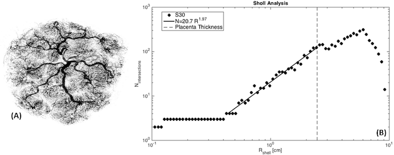
Recent advances have enabled a new wave of biomechanics measurements, and have renewed interest in selecting appropriate rheological models for soft tissues such as the liver, thyroid, and prostate. The microchannel flow model was recently introduced to describe the linear response of tissue to stimuli such as stress relaxation or shear wave propagation. This model postulates a power law relaxation spectrum that results from a branching distribution of vessels and channels in normal soft tissue such as liver. In this work, the derivation is extended to determine the explicit link between the distribution of vessels and the relaxation spectrum. The microchannel flow model explains the dramatic changes in tissue “stiffness” caused by changes in the vasculature, including rapid vasodilation or vasoconstriction.
A microchannel flow model for soft tissue elasticity
K. J. Parker
Phys Med Biol, vol. 59, no. 15, pp. 4443-4457 (2014). View Online
Journal Articles
- Mouse brain elastography changes with sleep-wake cycles, aging, and Alzheimer's disease
G. R. Ge, W. Song, M. J. Giannetto, J. P. Rolland, M. Nedergaard, and K. J. Parker
NeuroImage, vol. 295 , pp. 120662-1 -120662-10 (2024). View PDF - Brain elastography in aging relates to fluid-solid trendlines
K. J. Parker, I. E. Kabir, M. M. Doyley, A. Faiyaz, M. N. Uddin, and G. Schifitto
Phys Med Biol 69(11) , pp. 115037-1 -115037-16 (2024). View PDF - Limitations of curl and directional filters in elastography
K. J. Parker
Acoustics, vol. 5, no. 2 , pp. 575 -585 (2023). View PDF - Fluid compartments influence elastography of the aging mouse brain
G. R. Ge, J. P. Rolland, W. Song, M. Nedergaard, and K. J. Parker
Phys Med Biol, vol 68, no. 9 , pp. 095004-1 -095004-10 (2023). View PDF - Theory of sleep-wake cycles affecting brain elastography
G. R. Ge, W. Song, M. Nedergaard, J. P. Rolland, and K. J. Parker
Phys Med Biol, vol. 67, no. 22 , pp. 225013-1 -225013-16 (2022). View PDF - Comprehensive experimental assessments of rheological models’ performance in elastography of soft tissues
S. S. Poul, J. Ormachea, G. R. Ge, and K. J. Parker
Acta Biomaterialia, vol. 146 , pp. 259 -273 (2022). View PDF - A preliminary study of liver fat quantification using reported ultrasound speed of sound and attenuation parameters
J. Ormachea and K. J. Parker
Ultrasound Med Biol, vol. 48, no. 4 , pp. 675 -684 (2022). View PDF - The quantification of liver fat from wave speed and attenuation
K. J. Parker and J. Ormachea
Phys Med Biol, vol. 66, no. 14 , pp. 145011-1 -145011-10 (2021). View PDF - Fat and fibrosis as confounding cofactors in viscoelastic measurements of the liver
S. S. Poul and K. J. Parker
Phys Med Biol, vol. 66, no. 4 , pp. 04524-1 -04524-14 (2021). View PDF - Comprehensive viscoelastic characterization of tissues and the inter-relationship of shear wave (group and phase) velocity, attenuation and dispersion
J. Ormachea and K. J. Parker
Ultrasound Med Biol, vol. 46, no. 12 , pp. 3448 -3459 (2020). View PDF - Validations of the microchannel flow model for characterizing vascularized tissues
S. S. Poul, J. Ormachea, S. J. Hollenbach, and K. J. Parker
Fluids, vol. 5, no. 4 , pp. 228-1 -228-14 (2020). View PDF - Towards a consensus on rheological models for elastography in soft tissues
K. J. Parker, T. Szabo, and S. Holm
Phys Med Biol, vol. 64, no. 21 , pp. 215012-1 -215012-17 (2019). View PDF - Attenuation of shear waves in normal and steatotic livers
A. K. Sharma, J. Reis, D. C. Oppenheimer, D. J. Rubens, J. Ormachea, Z. Hah, and K. J. Parker
Ultrasound Med Biol, vol. 45, no. 4 , pp. 895 -901 (2019). View PDF - Shear wave propagation in viscoelastic media: validation of an approximate forward model
F. Zvietcovich, N. Baddour, J. P. Rolland, and K. J. Parker
Phys Med Biol, vol. 64, no. 2 , pp. 025008-1 -025008-13 (2019). View PDF - Group versus phase velocity of shear waves in soft tissues
K. J. Parker, J. Ormachea, and Z. Hah
Ultrason Imaging, vol. 40, no. 6 , pp. 343 -356 (2018). View PDF - The biomechanics of simple steatosis and steatohepatitis
K. J. Parker, J. Ormachea, M. G. Drage, H. Kim, and Z. Hah
Phys Med Biol, vol. 63, no. 10 , pp. 105013-1 -105013-11 (2018). View PDF - Analysis of transient shear wave in lossy media
K. J. Parker, J. Ormachea, S. Will, and Z. Hah
Ultrasound Med Biol, vol. 44, no. 7 , pp. 1504 -1515 (2018). View PDF - Are rapid changes in brain elasticity possible?
K. J. Parker
Physics in Medicine and Biology, vol. 62, no. 18 , pp. 7425 -7439 (2017). View Online - The microchannel flow model under shear stress and high frequencies
K. J. Parker
Physics in Medicine and Biology, vol. 62, no. 8 , pp. N161 -N167 (2017). View Online - Shear wave dispersion behaviors of soft, vascularized tissues from the microchannel flow, model
K. J. Parker, J. Ormachea, S. A. McAleavey, R. W. Wood, J. J. Carroll-Nellenback, and R. K. Miller
Physics in Medicine and Biology, vol. 61, no. 13 , pp. 4890 -4903 (2016). View Online - Biological effects of low frequency strain: physical descriptors
E. L. Carstensen, K. J. Parker, D. Dalecki, and D. Hocking
Ultrasound in Medicine and Biology, vol. 42, no. 1 , pp. 1 -15 (2016). View Online - Oestreicher and elastography
E. L. Carstensen and K. J. Parker
Journal of the Acoustical Society of America, vol. 138, no. 4 , pp. 2317 -2325 (2015). View PDF - Shear wave dispersion in lean versus steatotic rat livers
C. T. Barry, C. Hazard, Z. Hah, G. Cheng, A. Partin, R. A. Mooney, K. Chuang, and W. Cao
Journal of Ultrasound in Medicine, vol. 34, no. 60 , pp. 1123 -1129 (2015). View Online - What do we know about shear wave dispersion in normal and steatotic livers?
K. J. Parker, A. Partin, and D. J. Rubens
Ultrasound in Medicine and Biology, vol. 41, no. 5 , pp. 1481 -1487 (2015). View Online - Experimental evaluations of the microchannel flow model
K. J. Parker
Physics in Medicine and Biology, vol. 60, no. 11 , pp. 4227 -4242 (2015). View Online - Could linear hysteresis contribute to shear wave losses in tissues?
K. J. Parker
Ultrasound in Medicine and Biology, vol. 41, no. 4 , pp. 1100 -1104 (2015). View PDF - A microchannel flow model for soft tissue elasticity
K. J. Parker
Phys Med Biol, vol. 59, no. 15 , pp. 4443 -4457 (2014). View Online - Real and causal hysteresis elements
K. J. Parker
Journal of the Acoustical Society of America, vol. 135, no. 6 , pp. 3381 -3389 (2014). View Online - Physical models of tissue in shear fields
E. L. Carstensen and K. J. Parker
Ultrasound in Medicine and Biology, vol. 40, no. 4 , pp. 655 -674 (2014). View Online - Mouse liver dispersion for the diagnosis of early-stage fatty liver disease: a 70-sample study
C. T. Barry, Z. Hah, A. Partin, R. A. Mooney, K. Chuang, A. Augustine, A. Almudevar, W. Cao, D. J. Rubens, and K. J. Parker
Ultrasound in Medicine and Biology, vol. 41, no. 4 , pp. 704 -713 (2014). View Online - The Guassian shear wave in a dispersive medium
K. J. Parker and N. Baddour
Ultrasound in Medicine and Biology, vol. 40, no. 4 , pp. 675 -684 (2014). View Online - Shear wave dispersion measures liver steatosis
C. T. Barry, B. Mills, Z. Hah, R. A. Mooney, C. K. Ryan, D. J. Rubens, and K. J. Parker
Ultrasound Med Biol, vol. 38, no. 2 , pp. 175 -182 (2012). View Online - Congruence of imaging estimators and mechanical measurements of viscoelastic properties of soft tissues
M. Zhang, B. Castaneda, Z. Wu, P. Nigwekar, J. Joseph, D. J. Rubens, and K. J. Parker
Ultrasound Med Biol, vol. 33, no. 10 , pp. 1617 -1631 (2007). View PDF
