
Two eyes better than one in verifying unique phase of ice
Rochester researchers raise cautions about new approach to imaging biological samples
Ice occurs in at least 15 different “phases”—each with differing molecular geometries and densities. Of particular interest to biologists is amorphous (vitreous) ice, which lacks a regular pattern of molecules. Commonly scattered across vast distances in outer space, amorphous forms of ice created in labs on Earth have been used to preserve biological samples for imaging at cryogenic temperatures.
Especially exciting is a form of amorphous ice, called HDA, created at room temperatures by applying high pressures. The higher temperatures could greatly enhance the imaging of biological samples, by increasing the sharpness of the images and decreasing damage to the living samples.
However, researchers from six institutions, including Niaz Abdolrahim, assistant professor of mechanical engineering at the University of Rochester, and Ali Shargh, a PhD student in her lab, raise several flags of caution in a paper published in the Proceedings of the National Academy of Sciences (PNAS).
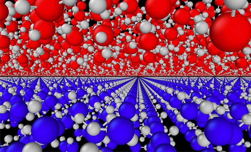
They discovered that the amorphous HDA ice they created at room temperature also included varying amounts of grains of a crystalline ice (Ice VI) as well, which could be detrimental to the biological samples.
Moreover, they determined that neither of the two imaging techniques commonly used in studying amorphous ice can, on its own, accurately determine the exact mix of HDA and Ice VI. Both Raman spectroscopy, which uses laser light to interact with molecular vibrations, and X-ray diffraction (XRD) spectroscopy, used to analyze crystalline structures, are required.
The findings were confirmed by molecular dynamics simulations conducted by Shargh, one of the lead authors.
“The take home message of the results, from both the experiments and simulations, is that this water is available as a coexistence of crystalline and amorphous states,” Shargh says. “We show that XRD can only detect the crystalline phases of this material, while Raman is able to capture the HDA.”
“If you stick with just one of these methods—either XRD or Raman—you end up with questionable results.”
The team was led by researchers at the University of Utah/Salt Lake City and also included researchers from the University of Nevada/Las Vegas, Argonne National Laboratory, University of Hawaii at Manoa, and the University of Illinois/Chicago.
“Major questions remain as to whether the structure of the biological membranes can survive in these conditions,” Shargh, Abdolrahim and their coauthors write in the paper. “Specifically, it remains to be investigated whether the small ice VI crystals... threaten the integrity of biological systems.”
Abdolrahim’s Advanced Computational Mechanics and Materials Laboratory also studies multiscale computational modeling and simulation of the nanomechanical properties of modern engineering materials where defects and interfaces control essentially the material’s macroscopic response.