
The New View of Tissue Scattering
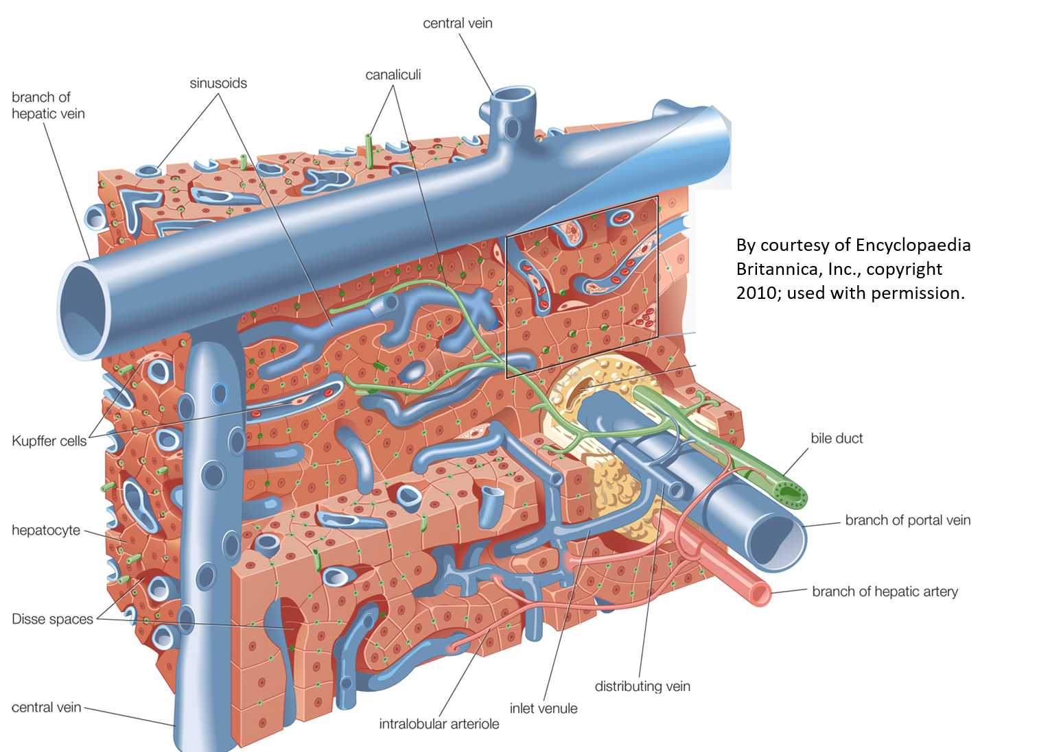
Do we really know what causes scattering of ultrasound from normal soft tissues such as the liver, thyroid, and prostate? Commonly, the answer is formulated around the properties of spherical scatterers, related to cellular shapes and sizes. However, our alternative view is that the closely packed cells forming the tissue parenchyma create the reference media, and the long cylindrical-shaped fluid vessels serve as the scattering sites. Assuming a weak scattering (Born approximation) for the vessels, in an isotropic, fractal branching pattern, some simple predictions can be made about the nature of backscatter as a function of frequency in soft tissues. We also have derived mathematical models for the first order statistics (the histogram) of echoes. These models are a major departure from the conventional views, but seem to capture the behavior seen in millions of B-scans obtained each year.
Journal Articles
- A power law reconstruction of ultrasound backscatter images
K. J. Parker
Acoustics, vol. 6, no. 3 , pp. 782 -791 (2024). View PDF - Burr distribution describes ultrasound speckle statistics of soft biological tissues
S. S. Poul, S. J. Hollenbach, and K. J. Parker
Proceedings, 2022 IEEE International Ultrasonics Symposium, doi: 10.1109/IUS54386.2022.9957933 , pp. 1 -4 (2022). View PDF - Power laws prevail in medical ultrasound
K. J. Parker
Phys Med Biol, vol. 67, no. 9 , pp. 09TR02-1 -09TR02-12 (2022). View PDF - Local Burr distribution estimator for speckle statistics
G. R. Ge, J. P. Rolland, and K. J. Parker
Biomed Opt Express, vol. 13, no. 4 , pp. 2334 -2345 (2022). View PDF - Generalized formulations producing a Burr distribution of speckle statistics
K. J. Parker and S. S. Poul
J Med Imaging, vol. 9, no. 2 , pp. 023501-1 -023501-15 (2022). View PDF - Burr, Lomax, Pareto, and logistic distributions from ultrasound speckle
K. J. Parker and S. S. Poul
Ultrasonic Imaging, vol. 42, no. 4-5 , pp. 203 -212 (2020). View PDF - Speckle from branching vasculature: dependence on number density
K. J. Parker and S. S. Poul
J Med Imaging, vol. 7, no. 2 , pp. 027001-1 -027001-12 (2020). View PDF - Liver backscatter and the hepatic vasculature's autocorrelation function
J. J. Carroll-Nellenback, R. J. White, R. W. Wood, and K. J. Parker
Acoustics, vol. 2, no. 1 , pp. 3 -12 (2020). View PDF - The first order statistics of backscatter from the fractal branching vasculature
K. J. Parker
J Acoust Soc Am, vol. 146, no. 5 , pp. 3318 -3326 (2019). View PDF - Shapes and distributions of soft tissue scatterers
K. J. Parker
Phys Med Biol, vol. 64, no. 17 , pp. 175022-1 -175022-15 (2019). View PDF - The 3D spatial autocorrelation of the branching fractal vasculature
K. J. Parker, J. J. Carroll-Nellenback, and R. W. Wood
Acoustics, vol. 1, no. 2 , pp. 369 -381 (2019). View PDF