
News & Events
A New High-Resolution Method for Imaging Below the Skin using a Liquid Lens
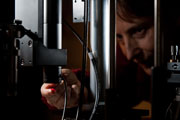 University of Rochester Optics and BME professor Jannick Rolland has developed an optical technology that provides unprecedented images under the skin's surface. The aim of the technology is to detect and examine skin lesions to determine whether they are benign or cancerous without having to cut the suspected tumor out of the skin and analyze it in the lab. Instead, the tip of a roughly one-foot-long cylindrical probe is placed in contact with the tissue, and within seconds a clear, high-resolution, 3D image of what lies below the surface emerges.
Rolland presented her findings at the 2011 annual meeting of the American Association for the Advancement of Science in Washington, D.C., on Feb. 19.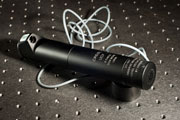
My hope is that, in the future, this technology could remove significant inconvenience and expense from the process of skin lesion diagnosis,
Rolland says. When a patient walks into a clinic with a suspicious mole, for instance, they wouldn't have to have it necessarily surgically cut out of their skin or be forced to have a costly and time-consuming MRIdone. Instead, a relatively small, portable device could take an image that will assist in the classification of the lesion right in the doctor's office.