Team:
- Amanda Adams
- Ahmet Gurcan
- Connor Heckman
- Gradi Bamfonga
- Ruyi (Lauren) Li
Mentor:
Dr. James McGrath
Customer:
Dr. Richard Burack
The Problem:
In histology, the process of mounting paraffin sections onto glass slides is one of the biggest limiting factors to allowing patients or researchers to receive their results in a timely manner. This is because the volume of tissue samples to be processed outweighs the capability of the technicians responsible for sectioning them. Thus, this has been deemed a rate-limiting step in the process of histology.
Our Goal:
This problem has led to the development of our device which aims to automate the mounting process in microtome histology. We hope to move the 5um thick section from the blade of the microtome onto a glass slide which is then ready for further processing such as staining. Additionally, we hope to achieve this with as little human intervention as possible.
Prototypes, Approach, and Challenges:
With the paraffin sections being around 5um thick on average, they are thinner than saran wrap. This poses a unique challenge in making them extremely delicate and sensitive to wrinkles. Also, because these tears and wrinkles greatly affect their viability for analysis, a method of transferring them without causing these issues had to be derived.

The above figure depicts the rough process outline involved with the device we intended to create.
Membrane Method:
The membrane method was one conclusion that our team came to when attempting to devise a solution to the problem of carefully transferring sections. This method involved the use of a water-soluble adhesive which we would adhere to the surface of the paraffin block. The block would then be cut with the paraffin adhered to the slice. The adhesive containing the section could then be stuck to a glass slide and immersed in water to remove the adhesive while keeping the section on the slide. It was believed that this would provide a stronger backing to the section and make it more durable.

The above figure depicts the rough process in which our team hoped to adhere the tape to the block, section it, and repeat.
Unfortunately, our team was unable to find an adhesive that could be completely removed from both the paraffin and glass slide’s surface. Therefore, the membrane method is not used in the final design.
Pre-wet Slide Method:
The pre-wet slide method is based on the observation that water is an integral factor in allowing the paraffin section to de-wrinkle. In this method, the glass slide would be loaded onto the white compartment shown in the figures below, water is applied onto the slide to catch the tissue section upon falling off from the blade.
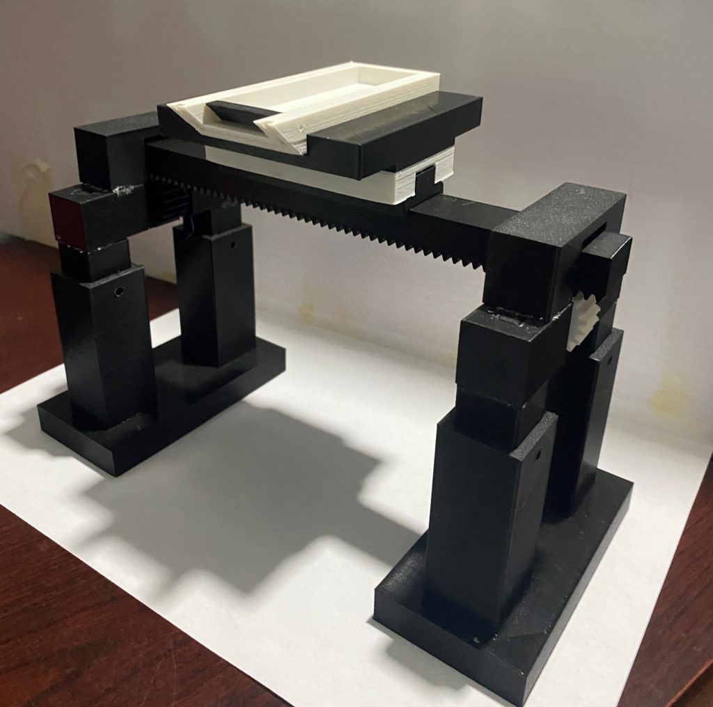
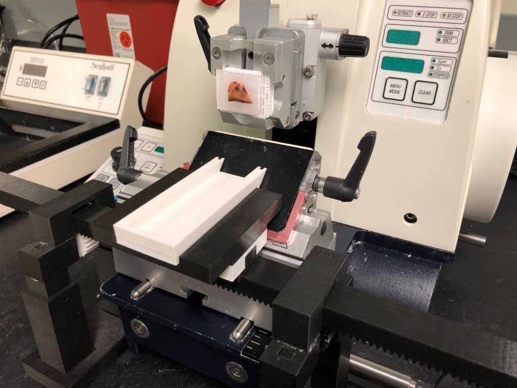
The figures above show our prototypes of the pre-wet slide method and its interaction with the microtome. The design was not used due to water leakage at the device interface with the microtome.
The Ramp:
The ramp is a crucial component of our pre-wet slide method. Its purpose is to not only move the section from the blade of the microtome into the boat but also to provide a smooth flow of water to prevent the section from curling or folding on itself.

The above depicts the first iteration of our ramp. The red block depicts the surface of the microtome which it interfaces with, while the gray part is our ramp. A hole in which water can be pumped into the ramp’s hollow cavity can be seen at the bottom. Additionally, the thin slit which can be seen at the top allows for water to smoothly flow out of the hollow compartment and down the surface of the ramp.
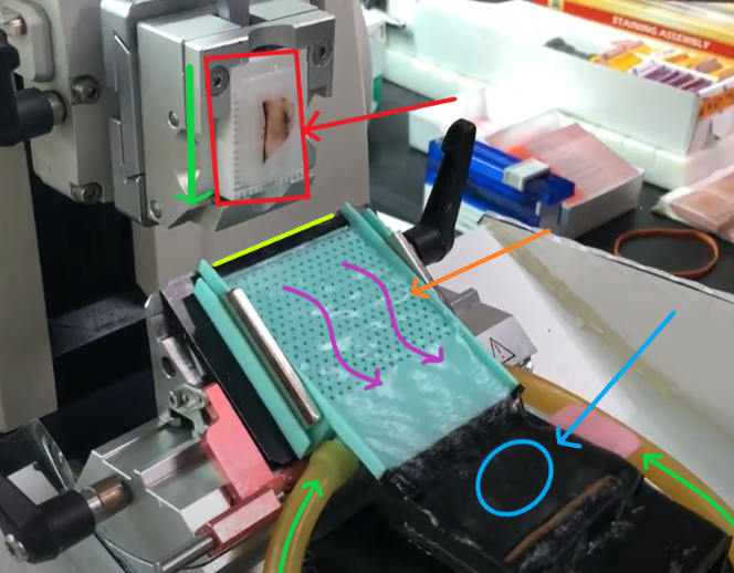
The above image shows our current turquoise-colored iteration of the ramp. It can be seen that we moved the water input to the bottom of the ramp to more evenly distribute the flow of water. Also added were magnets and slots for them to fit, which allow the ramp to seamlessly attach and detach to the metal surface of the microtome. In addition to the thin slit at the top of the ramp which was in the previous iteration, small holes in the ramp’s surface were added to more evenly distribute the flow of water.
The Boat:
What our team refers to as the boat component of our device is another integral part. This piece is responsible for holding the glass slide and catching the paraffin section after it flows down the surface of the ramp.

The above picture depicts the first prototype for our boat. The bottom of the boat would contain the glass slide and slits which allow the water to slowly drain and thereby delivering the section to the surface of the slide.

The above image shows our current boat. Slits in which the slide are inserted can be seen on both sides. Additionally, holes were made at the top to allow water to drain so as not to allow overflowing which could push the section over the top edges.
Our Current Device:
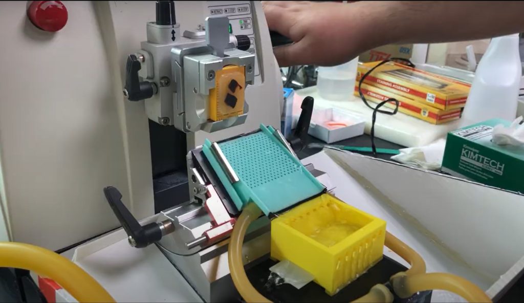
Our device currently consists of the waterslide (mint green), the boat (yellow), as well as rubber tubing and water collection box (white).
Future Directions:
For the future of our device we hope to further automate our design. This could potentially include adding a conveyor belt to move new boats into position while carrying boats which already contain a section to a heated element.
Additionally, our team envisions expansions into other markets. As it stands, research labs are only a small portion of the possible population that would benefit from our device. We are looking to adapt our device so that it may be used in clinical, clinical trial, and reference labs.
Acknowledgments:
TA:
Abbi Miller
Professors:
Dr. Amy Lerner
Dr. Scott Seidman
Special Thanks:
Philip Rock – Tissue Bank Technician, UR Medical Center
Mary Georger – Senior Technical Associate, Wilmot Cancer Institute Histopathology Core
Sandy Piampiano- Staff Accountant, UR Medicine Labs
Richard Burack, M.D., Ph.D.
Mert Gurcan – Edited teams Design Day video