Author
Henry Glover, University of Rochester, Undergraduate Student Class of 2026
Email: hglover@u.rochester.edu
Advisor
Dr. Terry-Ann Suer, Laboratory for Laser Energetics, Research Scientist
Collaborators
Dr. Samuel Crossley, University of Arizona, Research Scientist
Dr. Khanh Kieu, University of Arizona, Professor
Alexander Sentowski, University of Rochester Medicine, Graduate Student
Dr. Michael Giacomelli, University of Medicine, Professor
Summary
The use of Diamond Anvil Cell (DAC) is a well-established technique to apply high pressure to various materials. By utilizing DAC samples as targets for shot experiments at the OMEGA laser facility, this would help better understand various celestial objects like Jupiter or Stars by replicating similar high temperature and pressure conditions imposed on them in a laboratory setting. However, there are many interesting effects under high pressure inside Diamond Anvil Cell chambers that we cannot observe with a conventional scientific microscope.

The goal of this research is to address this problem by utilizing this powerful technique of multiphoton microscopy to produce high resolution 3D images of Diamond Anvil Cell samples, as well as better understand various localized material properties under pressure. By doing this, experiments were done with the goal of observing various pressure effects through multiphoton microscopy 3D images. So far, multiphoton microscopy is promising and has a lot of potential, as images from multiphoton microscopy experiments were successfully made for samples under pressure.
If you have any questions about this research or want to learn more about our research group, please do not hesitate to contact me.
Why Multiphoton Microscopy?
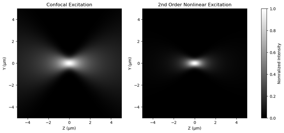
The benefit of multiphoton microscopy is that the excitation area is smaller compared to confocal microscopy as shown in the picture below. This enhances spatial and depth resolution by minimizing interference from off-focus scattering, in contrast to confocal microscopy, which is typically limited by the effective imaging depth less than 100 microns [1].
Recent research led by Sepehr Benis has shown that collecting emissions from three photon absorption can be used to characterize semiconductor bandgap energy [2]. The use of 3rd order nonlinear emissions allows the use of higher wavelength, near-infrared (NIR) laser source. This achieves greater depth, high resolution signal, compared to lower wavelength sources, as NIR laser sources are typically not affected by material bandgap absorption. Experimental results from this research has shown that there is a strong correlation between three photon absorption coefficient and bandgap energy.
One of the goals of this project is to see if images by multiphoton microscopy could contain information about the sample’s localized bandgap properties under high pressure inside diamond anvil cells by imaging 3rd order nonlinear emissions.
Experimental Setup
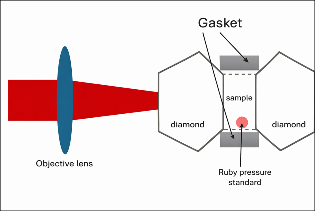
Multiphoton microscopy optical systems would split the image into multiple channels, each of which would collect second harmonic generation (SHG), two photon excitation fluorescence (2PEF), third harmonic generation (THG), and three photon excitation fluorescence (3PEF). As described in Figure 3, the objective lens in the optical system would focus the fundamental laser into the DAC chamber, where the sample would be located. The fundamental laser source is at near-infrared wavelength (1040 nm).
Results from Experiments
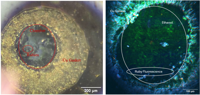
One of the samples selected for experiments at University of Arizona is Ethanol because it is widely available and well known under pressure. The goal with Ethanol is to observe pressure-induced phase change effects from liquid to solid through multiphoton microscopy, which occurs at around 2 GPa [3]. Images of the Ethanol sample were successfully captured at a pressure of 3 ± 1 GPa, which is beyond the Ethanol solidification pressure.
Red: SHG, Green: THG, Blue: 2PEF, Cyan: 3PEF
Another sample selected for experiments at University of Rochester Medical center is alpha-Quartz because it is also widely available and well known under pressure. Additionally, as a result of birefringent crystal properties, quartz has stronger SHG emissions. The goal with Quartz is to observer pressure-induced phase change effects from Quartz I to Quartz II, which occurs at around 22 GPa [4]. So far, images of Quartz inside Diamond Anvil Cell at ambient pressure was captured as a baseline before compression.
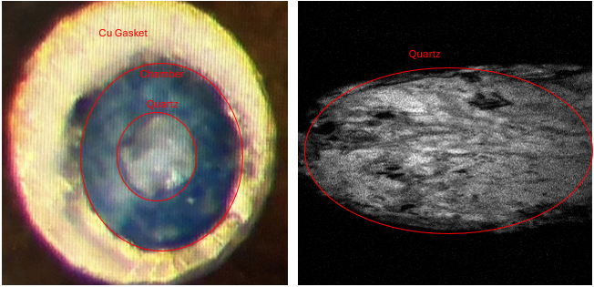
Note: Multiphoton microscopy image is distorted because of digital and alignment error
Acknowledgements
I would like to thank Dr. Terry-Ann Suer for the mentorship in this project. I would also like to thank the following collaborators from University of Arizona Wyant School of Optical Science (Samuel Crossley, Khanh Kieu) and University of Rochester Medicine (Alexander Sentowski, Michael Giacomelli), for allowing us to use their multiphoton microscopy system and for their assistance. Additionally, I would like to thank Prof. Wayne Knox for lending a microscope objective lens for this project and for his assistance in this senior thesis project.
This work is supported by the U.S. Department of Energy National Nuclear Security Administration under Award No. DE-NA0004144, the University of Rochester, and the New York State Energy Research and Development Authority.
References
[1] Hye Jin Cho, Hoon Jai Chun, Eun Sun Kim, and Bong Rae Cho, “Multiphoton microscopy: An introduction to gastroenterologists”, World Journal of Gastroenterology 17, no. 40 (2011): 4456–4460[2] Benis, S., Cirloganu, C. M., Cox, N., Ensley, T., Hu, H., Nootz, G., & Van Stryland, E. W. (2020). Three-photon absorption spectra and bandgap scaling in direct-gap semiconductors. Optica, 7(8), 888–899.
[3] Kondrin, M. V. (2014). Crystallization and vitrification of ethanol at high pressures. The Journal of Chemical Physics, 141(19), 194504.
[4] D. A. Pinnick et al., “Optical second-harmonic-generation study of quartz up to 31 GPa,” Physical Review B 55, no. 21 (1997): 14115. D. A. Pinnick et al., “Optical second-harmonic-generation study of quartz up to 31 GPa,” Physical Review B 55, no. 21 (1997): 14115