Abstract
Mold growth on silicone mouthpieces of water bottles is a frequently reported issue in consumer feedback. This study investigates the effects of silicone composition and zinc oxide (ZnO) nanoparticle additives on bacterial growth using Micrococcus luteus as a standardized model organism. Silicone coupons were fabricated from multiple commercial formulations and evaluated using a controlled “sandwich method,” with bacterial coverage quantified via image analysis.
Results show that baseline silicone composition significantly influences microbial adhesion, with Xiameter RBL-2004-40 exhibiting the lowest bacterial coverage. Contrary to expectations, higher ZnO concentrations increased bacterial growth, while low loading (0.1 wt%) consistently reduced coverage. This trend is attributed to nanoparticle agglomeration at higher concentrations, limiting effective antimicrobial activity.
Introduction
Project Objective and Motivation
Developing a mold-resistant silicone for Nalgene water bottle mouthpieces to eliminate microbial biofilm growth and meet food safety standards.
Voice of Customer complaints surrounding competing water bottles oftentimes city mold growth and hygiene as customers’ primary complaints, thus providing a motivation for exploring methods to prevent mold growth in this project.
The Substrate
The baseline material is a high-consistency silicone rubber that is hydrophobic but susceptible to mold growth on the surface. It is used by Thermo Fisher in their Nalgene Pop-top bottle mouthpieces and gaskets.
Bio-Film Formation Steps
1. Conditioning Film Formation
Upon repeated use, salivary proteins (such as mucins, cystatin, and statherin) adhere to the silicone surface. This creates a “conditioning film” that masks the silicone’s hydrophobicity and provides a nutrient-rich anchor for microbes.
2. Microbial Succession
Pioneer Species such as Candida albicans (yeast) typically colonizes the surface first, attaching to the protein layer, creating a structural scaffold.
This scaffold allows secondary colonizers, specifically Aspergillus brasiliensis (black mold), to embed and anchor onto the silicone
3. Biofilm Protection
The mature colony secretes Extracellular Polymeric Substances (EPS). This sticky matrix of DNA and polysaccharides shields the mold from standard cleaning, leading to the persistent “black spots” reported in consumer products.
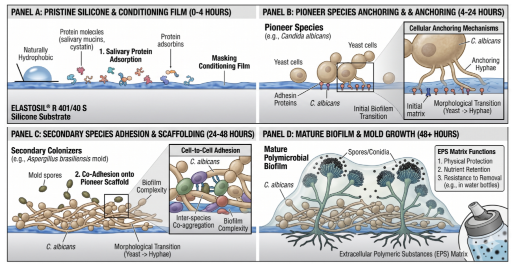
Figure 1 Mechanisms of Bacterial and Fungal Biofilm Development on Hydrophobic Silicone
Inoculation Methods
Pilot Phase
Baker’s Yeast (Saccharomyces cerevisiae) → surrogate for Candida albicans
Bread Mold (Rhizopus stolonifer) → surrogate for Aspergillus brasiliensis
- Mouthpieces dip-coated in sucrose/yeast nutrient solution
- Result: Proved biofilm formation possible; high variance observed
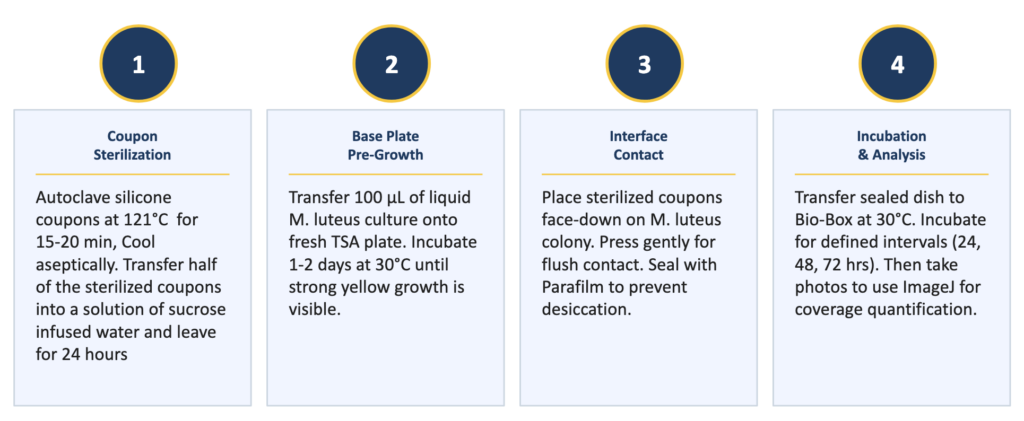
Figure 2 Experimental Phase Steps
Experimental Phase (Standardized Model)
For absolute quantitative control, we transitioned to a single, controlled strain: Micrococcus luteus (ATCC 4698). This non-pathogenic (BSL-1) strain is commonly found on human skin and in the mouth, making it a relevant representation of mouthpiece contamination.
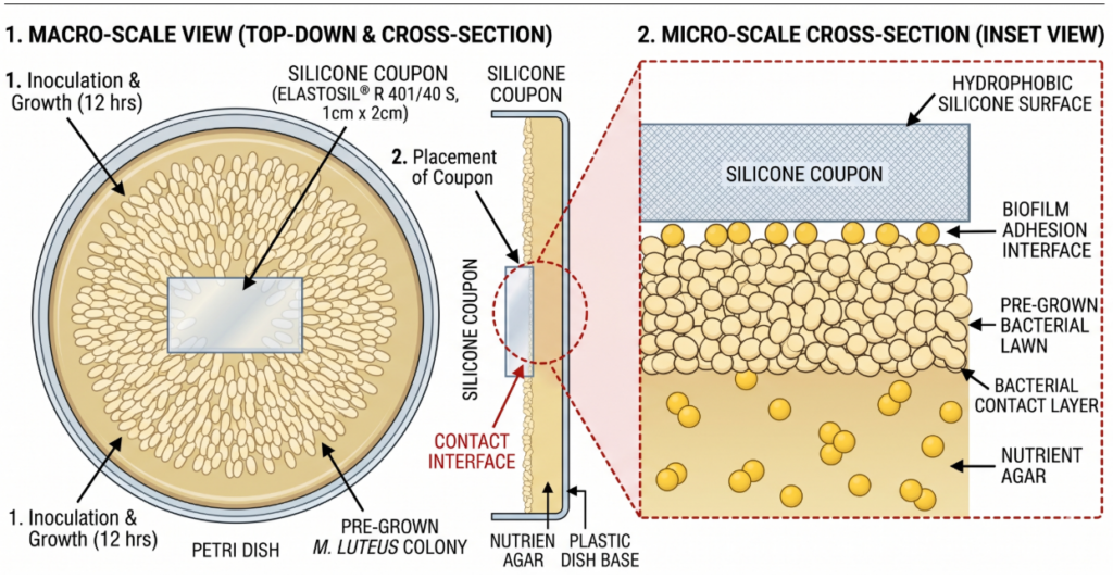
Figure 3: Schematic for Standardized Sandwich Method
The “Sandwich Method”
To improve reproducibility over liquid exposure, we implemented a face-to-colony contact method. M. luteus was pre-grown on agar, after which silicone coupons were placed directly onto the colonies to observe the growth onto the silicone surface.
Testing Methods
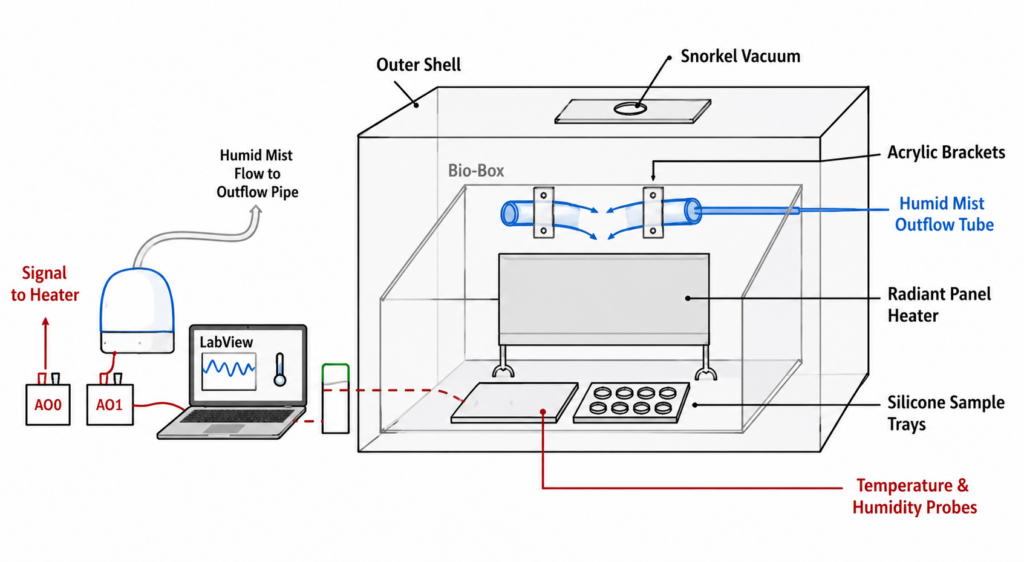
Figure 4 “Bio-Box” Accelerated Testing Chamber Setup
Accelerated Testing Chamber “Bio-Box”
- Long timelines in the pilot phase motivated the development of an accelerated testing environment
- This environment maintained high humidity (65%) and temperatures (30°C) to mimic the environment of a water bottle with its lid closed
- Primary features include P/I control, radiant heating, cool misting, and a snorkel vacuum for safety purposes
Silicone Fabrication Development

Figure 5 Vacuum Oven and Fabrication Schematic
- 1.5” square silicone coupons of the ELASTOSIL R 401/40S™, Silastic RBL-9200-5™ and Xiameter 2004-40™were produced for our testing
- Zinc oxide was incorporated at 0.1, 1, and 5 weight percent to test the effectiveness of anti-microbial additives.
- Silicone mixtures were spread into uniform 3 in x 4.5 inch layers on a baking sheet to produce 6 coupons per mixture. These flat sheets were degassed in a vacuum oven and thermally cured at 120°C for 15 minutes.
Coverage Analysis with ImageJ
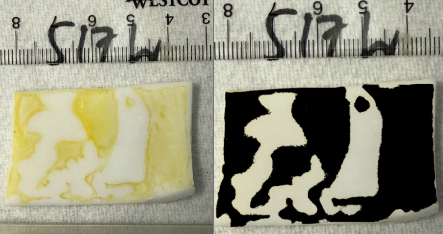
Figure 6: ImageJ process before (left) and after (right) showing 50.24% coverage.
- ImageJ software was used to measure the percent coverage on the samples.
- Sample images are uploaded with a known distance to calibrate the scale.
- Regions of the sample with bacterial coverage are outlined.
- ImageJ calculates the area of regions of interest.
Results and Discussion
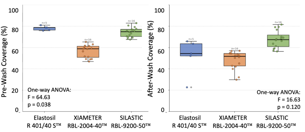
Figure 7 Pre- and After-wash % Bacterial Coverage of Undoped Silicone Samples
Silicone Type (one-way ANOVA)
- Significant effect of material (p = 0.03)
- Elastosil: Highest percent coverage (85% unrinsed, 75% rinsed)
- Silastic 9200: Variable coverage similar to Elastosil baseline. (80% unrinsed, 70% rinsed)
- Xiameter 2004: Lowest and most consistent percent coverage. (58% unrinsed, 49% rinsed)
Effect of ZnO (two-way ANOVA)
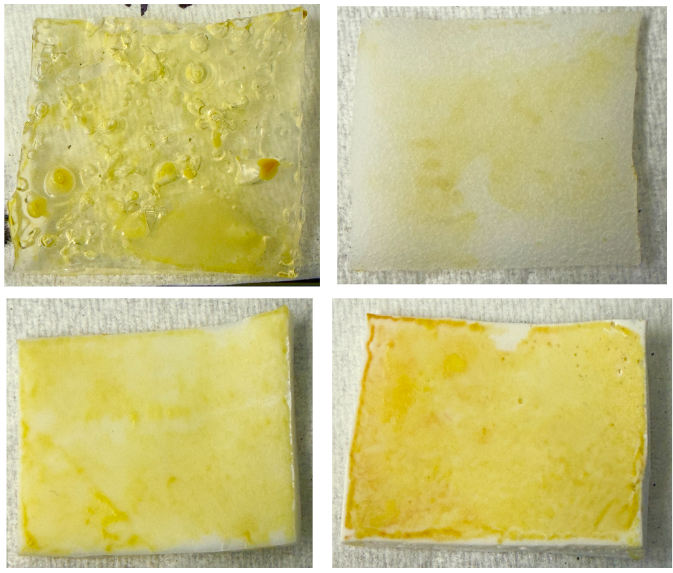
Figure 8 Baseline Silicone with 0%, 0.1%, 1%, and 5% wt. ZnO
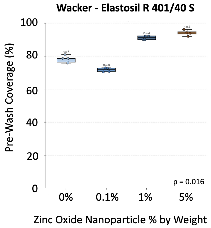
Figure 9 Elastosil™ % Coverage by Varying % wt. ZnO
- ZnO significantly reduces percent coverage (p = 0.042).
- Effect is beneficial overall but not uniform in magnitude.
Primary Considerations
Across all materials, rinsing lowered the percent coverage but also added statistical variation that needs to be further explored. For the unmodified silicones, Elastosil and Silastic 9200 showed relatively high coverage, while Xiameter 2004 demonstrated the best mold resistance due to the lowest coverage of M. Luteus.
- With ZnO additives, 0.1 % wt. showed the best performance, but increasing concentrations beyond that negatively impacted the mold resistance.
Future Work
Our preliminary data suggest a relationship between increased surface roughness and greater mold growth, indicating surface topography may play a meaningful role in microbial adhesion
- In particular, the rougher samples exhibit pronounced valleys and micro-scale features that can:
- Promote liquid pooling
- Reduce exposure to shear forces
- Provide protected sites for spore attachment, growth
Future work should focus on engineering uniform surface structures that mitigate these effects. Approach:
- Controlled nanotexturing to shift the surface toward a Cassie-Baxter wetting regime
- Use of a silicon wafer with nanotexture as a “soft lithography” methodology
- AFM and EDAX for better understanding of surface roughness and distribution
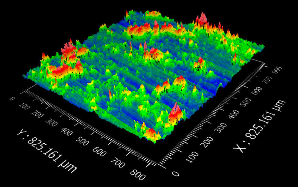
Figure 10: 2.5 wt% ZnO Profilometery
Acknowledgments
We would like to thank:
Mr. Henry Davignon, Thermo Fisher Scientific
Dr. Martin Pavelka, Department of Microbiology and Immunology, UR Medicine
Dr. Sonia Rosenberger, Environmental Health and Safety
Prof. Gang Fan, University of Rochester Department of Chemical Engineering
Tarek Rafeedi, Dominic Lippa, and Elena Perez
Prof. Doug Kelley, Prof. Mark Juba, Prof. Wayne Griffin, Lena Lederman, Dominic Lippa, Clair Cunningham, Mason Garlatti, and Elena Perez
Wacker Chemie, RD Abbott, and Dow Chemical
and our advisors, Prof. Melodie Lawton and Lena Lederman