Team Zeiss: Allen Wu, Zehui Yuan, Yepeng Zhou
Customer: Annie Isaac (Zeiss)
Faculty Advisor: Len Zheleznyak
Senior Design, Institute of Optics, University of Rochester
PROBLEM STATEMENT
This project aims to develop and model solutions to improve peripheral image quality in wide-field retinal systems. The goal of this project is to ensure high image quality over a wide range of human eyes with varying anatomical geometries. The outcome will provide modeling tools and recommendations for ZEISS to improve wide-field imaging quality in an OCT retinal imaging system.
BACKGROUND
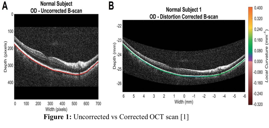
OCT: An Optical Coherence Tomography (OCT) system is used in this project for wide field imaging on the human eye. OCT uses near infrared light to collect scattered wavelengths of light for image resolving on biological tissue, such as the eye. While a system may have aberration, it is possible to correct some of the aberration, such as distortion after data collection, allowing for a clearer image.
OCT is typically low coherence broadband light, with light splitting and hitting both a sample (sample arm) and a mirror (reference arm), before recombining. This allows for equal beam path length, resulting in an interference pattern, which can be detected.
Aberrations: The human eye has inherent aberrations, with different aberrations having different levels of effect on the eye. While most aberrations get worse with a larger field, for the wide-angle measurements of the eye (past 40 degrees), typically astigmatism becomes the worst aberration affected by field. Aberrations such as spherical and defocus are not affected by field, and so they stay constant regardless of the field of view. On the other hand, aberrations such as coma, astigmatism, petzval, and distortion get worse with a larger field. While there are many studies on the aberrations of the eye in a small field, as the field gets larger, there are less studies done, resulting in less data, less models, and lower accuracy. Therefore, while modelling and optimizing a system on a wider field on the eye is possible, it may not be exactly true to the real eye at a larger field.
METHODS
Our workflow starts with the original OCT design and replaces the eye model in Zemax. After setting up different field configurations, we optimize the OCT optics for the target FOV while keeping the retinal imaging performance as the main evaluation metric. The optimized designs are then compared performance.
EYE MODELS
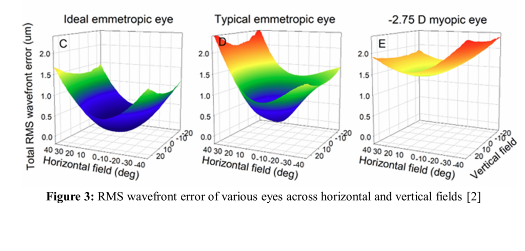
Differences in people’s eyes results in difficulty in modeling eyes, since each one will have different variables. In addition to this, there is very limited amounts of data available for wide field eye modeling and aberrations (past 40 degrees). The result of this is higher difficulty in modelling the eye correctly at larger angles. Within 40 degrees, however, there is more data available, such as in a paper published by Hastings et al. (2024) on wide-field optical eye models, where they discuss wavefront error and aberrations of myopic and emmetropic eyes for a horizontal field of -40 to 40 degrees.
RESULTS
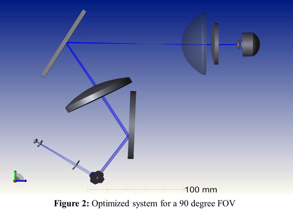
Two designs were developed in this project: a 90° FOV optimized design for improved RMS spot radius over various eye models, and a 110°+ FOV design for extended wide-field performance. The comparison highlights the trade-off between reducing the spot size at a specific field angle and maintaining image quality at higher FOV.
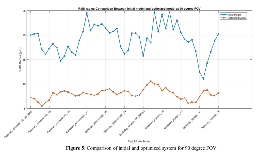
The 90° optimized design reduced the average RMS spot radius at 0° to 90° FOV from 16.9 μm to 7.8 μm, while the 110°+ optimized design maintained acceptable average spot radius performance of 20μm at higher field angles up to 120°. This comparison shows the trade-off between minimizing spot size at the target field and maintaining robust wide-field performance.
CONCLUSIONS
This project demonstrates a Zemax-based modeling approach for evaluating and optimizing wide-field retinal imaging performance using anatomical eye models. Two optimized designs were compared to the initial design a 90° FOV design that significantly reduced RMS spot radius over the various eye models, and a 110°+ FOV design that maintained reliable imaging performance. The results show a clear trade-off between minimizing the spot size at field angles and maintaining robust imaging quality across wider field of views. Future work should focus on improving high-FOV corrections across more anatomical eye models, and further correcting the peripheral eye aberrations while preserving ray transmission to the retina in this optical system.
REFERENCES
[1] McNabb, Ryan P et al. “QUANTITATIVE TOPOGRAPHIC CURVATURE MAPS OF THE POSTERIOR EYE UTILIZING OPTICAL COHERENCE TOMOGRAPHY.” Retina (Philadelphia, Pa.) vol. 41,4 (2021): 804-811.
[2] Hastings GD, Tiruveedhula P, Roorda A. Wide-field optical eye models for emmetropic and myopic eyes. J Vis. 2024 Jul 2;24(7):9.
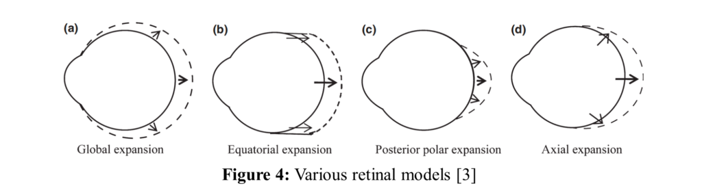
[3] Verkicharla PK, Mathur A, Mallen EA, Pope JM, Atchison DA. Eye shape and retinal shape, and their relation to peripheral refraction. Ophthalmic Physiol Opt. 2012 May;32(3):184-99. doi: 10.1111/j.1475-1313.2012.00906.x. Epub 2012 Apr 9. PMID: 22486366.