Introduction
A patient is eligible for a prostatectomy when their prostate is in an enlarged or cancerous state. The gold-standard for prostate removal is a nerve sparing prostatectomy. Laparoscopic prostatectomies require precise manipulation of the prostate while preserving delicate neurovascular structures that are essential for continence and sexual function. Often during surgery, the neurovasuclar bundles are damaged and cause harm to the patient. The neurovascular bundles are located posterolaterally to the prostate and are surrounded by adipose tissue.

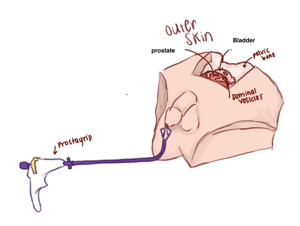
To avoid nerve damage, LSI Solutions® has developed the PROSTAGRIP™ device. This device travels transurethrally to provide the surgeon with precise manipulation of the prostate, which allows for better visualization of the neurovascular bundles. There is currently no dedicated physical simulator that clearly demonstrates how the PROSTAGRIP™ functions within relevant anatomy, limiting effective training and device demonstration.
The PROSTAGRIP™ device travels through the external urethral meatus through the urethra into the prostate. Once in the prostate, the grippers are expanded and the prostate can be rotated.
Problem Statement
The goal of this project is to create a functional surgical simulator to demonstrate LSI Solutions®’ PROSTAGRIP™’s transurethral manipulation of the prostate during prostatectomies to increase exposure of the neurovascular bundles. This phantom accurately represents the male pelvic anatomy and allows medical professionals to visualize key procedural steps involving the PROSTAGRIP™ device in a controlled, non-clinical environment.
Use Scenario
The surgical simulator is a desktop model that depicts a complete laparoscopic prostatectomy. The model is anatomically accurate and has organs that are representative of the average adult male, but it does not need to have all the tissues that are present in the human body. The surgical model will be used in a “dry” environment and does not require a sterile surgical field.
To use the surgical simulator, the PROSTAGRIP™ is inserted through the external urethral meatus and travel through the urethra to the prostate. Then, the grippers are expanded to hold the prostate in place as it is excised from the simulator. Once the prostate is removed, LSI Solutions®’ RD® and Ti-Knot® devices are used for the anastomosis between the urethra and bladder. The surgical simulator can be used by potential PROSTAGRIP™ users to experience how the device streamlines a prostatectomy.
Our Surgical Simulator

Our design for the surgical simulator includes external and internal systems. The external system consists of the outer shell of the mannequin, penis, and viewing window/laparoscopic cover. This system is durable so it can withstand repeated simulations during demonstrations. The internal system includes the anatomy of the male pelvic region: prostate, bladder, seminal vesicles, urethra, and pelvis. The internal system is supported by expandable foam which allows for the organs to be supported and held in place.
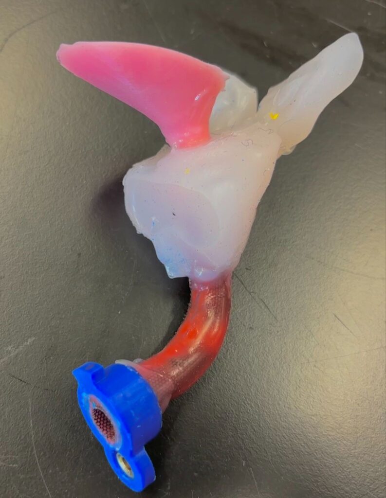
The main subsystem is the interchangeable system. This system includes the bladder, prostate, seminal vesicles, and urethra. The surgical site is supplemented with power mesh to ensure the silicone can hold a suture. To replace this system after a simulation, there are two quick connection points: one between the bladder and pelvis and the other between the urethra and penis. These connection points allow for an easy and fast reset process between simulations.
Prototyping process
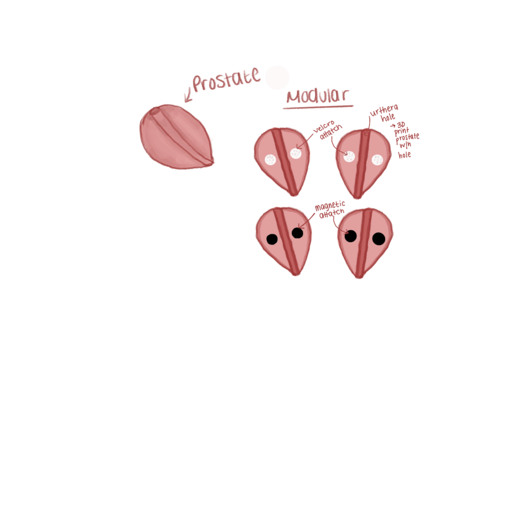

Prostate-Urethra Connection
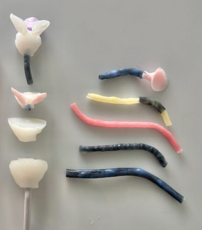
The prostate must be modular and removable after anastomosis. The first prototype of the prostate was a magnetic connection between the halves of the prostate. The next development used silicone epoxy to adhere the prostate and urethra. Currently, the prostate and urethra are adhered with silicone during the prostate curing process. This allows for the Ti-KNOT® and RD® devices to perform the anastomosis. By connecting the urethra and prostate while the prostate is curing, an assembly step is simplified.
Urethra Tube Suturability
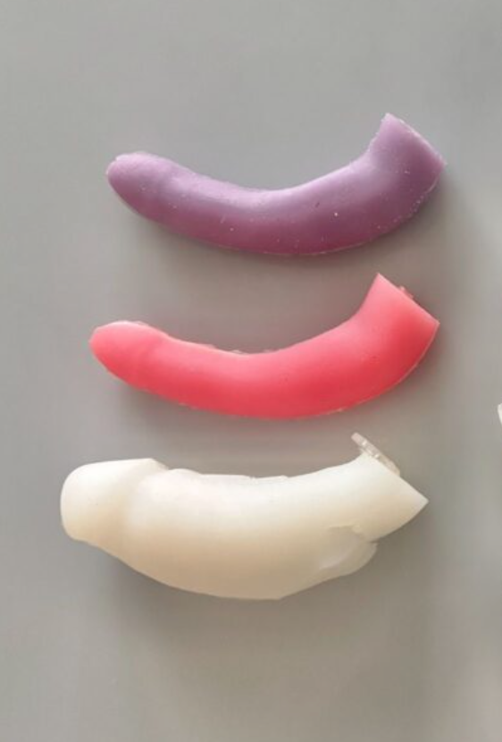
The urethral tube needs to be able to hold a suture to perform an anastomosis. Our first urethra was a silicone straw that was purchased. This first prototype allowed for the PROSTAGRIP™ to travel to the prostate, but it did not allow the PROSTAGRIP™ to expand. Next, we used a mesh tube and heat shrink fabric coated in Dragon SkinTM FX-ProTM. Both of these options did not allow for the PROSTAGRIP™ to expand, but were able to hold a suture. Our final prototype is a power mesh tube layered with the Dragon SkinTM FX-ProTM. This version allows for the urethra to expand and hold a suture.
Bladder Connection
First, the whole silicone bladder was attached to the prostate via silicone epoxy. Then, we split the silicone bladder in half with one side attached to the prostate via silicone epoxy, and other side suspended to the pelvis. Our third iteration, had the superior half of the bladder 3D printed and the inferior half molded out of silicone. These pieces were connected via velcro, which allowed for a consistent connection between the parts. However, the connection took up too much space in the pelvic cavity, so the current model is a hollow silicone bladder with an elastic embedded to attach to the pelvis.
Penis-Urethra Connection
The penis and the urethra were connected via magnets, then an acrylic plate with screws. The final design uses a 3D printed magnetic twist lock.
Testing and Validation
In order to create a urethra that can withstand anastomosis, we tested the mechanical properties of Dragon Skin™ FX-Pro™ and Dragon Skin™ FX-Pro™ with power mesh embedded. These tests prove that power mesh increases the yield stress of the silicone. We performed tensile tests to compare the stress/strain curves of the silicone with and without power mesh embedded. Tensile testing was performed on the samples with and without sutures.

Figure 1. Figure 1 depicts the stress versus strain graphs of the Dragon Skin™ FX-Pro™ with and without power mesh embedded. The results are inconclusive due to slipping.
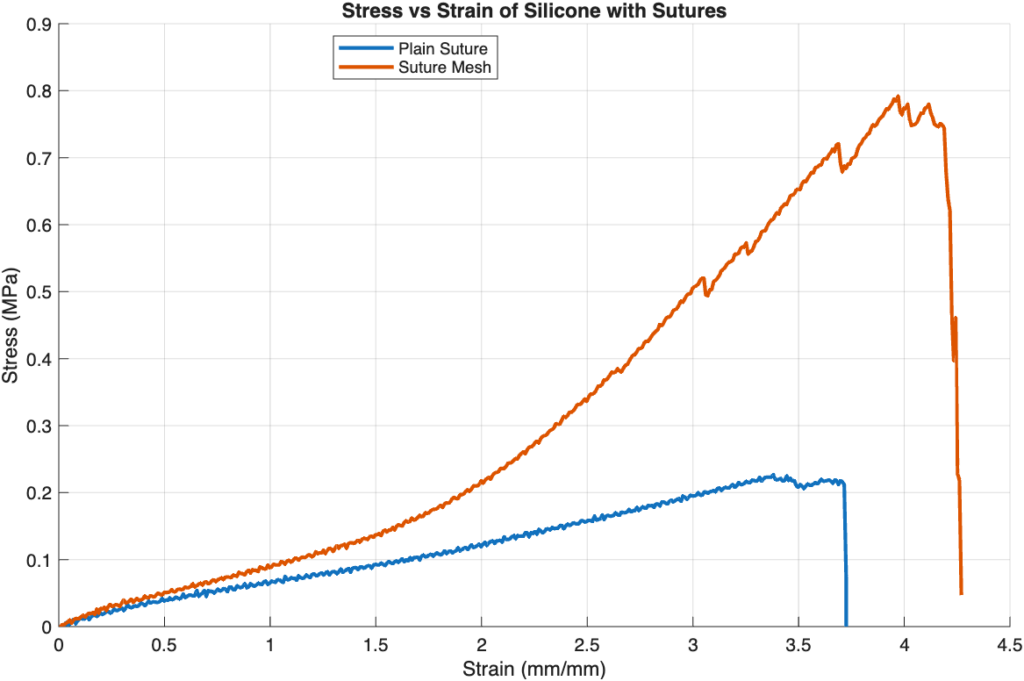
Figure 2. Figure 2 compares the stress and strain of the samples with four sutures embedded in each. This demonstrates a much higher yield stress for the sample with power mesh embedded.
Final Output
Interchangeable Surgical Site System:
This model includes a removable surgical site that includes portions of the urethra, prostate and bladder. This allows for the prostate to be modular in size for different clinical scenarios and enables the use of the Ti-KNOT® and RD® devices. These devices along side laparoscopic scissors perform an anastomosis. After each customer executes this simulation, the system can be fully replaced.
Aspects of this system: (superficial to deep)
- Magnetic lock
- Suturable Urethra
- Prostate & Seminal Vesicles
- Bladder
External System:
The exterior of the phantom depicts the male lower torso, set with the laparoscopic ports, a viewing window, and external male anatomy. This allows for the RD® and Ti-KNOT® devices, a laparoscopic camera, laparoscopic scissors, and laparoscopic graspers to be used in the simulator.
Final Assembly:
The final design assembly started with adding foam as internal support and placing the pelvis in the foam. Then, the penis is threaded through a hole in the mannequin. From here, the interchangeable system is connected to the permanent system via the penis and pelvis to complete the simulator.
Video of Surgery:
Design Team

Maddie Weigle
Concentration:
Cell & Tissue Engineering
Sophie Weigle
Concentration:
Cell & Tissue Engineering
Sofia Heferle
Concentration:
Biosignals & Biosystems
Kai Hua Liu
Concentration:
Biomechanics
Acknowledgments
We would like to thank Dr. Greg Gdowski, Michelle Eldred, Mitchell Hoestermann, Martin Gira, Jim Alkins, Dr. Scott Seidman, Dr. Benjamin Castañeda, John Moyer, Tessa Ooyama, and the rest of the LSI Solutions® Team for lending their support for this project.